About this course:
This course reviews the pathophysiology, clinical manifestations, diagnosis, complications, and treatment of various throat conditions, including group A Streptococcus (GAS).
Course preview
This course reviews the pathophysiology, clinical manifestations, diagnosis, complications, and treatment of various throat conditions, including group A Streptococcus (GAS).
Upon completion of this course, learners will be prepared to:
- identify common causes of acute pharyngitis
- examine the occurrence of pharyngitis in the United States
- discuss the risk factors for the various types of pharyngitis
- describe the current diagnostic standards for the various types of pharyngitis
- discuss the appropriate treatment and follow-up for various types of pharyngitis
- summarize the best practices for preventing various types of pharyngitis in pediatric and adult populations
- discuss the potential complications for the various types of pharyngitis
Pharyngitis is an acute inflammation or infection of the mucous membranes of the oropharynx that is typically caused by a bacterial or viral infection. Acute pharyngitis is one of the most common conditions healthcare providers (HCPs) encounter in the outpatient setting, ranking among the top 20 most common primary diagnoses. Approximately 12 million outpatient health care visits annually in the United States are attributed to acute pharyngitis. In addition, acute pharyngitis peaks in childhood/adolescence, with about half of all cases occurring before age 18. The incidence among adult patients declines after age 40. Several viruses and bacteria can lead to acute pharyngitis, but a common cause is Streptococcus pyogenes (S. pyogenes), also known as group A Streptococcus (GAS) or group A strep. This bacterium can live on human skin and the throat without causing illness, also known as colonization. Infected or colonized individuals are the source of nearly all infections. GAS is a significant source of community-acquired strep throat. In addition, GAS is the most common bacterial cause of acute pharyngitis, responsible for approximately 5% to 15% of adult and 20% to 30% of pediatric cases of acute pharyngitis. In the US, the estimated costs of GAS infections are between $224 and $539 million annually. The direct and indirect costs of recurrent infections include medical visits, medications, and the loss of work and school for the child and their caregiver(s). Misuse of antibiotics for viral pharyngitis or overuse of antibiotics for GAS can lead to resistant organisms. Many children with recurrent episodes of GAS pharyngitis may have indications for a tonsillectomy, as this is one of the two most common reasons for this procedure (Buensalido, 2023a; Centers for Disease Control and Prevention [CDC], 2025c; Chow & Doron, 2026; Goldin & Graber, 2024).
Anatomy and Pathophysiology
The respiratory system comprises the upper and lower respiratory tracts and is responsible for moving air in and out of the lungs. The upper respiratory tract warms and filters inspired air, while the lower respiratory tract is responsible for gas exchange. The upper respiratory tract includes the nose, paranasal sinuses, pharynx, tonsils, adenoids, larynx, and trachea. The nose provides a passageway for air to move to and from the lungs, filtering impurities and humidifying and warming the air during inhalation. As air enters the nostrils, the nasal mucosa (i.e., the large surface of moist, warm, highly vascular, ciliated mucous membranes) traps dust and organisms in the inhaled air. The paranasal sinuses are four pairs of bony cavities (i.e., frontal, ethmoid, sphenoid, and maxillary) connected by a series of ducts that drain into the nasal cavity. The pharynx (throat) is a tubelike structure connecting the oral and nasal cavities to the larynx. The tonsil and adenoids encircle the throat and help guard the body against the invasion of organisms. The pharynx is a passageway for the respiratory and digestive tracts. Gas exchange involves delivering oxygen through the bloodstream to the tissues and eliminating carbon dioxide during expiration (Hinkle et al., 2025; Hoagland, 2025).
Viral Pharyngitis
Approximately 25% to 45% of acute pharyngitis cases are caused by respiratory viruses. Several different viruses can cause pharyngitis, including rhinovirus, coronavirus, Epstein-Barr virus (EBV; causing mononucleosis), influenza A or B (flu), HIV, respiratory syncytial virus (RSV), adenovirus, cytomegalovirus (CMV), coxsackie A viruses, enteroviruses, and herpes simplex (Aung, 2023c; Chow & Doron, 2026; Fried, 2025).
Pathophysiology
Viruses are spread easily from sneezes or coughs, which cause the expulsion of viral droplets into the surrounding air. The droplets from an infected person’s nose or mouth can directly contact mucous membranes or adhere to unwashed hands, making their way to the mouth, nose, or eyes when the individual touches them. The virus invades the pharyngeal mucosa along the back of the tongue, the roof of the mouth, and the tonsils. The localized tissue invasion causes inflammation and excess secretions, leading to throat irritation, which is worsened by nasal secretions. There are approximately 100 different serotypes of rhinovirus that cause 20% of cases of acute pharyngitis. As this virus enters the nose, it causes edema of the nasal mucous membranes and swelling of the pharynx. Bradykinin and lysyl-bradykinin are generated in the nasal passages, stimulating pain at the nerve endings. Rhinoviruses do not invade the pharyngeal mucosa. In contrast, adenovirus directly invades the pharyngeal mucosa, leading to acute pharyngitis (Aung, 2023c; Chow & Doron, 2026; Fried, 2025).
Approximately 50% of patients with influenza A will experience pharyngitis, which is less common with influenza B. The virus invades the respiratory epithelium, leading to necrosis and predisposing the patient to a secondary bacterial infection. Severe pharyngitis can occur with influenza A. Parainfluenza virus presents as symptoms of the common cold, including pharyngitis. These viral infections tend to occur in the fall and winter months. Similarly, coronavirus may manifest as a common cold with pharyngitis without other symptoms of COVID-19. The SARS-CoV-2 enters the host through the spike (S) protein, binding to the angiotensin-converting enzyme 2. The transmission of RSV occurs through formites, or large particle aerosols produced by coughing or sneezing. Although the exact mechanism is unknown, RSV can also cause pharyngitis (Aung, 2023c; Chow & Doron, 2026; Fried, 2025).
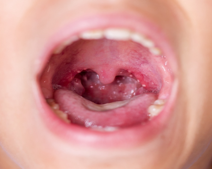
Enteroviruses, specifically coxsackie and echovirus, can also cause pharyngitis (Figure 1). The fecal-oral route is...
...purchase below to continue the course
Figure 1
Coxsackie Pharyngitis
Shutterstock ID 1151828390
Risk Factors
Viral pharyngitis causes between 50% and 80% of acute pharyngitis cases annually among children and adults. The peak season for all types of pharyngitis is usually late fall, winter, and early spring. Most types of pharyngitis have similar risk factors, including age (children and adolescents at highest risk), close living quarters (viral and bacterial infections spread more readily in areas where people gather, such as schools, daycare centers, offices, or airplanes), compromised immunity (due to HIV, diabetes, steroid therapy, chemotherapy, stress, poor diet, or fatigue), and irritants such as:
- tobacco (both smoking and secondhand smoke increase the risk of viral or bacterial infection)
- allergies (seasonal allergies or allergies to animals, dander, or molds)
- chemicals (particles from burning fossil fuels and common household chemicals)
- chronic sinus infections (drainage from the nose; Aung, 2023c; Chow & Doron, 2026)
Any irritation leaves the pharyngeal mucosa vulnerable to invasion by viruses and other microorganisms. Recent immigration from an underdeveloped country or lack of vaccination is a risk factor for diphtheria infection. Protective factors should include decreasing stress, eating a healthy diet, getting adequate rest, and avoiding exposure to smoke and chemicals. Frequent hand hygiene, limiting human contact, respiratory etiquette, and using disposable tissues are preventive measures for the common cold and other causes of viral pharyngitis. In addition, the influenza vaccination is another preventive measure that can be employed against the influenza virus (Acerra, 2024; Aung, 2023b, 2023c; Chow & Doron, 2026).
Signs and Symptoms
The HCP should conduct a history and physical examination to look for findings of acute pharyngitis and exclude other potentially serious illnesses. Uncomplicated viral pharyngitis is typically self-limited to 5 to 7 days, is not progressive, is bilateral, and is not associated with airway obstruction. Symptoms associated with viral pharyngitis may include:
- itchy or painful throat
- pain that increases with swallowing (odynophagia)
- swollen, red tonsils
- hoarse or muffled voice
- fever (low grade, except in influenza)
- cough
- rhinitis or rhinorrhea
- diarrhea
- conjunctivitis
- discrete ulcerative stomatitis (painful, red, and swollen mucosa with open ulcers)
- viral exanthem (skin rash; CDC, 2024c; Chow & Doron, 2026; Fried, 2025; Goldin & Graber, 2024)
Although most signs and symptoms associated with viral pharyngitis make the underlying viral cause difficult to identify, some characteristic signs and symptoms may help identify a particular viral cause for pharyngitis. For example, in addition to pharyngitis, adenovirus tends to cause benign follicular conjunctivitis, fever, and cervical lymphadenopathy. Concurrent conjunctivitis and fever (pharyngoconjunctival fever) occur in 33% to 50% of affected patients with adenovirus. Coxsackie A viruses are most often seen in infants and young children, with incidence rates decreasing with age. This virus typically presents with vesicles in the posterior pharynx and/or on the soles of the feet and the palms of the hands. Pharyngeal edema occurs in approximately half of all children diagnosed with COVID-19. The stomatitis caused by herpes simplex typically occurs in the anterior buccal mucosa but may extend to the tonsillar pillars. However, vesicles could be present within the mouth, lips, or throat. The blisters and pain may last several days to a few weeks, and the vesicles make it difficult for the patient to eat or drink. Lymph nodes may be swollen and tender (Aung, 2023a; Chow & Doron, 2026; Workowski et al., 2021).
Mononucleosis is most often seen in adolescent patients and may present with significant tonsillar hypertrophy; this can cause respiratory distress and should be monitored closely due to the risk of airway obstruction. Symptoms typically develop gradually over days or weeks, including splenomegaly, extreme fatigue that may last more than a month, large and mildly tender anterior and posterior cervical lymph nodes, headache, fever, and body aches. Symptoms may take 4 to 6 weeks to appear, during which the patient may be contagious, and symptoms may persist for up to 4 months. Influenza typically causes a fever and may also cause headaches, muscle/body aches, and fatigue. Symptoms of an early HIV infection may also include headaches and muscle and joint pain lasting approximately 2 weeks (Aung, 2023a; Auwaerter, 2026; Chow & Doron, 2026).
Diagnosis
The differential diagnosis will be dependent on associated symptoms due to the existence of various potential viral causes. Refer to Table 1 for diagnostics related to other common causes of acute pharyngitis.
Table 1
Diagnostics for Other Causes of Acute Pharyngitis
Cause | Diagnostics |
EBV |
|
Influenza |
|
HIV |
|
HSV |
|
If concern exists for a retropharyngeal abscess, as evidenced by significant difficulty swallowing or refusal to move the neck in an ill-appearing patient, a soft tissue radiograph of the lateral neck should be done. This radiograph should be done in full extension during inspiration as a true lateral image. A retropharyngeal abscess will typically result in a prevertebral space that is greater than half the thickness of the anteroposterior measurement of the adjacent vertebral body from C1 to 4, or 7 mm at C2, or greater than the full thickness of the vertebral body from C5-C7 and 14 mm at C6 (Fine, 2026; Wald, 2026b).
Treatment
Viral pharyngitis typically lasts 5 to 7 days and is treated supportively. Most patients who are seen for viral pharyngitis are looking for relief from throat pain. Effectively managing symptoms can enhance comfort and improve the patient’s ability to hydrate. Antibiotics are neither effective nor recommended. The best treatment option should consider patient factors (i.e., symptom severity, presence of accompanying symptoms, and comorbidities) and therapy-related factors (i.e., onset of action, duration of effectiveness, and side effects; Aung, 2023d; Fried, 2025; Stead, 2025). The following are suggested for decreasing throat pain in all types of viral pharyngitis:
- Acetaminophen (Tylenol; start at 325 mg or dose by weight in pediatric patients) or a nonsteroidal anti-inflammatory drug (NSAID) such as ibuprofen (Motrin; start at 200 to 400 mg or dose by weight in pediatric patients)
- Oral rinses with salt water (1 cup of water with ¼ to ½ teaspoon of table salt)
- Oral anesthetic sprays such as phenol (Chloraseptic)
- Lozenges (cough drops) containing topical anesthetics such as benzocaine/menthol (Cepacol, Chloraseptic)
- Sipping honey/lemon tea, chicken soup, or other warm beverages, cold beverages, or frozen desserts such as ice cream or popsicles (children under 12 months should not be given honey due to the risk of botulism poisoning)
- Alternative therapies include chamomile tea, peppermint, fenugreek, marshmallow root, licorice root, slippery elm, apple cider vinegar, garlic, or cayenne pepper
- Humidifying the ambient air through a humidifier or spending a few minutes in a closed bathroom with a hot shower several times throughout the day may relieve throat pain
- Avoiding exposure to tobacco smoke
- Avoid multisymptom preparations if the patient’s only symptom is throat pain (Aung, 2023d; Fried, 2025; Stead, 2025)
A short course of corticosteroids (i.e., 10 mg dose of dexamethasone [Decadron]) may be justified in severe cases of throat pain associated with difficulty swallowing. However, routine oral glucocorticoids are not recommended for acute pharyngitis due to the increased risk of mood changes and hyperglycemia. The treatment goal for viral pharyngitis is to maintain rapport with patients, manage symptoms, and avoid over or inappropriate prescribing of antibiotics. In several studies evaluating why patients sought care for pharyngitis, patient education regarding their options for pain relief, reassurance, and information about their illness was considered a higher priority for patients. Only about 38% of patients were specifically seeking an antibiotic prescription. In addition, it is vital to ensure that patients and family members understand how to avoid spreading the infection. Individuals infected with a viral illness should practice good handwashing and avoid sharing utensils, drinks, or kissing. Follow-up for any of these illnesses depends on unresolved symptoms interfering with activities of daily living (Aung, 2023d; Fried, 2025; Stead, 2025).
Mononucleosis
The treatment goal for mononucleosis is to ease the impact of symptoms and allow the immune system to contain the virus. Antiviral therapy is not proven to treat or cure EBV effectively. Antivirals have shown short-term suppression of viral shedding, but significant clinical benefits of antivirals have not been demonstrated (Aung, 2023d; Auwaerter, 2026; Mohseni & Jenks, 2026). In addition to the preceding supportive treatments for odynophagia, mononucleosis should be treated with the following:
- Rest is vital for patients with mononucleosis due to fatigue
- Diet may need to be modified due to a loss of appetite
- Increase fluids, particularly if taking ibuprofen (Motrin) for pain, to avoid dehydration, as kidney damage can occur with extended use of NSAIDs
- Acetaminophen (Tylenol) and ibuprofen (Motrin) should be used for pain relief and fever (Aung, 2023d; Auwaerter, 2026; Mohseni & Jenks, 2026)
In severe cases, systemic oral corticosteroids may be indicated for concerns regarding excessive swelling in the throat or tonsils. Additionally, patients diagnosed with mononucleosis should be encouraged to rest until they fully recover. Returning to their former level of activity may take several weeks to months. The patient should also avoid activities that could lead to injury due to the risk of splenic rupture, a medical emergency resulting in severe bleeding. Adolescents with mononucleosis who cannot participate in school, activities, and sports may have difficulty with isolation. Consider psychological referrals for depression or anxiety as needed (Aung, 2023d; Auwaerter, 2026; Mohseni & Jenks, 2026).
Influenza
Influenza is primarily treated symptomatically, but antiviral treatment may be used to reduce the severity and/or duration of symptoms. The decision to use antivirals should be based on how ill the patient is and/or the presence of risk factors for developing complications, such as those over 65 years of age, young children under age 4, those with a weakened immune system, those with diabetes, or those with a history of heart or lung disease. Individuals who have no risk factors or who are only mildly ill should be given antivirals only if they have had symptoms for less than 48 hr after discussing the risks and benefits. The priority for antivirals should be given to anyone with suspected or confirmed influenza who is hospitalized; has severe, complicated, or progressive illness; or is at high risk due to complications, as listed previously (Aung, 2023d; CDC, 2026a, 2026c; Zachary, 2025). For common antivirals used with influenza, refer to Table 2.
Table 2
Common Antivirals for Influenza
Drug | Dose | Duration |
Oseltamivir (Tamiflu) | Infants 2 weeks of age up to 1 year: 3 mg/kg/dose PO BID Children 1 to 12 years: 30 to 75 mg or 5 to 12.5 mL PO BID (weight-based) Adults and adolescents: 75 mg or 12.5 mL PO BID | 5 days |
Zanamivir (Relenza) | Adults, adolescents, and children 7 years and older: 2 puffs BID (inhalation route) | 5 days |
Peramivir (Rapivab) | Children 6 months to 12 years: 12 mg/kg intravenous (IV), up to a maximum of 600 mg Adults and adolescents 13 years or older: 600 mg IV | Single dose |
Baloxavir marboxil (Xofluza) | Adults and children 12 years or older, between 20 and 80 kg: 40 mg tablet Adults and children 12 years or older, 80 kg or more: 80 mg tablet | Single dose |
(Dolin, 2026; Genentech, Inc., 2025; Rapivab, n.d.; Zachary, 2025)
Antibiotics are not used in treating viral illnesses, including influenza, unless there is a secondary bacterial infection, such as sinusitis, otitis media, or pneumonia (Dolin, 2026; Zachary, 2025).
HIV
The patient diagnosed with HIV will have long-term complex health maintenance concerns. Life-long antiretroviral therapy should be started immediately upon diagnosis to decrease the risk of transmitting HIV to others and to decrease the complications of infection for the individual. For sore throats related to HIV infection, the patient should use symptomatic treatments as listed previously (Aung, 2023d; Wood, 2025). For additional information on managing chronic HIV/AIDs, please refer to the NursingCE course: HIV/AIDs.
Oral Herpes Simplex
Oral herpes treatment may include a topical anesthetic such as viscous lidocaine (Xylocaine) and PO antiviral medications such as acyclovir (Zovirax), valacyclovir (Valtrex), and famciclovir (Famvir). Initial outbreaks are typically treated for 7 to 10 days. Acyclovir (Zovirax) should be dosed at 400 mg three times daily (TID) or 200 mg five times daily. Valacyclovir (Valtrex) is typically dosed at 1 g twice daily and famciclovir (Famvir) at 250 mg TID or 500 mg BID. The dosage of PO antiviral medications may vary depending on the patient’s HIV status, age, and underlying renal dysfunction. They can be nephrotoxic, and a dose reduction may be needed with acute or chronic kidney dysfunction. Patients should be educated that these medications do not eradicate the virus or reduce the risk, frequency, and severity of recurrences. Suppressive therapy can reduce outbreak frequency by 70% to 80%. Safety has been established for the use of acyclovir (Zovirax) for as long as 6 years and valacyclovir (Valtrex) and famciclovir (Famvir) for up to 1 year. Acyclovir (Zovirax) should be dosed at 400 mg BID for long-term suppression. Valacyclovir (Valtrex) can be dosed at 500 mg or 1 g daily, and famciclovir (Famvir) at 250 mg BID. Valacyclovir (Valtrex) 500 mg daily has been shown to reduce sexual transmission. Famciclovir (Famvir) may be less effective than the other regimens for suppressing viral shedding. Episodic treatment can effectively reduce the severity and duration of outbreaks if initiated within 24 hr of lesion onset. Acyclovir (Zovirax) should be dosed at 400 mg TID or 800 mg BID for 5 days or 800 mg TID for 2 days. Valacyclovir (Valtrex) can be dosed at 500 mg BID for 3 days or 1 g daily for 5 days. Famciclovir (Famvir) can be dosed at 125 mg BID for 5 days, 1 g BID for 1 day, or 500 mg once, followed by 250 BID for 2 days. Topical therapies offer minimal clinical benefit and are not encouraged. Herpes is incurable, and patients are subject to lifelong periods of exacerbation/flare-ups and a need for treatment; some patients are on prophylactic antivirals (Aung, 2023d; Wald & Johnston, 2026; Workowski et al., 2021).
Complications
Viral pharyngitis may have disease-specific complications and implications, making follow-up vital to the avoidance or early identification of complications. Refer to Table 3 for specific complications associated with these infections.
Table 3
Complications Related to Viral Pharyngitis
Type of Pharyngitis | Complications |
Mononucleosis |
|
Influenza |
|
HIV |
|
(Auwaerter, 2026; Dolin, 2026; Wood, 2025)
GAS
The CDC estimates several million cases of noninvasive GAS annually in the United States, resulting in 5.2 million outpatient visits and 2.8 million antibiotic prescriptions. Globally, the burden of GAS is even more significant, with 100 million children developing impetigo, 470,000 new cases of acute rheumatic fever (ARF), and 282,000 new cases of rheumatic heart disease annually. The CDC has been tracking an increased incidence of invasive GAS within the last 5 years. Approximately 20,000 to 27,000 new cases of invasive GAS occur annually, resulting in 1,800 to 2,400 deaths annually (CDC, 2024a).
Pathophysiology and Risk Factors
S. pyogenes are gram-positive cocci that grow in chains and belong to group A in the Lancefield classification system. For a GAS infection to develop, exposure through saliva or nasal secretions from an infected individual through person-to-person contact must occur. Those with acute, symptomatic infection are more likely than asymptomatic carriers to transmit the disease to others. In rare occurrences, exposure to the bacteria can result from contaminated food, particularly milk or milk products. Rarely can infections be transmitted via food handling or fomites via toys or shared kitchen items (i.e., plates, glasses, or utensils). Humans appear to be the primary source of the infection, and pets are not considered potential reservoirs that transmit the bacteria to humans. Following exposure, the bacteria adhere to the pharyngeal mucosa through adhesions on the organism’s surface and invade the mucosal tissue. They produce proteases and cytolysins that cause inflammation, manifesting as the signs and symptoms of pharyngitis. An M protein on the surface of the bacteria plays a role in developing rheumatic fever and other complications from a GAS infection, discussed later in this section. In addition to the previously listed risk factors related to viral pharyngitis, close quarters promote infection, such as schools, daycare centers, or military training facilities (Ashurst et al., 2025; Buensalido, 2023a; Bush, 2025; CDC, 2025a).
Signs and Symptoms
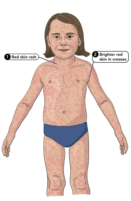
The incubation period for GAS is 2 to 5 days. GAS infections are most commonly present in children and adults as acute pharyngitis accompanied by the sudden onset of fever, tonsillar inflammation, odynophagia, and enlargement of the cervical lymph nodes. Less common symptoms may include headaches, nausea, vomiting, or abdominal pain. Close examination of the throat may show tonsillar erythema with or without exudate (refer to Figure 1), strawberry tongue, and petechiae along the palate. Children under 3 years of age with GAS infection rarely present with acute pharyngitis but typically develop mucopurulent rhinitis (inflammation or swelling of the mucous membranes of the nose, causing the production of mucous and pus). These children may also present with high fever, anorexia (appetite loss), and irritability. These symptoms should be carefully delineated from the common presentation seen in viral pharyngitis, which is often accompanied by cough, rhinorrhea, hoarseness, and conjunctivitis. A scarlatiniform rash (refer to Figure 2) could be present and indicates a syndrome known as scarlet fever or scarlatina. The rash is typically erythematous, blanches when pressure is applied, and has a characteristic sandpaper feel. It typically starts on the trunk and then spreads to the extremities, but it is not commonly found on the face, palms, or soles. It may be accentuated in the flexor creases (groin, underarm, inside the elbow, behind the knee) and described as Pastia’s lines. The rash typically lasts for 1 week and may be followed by desquamation. The face may appear flushed, and a white/yellow coating with red papillae may develop on the tongue, followed by a “strawberry tongue” (Ashurst et al., 2025; Buensalido, 2023a; Bush, 2025; CDC, 2025a, 2025g).
Figure 2
Scarlet Fever with Characteristic Rash
(CDC, 2025g)
Diagnosis and Treatment
An accurate diagnosis by symptoms alone is challenging since viral and bacterial pharyngitis present similarly (CDC, 2025c). Since most pharyngitis cases are due to GAS or a virus, initial screening should be focused on these two primary etiologies. The Centor criteria to identify patients at risk for GAS assigns one point for each of the following criteria:
- fever greater than 100.5°F
- absence of cough
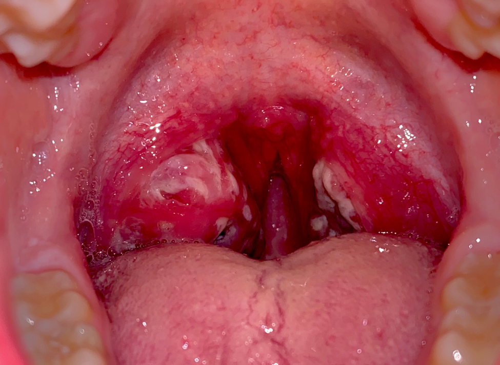
- tonsillar exudate (refer to Figure 3)
- tender anterior cervical lymphadenopathy (Buensalido, 2023b; Chow & Doron, 2026; Wald, 2025c)
Figure 3
Streptococcal Pharyngitis with Exudate
If the patient is between 3 and 14 years of age, one point is added; if the patient is an adult greater than 45 years of age, one point is subtracted. Patients with one or fewer of the four criteria listed previously are low risk and should not have additional testing. Patients with a score of 3 or above should be tested for GAS, although some will test with a score of 2. These criteria should not be used as a replacement for testing but as a decision-making tool regarding which patients should be tested. The criteria are endorsed by the European Society of Clinical Microbiology and Infectious Diseases and the Infectious Disease Society of America (IDSA; Buensalido, 2023b; Chow & Doron, 2026; Wald, 2025c). The CDC and the American College of Physicians (ACP) make the following testing and treatment recommendations based on the Centor score, refer to Table 4.
Table 4
CDC/ACP Testing and Treatment Recommendations Based on the Centor Score
Centor Score | Recommendation |
0 | Do not test and do not treat |
1 | Do not test and do not treat |
2 | Treat if the rapid test is positive for GAS |
3 | Treat if the rapid test result is positive for GAS or Treat empirically |
4 | Treat empirically |
(Buensalido, 2023b; Chow & Doron, 2026; Wald, 2025c)
The American Academy of Family Physicians and the National Institute for Health and Care Excellence in the UK recommend using either the Centor score or the FeverPAIN score. For the FeverPAIN score, a point is given for each: fever within the past 24 hr, markedly inflamed tonsils, no coryza or cough, presented within 3 days of symptom onset, and purulence of tonsils. No testing or treatment is recommended for a score of 0 to 1. However, some clinicians may test with a throat culture for children ages 3 to 15. A rapid antigen detection test (RADT) is recommended for scores of 2 or 3, and empiric treatment is recommended for a score of 4 or 5 (Buensalido, 2023b; Chow & Doron, 2026; Hamilton & McCrea, 2024; Wald, 2025c).
Current US guidelines for GAS include a RADT and a bacterial culture using a pharyngeal swab. RADTs are easy to use and have less than 10-min turnaround times. These tests have an 88% to 99% specificity but a sensitivity of only 70% to 90%, increasing the risk of false negatives. Due to the low sensitivity, a negative RADT requires a bacterial culture in specific patient populations for confirmation. Cultures have a sensitivity between 90% and 95% and a specificity between 95% and 99%. This is recommended for pediatric patients over 3 years of age (unless they have a sibling with known GAS pharyngitis), symptomatic household members of patients with GAS pharyngitis, or those at increased risk of complications. Cultures typically take 24 to 48 hr to return, and empirical treatment while awaiting culture results is unnecessary, as short delays in treatment are not associated with an increased risk for complications. Positive RADTs do not require a follow-up culture due to the high specificity of the test (Boyanton et al., 2024; Buensalido, 2023b; Bush, 2025; Chow & Doron, 2026).
Several NAAT assays for GAS pharyngitis diagnosis have been approved by the US Food and Drug Administration since 2016. NAAT assays provide sensitivity and specificity similar to bacterial culture and higher sensitivity than RADTs. This improved diagnosis with a single assay has led many health care facilities to implement this type of testing. With expedited transport and reporting, results can be obtained in hours. The rapid result and higher accuracy allow providers to decide on antibiotic use faster. Currently, the IDSA guidelines recommend the use of RADTs and cultures and have been slow to endorse the use of NAATs. Although NAATs may replace RADTs in the future, many concerns have been noted that have prevented widespread adoption. The barriers to NAAT adoption for GAS include higher testing costs and the need for specialized equipment. In addition, due to the high sensitivity of NAATs, they are more likely to detect GAS in colonized individuals who do not have an active infection and nonviable GAS DNA, which can remain in the pharynx for 2 to 6 weeks after infection (CDC, 2025c; Hamilton & McCrea, 2024; Wald, 2025c).
Due to the very low risk of ARF, the CDC recommends that children younger than 3 years and adults not be tested routinely. GAS treatment is a priority due to potential complications, including ARF, rheumatic heart disease, or poststreptococcal glomerulonephritis (PSGN). Treatment within 9 days of the onset of symptoms has been shown to prevent ARF. The CDC recommends antibiotic treatment for GAS pharyngitis to shorten the duration of the symptoms, prevent complications, and decrease the risk of transmission to others. The treatments of choice are penicillin (PCN, Pen-V) or amoxicillin (Amoxil). There are no confirmed reports of resistance to these antibiotics. A narrow-spectrum cephalosporin such as cephalexin (Keflex) or azithromycin (Zithromax) is recommended for patients with a PCN allergy. Azithromycin (Zithromax) and clarithromycin (Biaxin) should not be utilized as first-line treatment for GAS pharyngitis, as there have been reports of resistance against these in some communities (Ashurst et al., 2025; Bush, 2025; CDC, 2025c; Pichichero, 2026c). Refer to Table 5 for suggested antibiotic treatment regimens for GAS pharyngitis.
Table 5
Suggested Antibiotic Treatment for GAS Pharyngitis
Drug | Dose | Duration |
Penicillin (PCN, Pen-V) | Patients under 27 kg: 250 mg PO BID or TID Patient over 27 kg: 500 mg PO BID or TID | 10 days |
Amoxicillin (Amoxil) | Children or adolescents: 50 mg/kg PO daily or 25 mg/kg PO BID (1,000 mg/day maximum) Adults: 500 mg PO BID | 10 days |
Penicillin G benzathine (Bicillin L-A) | Patients under 27 kg: 600,000 units intramuscular (IM) Patients over 27 kg: 1,200,000 units IM | Single dose |
For individuals with a PCN allergy | ||
Cephalexin (Keflex) | Children or adolescents: 20 mg/kg/dose PO BID (500 mg/dose maximum) Adults: 500 mg PO BID | 10 days |
Cefadroxil (Duricef) | Children or adolescents: 30 mg/kg PO daily or in divided doses every 12 hr (1,000 mg/day maximum daily) Adults: 1 g PO daily | 10 days |
Clindamycin (Cleocin) | Children or adolescents: 7 mg/kg/dose PO TID (300 mg/dose maximum) Adults: 300 mg PO TID | 10 days |
Azithromycin (Zithromax) | Children or adolescents: 12 mg/kg PO daily (500 mg/dose maximum) Adults: 12 mg/kg PO daily (500 mg/dose maximum) | 5 days for children and adolescents and 3 days for adults |
Clarithromycin (Biaxin) | Children or adolescents: 7.5 mg/kg/dose PO BID (250 mg/dose maximum) Adults: 250 mg PO BID | 10 days |
(Pichichero, 2026c)
Adjunctive therapy with analgesics or antipyretics such as acetaminophen (Tylenol) or ibuprofen (Motrin) should be used for fever associated with GAS pharyngitis. Acetylsalicylic acid (Aspirin) should be avoided in children with GAS pharyngitis due to the risk of Reye syndrome. Increased fluids and a diet as tolerated should be encouraged. Routine follow-up RADT or cultures are not recommended except in special circumstances, such as an unresolved infection (CDC, 2025c; Pichichero, 2026c).
Clinicians should ensure that children and adults diagnosed with GAS pharyngitis are educated on infection control measures. They should remain home from school, work, or daycare until they are afebrile and have been on antibiotic therapy for at least 24 hr. Optimal methods for infection prevention include proper handwashing and respiratory etiquette. Handwashing should occur after a cough or sneeze and before eating or preparing food. Alcohol-based hand rubs are an equal alternative to handwashing if soap and water are unavailable. Respiratory etiquette includes covering the mouth or nose during sneezes and coughs or coughing into the elbow. Droplet precautions should be implemented in hospitalized patients with GAS pharyngitis. Ensure the patient and caregiver understand their medication regimen and the importance of taking all antibiotics as prescribed to avoid complications (CDC, 2025c; Pichichero, 2026c; Wald, 2025c). The clinician should educate caregivers about symptoms to monitor for that require immediate intervention, as they may indicate upper airway obstruction or worsening infection, including the following:
- difficulty swallowing or breathing (stridor, tachypnea, dyspnea, retractions)
- excessive drooling in an infant or young child
- temperature higher than 101°F
- swelling or stiffness of the neck
- muffled, “hot potato” voice or hoarseness
- “sniffing” or tripod positioning to help maintain airway patency
- difficulty opening their mouth (Chow & Doron, 2026; Pichichero, 2026c; Wald, 2025c)
Complications
As previously mentioned, recurrent GAS infections may be an indication for a tonsillectomy in children. Research has shown that children with recurrent throat infections who undergo a tonsillectomy show a reduction in infections and a decrease in missed school days during the first year. Unfortunately, the benefits are not consistent after this initial time frame. Parents must consider the potential harms of a tonsillectomy, including the risks of anesthesia and postoperative bleeding, combined with the prolonged throat pain and financial costs. Other possible intraoperative complications include damage to the teeth, pharyngeal wall, or soft palate; laryngospasm; aspiration; respiratory compromise; and cardiac arrest. Postoperative complications include nausea, vomiting, dehydration, and postobstructive pulmonary edema. Most cases of GAS pharyngitis resolve quickly with antibiotic therapy. However, there are rare complications that can result in severe morbidity, such as suppurative (inflammation accompanied by pus formation or discharge) local infections (i.e., peritonsillar abscess, cervical lymphadenitis, mastoiditis) or nonsuppurative sequelae (i.e., ARF or PSGN; CDC, 2025b; Pichichero, 2026a).
A peritonsillar abscess typically presents with the aforementioned signs/symptoms of acute pharyngitis, in combination with dysphagia (which may present as drooling), trismus (lockjaw), and a muffled or “hot potato” voice. Patients may also report ear pain on the ipsilateral side, and the swelling may cause contralateral deviation of the uvula. Inspection of the pharynx reveals swelling and erythema unilaterally in the anterior tonsillar pillar and the overlying soft palate. It is most common in adolescents and young adults between 20 and 40. There is consensus that treatment should consist of drainage of the abscess with needle aspiration, followed by antibiotic therapy and symptomatic management of pain and/or fever if applicable. Referral to an otolaryngologist should occur if peritonsillar or another abscess is suspected or if tonsillar hypertrophy persists. Most can be managed as an outpatient, but hospitalization should be considered if symptoms do not improve within 4 hr of needle drainage. When deciding on the antibiotic regimen, coverage of GAS, Staphylococcus aureus (S. aureus), and respiratory anaerobes is important as most peritonsillar abscesses are polymicrobial. Oral antibiotic regimens for adults can include penicillin V (PCN, Pen-V) 500 mg PO every 6 hr plus metronidazole (Flagyl) 500 mg PO every 6 hr for 10 to 14 days. An alternative regimen can include amoxicillin/clavulanate (Augmentin) 875 mg PO every 12 hr for 14 days or a third-generation cephalosporin (cefdinir [Omnicef] 300 mg PO every 12 hr) plus metronidazole (Flagyl) 500 mg PO every 6 hr. For patients with suspected methicillin-resistant S. aureus (MRSA) infection, linezolid (Zyvox) 600 mg PO BID for 14 days or clindamycin (Cleocin) 300 mg to 450 mg PO TID for 14 days are recommended. For hospitalized patients, the preferred regimen is IV penicillin G (Bicillin L-A) 10,000,000 units combined with metronidazole (Flagyl) 500 mg every 6 hr. Alternatives include ampicillin/sulbactam (Unasyn), ceftriaxone (Rocephin) combined with metronidazole (Flagyl), piperacillin/tazobactam (Zosyn), or clindamycin (Cleocin) if allergic to PCN (Pichichero, 2026a; Wald, 2026a).
Cervical lymphadenitis involves enlarged, inflamed, and tender lymph nodes within the neck. This condition is to be differentiated from lymphadenopathy, which is the presence of enlarged lymph nodes. GAS infections typically present with acute bilateral lymphadenitis accompanied by exudative pharyngitis, although some may present with unilateral findings. Differential diagnoses include Arcanobacterium hemolyticum (A. hemolyticum) and EBV, especially in adolescents. The lymph nodes should gradually diminish in size and tenderness with appropriate antibiotic treatment for the underlying GAS infection, and a follow-up appointment may be scheduled for 2 to 3 weeks later to confirm resolution (Healy, 2026).
Although more commonly associated with acute otitis media, mastoiditis is a suppurative infection of the mastoid air cells. It is typically diagnosed clinically based on the presence of postauricular tenderness, erythema, swelling with protrusion of the auricle, and fever. Patients will report ear pain and lethargy/malaise. Due to its proximity to the facial nerve, semicircular canals, sternocleidomastoid muscle, jugular vein, internal carotid artery, sigmoid sinus, brain, and meninges, this condition carries a high risk for potentially serious complications. These patients should be referred to an otolaryngologist early. Treatment typically consists of aspiration and drainage of the mastoid and middle ear (myringotomy) and IV antibiotics. Complications may require additional surgical intervention, including a mastoidectomy (Wald, 2025a, 2025b).
The immune response to GAS pharyngitis, rather than a direct result of the infection itself, is thought to be primarily responsible for ARF and PSGN. ARF is a delayed sequela of GAS pharyngitis that may affect the heart, joints, or nervous system. It typically presents 1 to 5 weeks after the onset of pharyngitis. The primary clinical feature is a fever; most patients (50% to 70%) also present with carditis with or without valvulitis. Physical indications of carditis at presentation are typically a new-onset heart murmur, cardiomegaly, pericardial friction rub, pericardial effusion, or congestive heart failure. An electrocardiogram (ECG) may indicate a prolonged PR interval. A migratory polyarthritis affecting the elbows, wrists, knees, and ankles may develop. Firm, painless subcutaneous nodules and erythema marginatum (nonpruritic, nonpainful transient macular lesions typically found on the trunk or proximal extremities with outward extension and central clearing) may be evident (CDC, 2025b; Steer & Gibofsky, 2026; Pichichero, 2026a).
Chorea is the central nervous system manifestation most often found in patients with ARF. Chorea is nonrhythmic, involuntary, sudden movements typically combined with emotional lability and muscle weakness. Inadequate antibiotic treatment for GAS pharyngitis increases the risk of ARF; approximately one-third of ARF cases follow a subclinical case of pharyngitis or one for which medical attention was not obtained. A prior history also increases the risk, especially for the first few years after the initial occurrence of ARF. Children between the ages of 5 and 15 years are at the highest risk, and ARF is extremely rare in adults and children under 3. Differential diagnoses may include rheumatoid arthritis, juvenile idiopathic arthritis, septic arthritis, systemic lupus erythematosus, serum sickness, Lyme disease, infective endocarditis, viral myocarditis, Henoch-Schonlein purpura, gout, sarcoidosis, leukemia, and Hodgkin’s disease. ARF is diagnosed clinically based on the Jones Criteria, in combination with evidence for previous GAS infection based on throat culture, RADT, or antibody titer. For the initial diagnosis of ARF, a patient must satisfy at least two major manifestations or one major and two minor manifestations. If one major manifestation is cardiac or joint-related, the other manifestations must be from a separate clinical category. Recurrent ARF can be diagnosed based on the presence of three minor manifestations (CDC, 2025b; Steer & Gibofsky, 2026). Refer to Table 6 for Jones Criteria.
Table 6
Jones Criteria for ARF Diagnosis
Low-risk populations (ARF incidence £2 per 100,000 children/£1 per 1,000 population per year) | Moderate and/or high-risk populations (all other communities not defined as low risk) |
Major Manifestations | |
Cardiac: clinical or subclinical carditis | |
CNS: Chorea | |
Integumentary: erythema marginatum or subcutaneous nodules | |
Joint: Polyarthritis | Joint: poly- or monoarthritis; polyarthralgia if other etiologies have been excluded |
Minor Manifestations | |
Cardiac: prolonged PR interval on ECG after accounting for age (unless carditis is used as a major criterion above) | |
Fever (³ 101.3°F/38.5°C) | |
Joint: Polyarthralgia (unless polyarthritis is used as the major criterion above) | Joint: Monoarthralgia (unless arthritis or arthralgia is used as the major criterion above) |
Erythrocyte sedimentation rate (ESR) ³ 60 mm in the first hour or C-reactive protein (CRP) ³ 3.0 mg/dL | ESR ³ 30 mm/hr or CRP > 3.0 mg/dL |
(CDC, 2025b; Steer & Gibofsky, 2026)
Routine echocardiography/Doppler is recommended for patients with ARF, suspected or confirmed. Treatment consists of symptomatic management, including salicylates (aspirin, ASA) or anti-inflammatory medications to alleviate inflammation and treat the fever, as well as diuretics and antihypertensives. Antibiotic treatment should resume (as in Table 5) to eradicate any residual GAS regardless of whether or not pharyngitis is present. Long-term complications are typically related to rheumatic heart disease and vary based on the extent or severity of cardiac involvement (CDC, 2025b; Steer & Gibofsky, 2026).
Nephritogenic strains of GAS cause PSGN. It typically occurs 10 days after GAS pharyngitis or 21 days after a GAS skin infection, and clinical features of PSGN include:
- facial or periorbital edema, particularly upon awakening
- hypertension
- proteinuria
- hematuria, with cola-colored urine (reddish-brown, dark)
- lethargy, anorexia, and generalized weakness (CDC, 2025f; Niaudet & Somers, 2026)
Treatment for PSGN should include the management of any hypertension and/or edema. Antibiotics should be given as described in Table 5, with a preference for Penicillin G benzathine (Bicillin L-A). While over 90% of children with PSGN will fully recover, some adults may develop long-term renal function impairment (CDC, 2025f; Niaudet & Somers, 2026).
Children are often reluctant to drink fluids due to odynophagia and are at risk for dehydration. Parents should be educated on monitoring for the signs of dehydration and encouraging fluid intake. Infants should produce at least one wet diaper every 6 hr. Early signs of dehydration include dry mouth, thirst, decreased urination, or darkening of the urine. Signs of moderate to severe dehydration include excessive thirst, a lack of tears when crying, sunken eyes and fontanelles, irritability, listlessness, lightheadedness, tachycardia, low blood pressure, and tachypnea (Somers, 2025).
GAS pharyngitis in children may be associated with the development of autoimmune neuropsychiatric symptoms such as obsessive thoughts, compulsive behaviors (commonly seen in patients with obsessive-compulsive disorder), and tics (a habitual spasmodic contraction of the muscles); this is known as pediatric autoimmune neuropsychiatric disorders associated with streptococcal infections (PANDAS). PANDAS typically presents with an abrupt onset of the aforementioned symptoms associated with a recent GAS infection. The condition is somewhat controversial, as some children diagnosed with PANDAS have underlying comorbidities such as anxiety or attention-deficit/hyperactivity disorder, making it difficult to differentiate between additional/worsening neuropsychiatric symptoms or an autoimmune response to GAS. Most cases of PANDAS resolve with antibiotic therapy. A tonsillectomy may decrease the number of GAS infections and reduce the symptoms of PANDAS (Pichichero, 2026a, 2026b).
In addition to pharyngitis, GAS can also cause impetigo or invasive infections, including streptococcal toxic shock syndrome (STSS), necrotizing fasciitis (NF), bacteremia, osteomyelitis, septic arthritis, and pneumonia. STSS involves bacterial exotoxins within human tissues and the bloodstream, inducing a cytokine cascade and leading to sudden shock and organ failure. The initial presentation for patients with STSS typically includes fever, chills, myalgia, nausea, and vomiting; this may progress quickly to hypotension, tachycardia, tachypnea, and signs/symptoms of organ failure suggestive of sepsis. Risk factors for STSS include age over 65, recent surgery, open wounds, or chronic illness such as alcohol use disorder or diabetes. It is typically diagnosed clinically based on the presence of hypotension (systolic blood pressure less than 90) and two or more indications of multiorgan involvement (i.e., renal impairment as evidenced by elevated creatinine, coagulopathy as evidenced by decreased platelet count, elevated liver enzymes, hypoxemia with acute diffuse pulmonary infiltrates, a generalized erythematous macular rash, or soft tissue necrosis); a positive culture for GAS is typically used for confirmation. These patients require hospitalization with immediate fluid resuscitation and IV antibiotics, typically a combination of penicillin G (Bicillin LA) and clindamycin (Cleocin). Severe cases may also require surgical debridement and the use of IV immunoglobulin. The mortality rate for STSS is currently 30% to 70% (CDC, 2025d; Pichichero, 2026a; Stevens, 2025).
NF destroys the fascia and adipose tissue of the body. It typically occurs following trauma or surgery or as a complication of a varicella lesion. NF most commonly affects the extremities, especially the legs. Adults with an immune disorder, on immunosuppressive medications, or chronic diseases such as diabetes have compromised immune systems and are at increased risk for NF. The presenting signs/symptoms in the affected limb include pain, warmth, swelling, erythema, and tenderness. The bulk of the tissue damage may not be readily apparent, as NF often spares the overlying skin. If untreated, the swelling can progress to brawny edema followed by dark red induration. The overlying skin may become dusky as cutaneous thrombosis and ischemia develop. Bullae, or fluid-filled sacs, may develop and become hemorrhagic. Tissue may appear progressively darker, from red to black, if untreated. The skin may become anesthetized in severe cases as superficial nerves are destroyed. Eventually, the skin will slough, and necrotic eschar forms; this may appear similar to a third-degree burn (CDC, 2025e; Pichichero, 2026a; Stevens & Baddour, 2024).
When NF occurs in an extremity, it may lead to compartment syndrome, necessitating emergency fasciotomy. If left untreated, NF can progress to sepsis, shock, organ failure, and death. Treatment typically involves surgical debridement/exploration or biopsy for Gram stain and culture. Incisions are typically large and should be left open for at least 24 hr for observation. Amputation may be required, depending on the location of the infection and the severity/extent of the tissue damage. Imaging studies, including computed tomography or magnetic resonance imaging, may be helpful early but may also delay diagnosis and are not definitive. Patients typically present with abnormal laboratory findings, including leukocytosis (elevated white blood cell count), thrombocytopenia (decreased platelet count), and azotemia (elevated blood urea nitrogen). Broad-spectrum parenteral antibiotics should be given initially, followed by high-dose penicillin G (Bicillin LA) and clindamycin (Cleocin) once the Gram stain and culture results have identified S. pyogenes. Although not proven effective, IV immunoglobulin may be considered for severe cases. The mortality rate for NF is 24% to 34%. When coinciding with STSS, the mortality rate is 60% (CDC, 2025e; Pichichero, 2026a; Stevens & Baddour, 2024).
Other Causes
Pathophysiology
Fungal pharyngitis (oropharyngeal candida infection or thrush) is common in breastfeeding infants and will cause significant pain. Otherwise, this is typically an opportunistic infection not seen in immunocompetent patients. Haemophilus influenzae (H. influenzae) type b (Hib) was once a common cause of febrile illness, especially in children. This typically presents with pharyngitis, high fever, stridor, drooling, and an ill-appearing child. Epiglottitis is also a concern as it could lead to life-threatening upper airway obstruction with an abrupt onset. However, the incidence has been dramatically reduced with the advent of routine vaccination against Hib in this country. Arcanobacterium haemolyticum (A. haemolyticum), Mycoplasma pneumoniae (M. pneumoniae), Chlamydophila pneumoniae (C. pneumoniae), or other groups of streptococci (groups C or G) are among the bacteria that may cause acute pharyngitis. They are more commonly found in adolescent and young adult patients. A. haemolyticum accounts for only 1% to 2.5% of acute pharyngitis cases and is most common in adolescents or young adults. In addition to causing acute pharyngitis, Fusobacterium necrophorum (F. necrophorum) infection can lead to Lemierre syndrome, a rare infection associated with jugular thrombophlebitis and the formation of septic emboli. Several less common sexually transmitted infections can cause pharyngitis independently or in conjunction with a comorbid infection. Oral Neisseria gonorrhea (N. gonorrhea), Chlamydia trachomatis (C. trachomatis), and Treponema pallidum (T. pallidum; the bacteria responsible for syphilis) are typically contracted sexually but can also cause oral infections presenting with pharyngitis following unprotected oral sexual contact with an infected individual (Bush, 2024; CDC, 2026d; Chow & Doron, 2026; Fine, 2026).
Risk Factors
The risk factors for most of these infections include those listed in the viral pharyngitis section. The most significant risk factor for sexually transmitted infections is unprotected sexual activity (Chow & Doron, 2026; Fine, 2026).
Signs and Symptoms
With most of the aforementioned organisms, the patient typically presents with symptoms similar to those of other viral infections, such as redness, irritation, odynophagia, and a cough. Oral candidiasis (fungal pharyngitis) typically presents with white patches along the buccal mucosa, tongue, soft palate, and pharynx with underlying erythema and tenderness. The patient may report a cotton sensation within the mouth and loss of taste. It is typically diagnosed clinically. A. haemolyticum often presents similarly to a GAS infection, with a scarlatiniform rash in about half of cases. Pharyngitis related to M. pneumoniae or C. pneumoniae is typically associated with lower respiratory tract symptoms. F. necrophorum should be considered in very ill patients with neck pain, severe pharyngitis, and respiratory distress. Oral chlamydia or gonorrhea can be asymptomatic or cause acute pharyngitis associated with oral-genital contact (CDC, 2026d; Chow & Doron, 2026; Fine, 2026; Workowski et al., 2021).
Diagnosis
History and physical exam may indicate the most likely causes of acute pharyngitis. History should inquire about recent travel, sick contacts, sexual exposure, recent immunizations, current medications, and any history of immunocompromising conditions. An inflamed eardrum likely indicates that the pathology is nonoropharyngeal and that the reports of throat pain are likely due to referred pain. Similarly, an inflamed area surrounding a tooth indicates a potential abscess and warrants referral to a dentist. If testing has ruled out GAS infection in a patient with acute pharyngitis, supportive treatment should be attempted for at least 5 to 7 days before additional diagnostic testing is explored. Most other etiologies of acute pharyngitis can be diagnosed via throat culture. While different strains of streptococci and A. haemolyticum can be diagnosed using a routine aerobic culture, F. necrophorum requires an anaerobic culture. There are commercially available real-time PCR testing kits for M. pneumoniae and C. pneumoniae, which are much faster than culture or serology enzyme immunoassay; these tend to be expensive. NAATs for C. trachomatis and N. gonorrhoeae are available as oropharyngeal swabs; certain labs have met the regulatory requirements and established specifications for using these tests with oropharyngeal samples. These swabs are often done to detect both infections from a single specimen. The sensitivity of these NAAT tests for C. trachomatis and N. gonorrhoeae is superior to culture but varies by manufacturer. Diagnosis of infection with T. pallidum is confirmed via two separate tests: first, a nontreponemal test (i.e., Venereal Disease Research Lab or the Rapid Plasma Reagin), followed by confirmation with a treponemal test (i.e., fluorescent treponemal antibody absorbed, the passive particle agglutination assay, an enzyme immunoassay, chemiluminescence immunoassay, immunoblot, or rapid treponemal assay; CDC, 2026b; Chow & Doron, 2026; Fine, 2026; Workowski et al., 2021).
Treatment
Mild to moderate oral candidiasis is typically treated for 7 to 14 days with antifungal medications such as clotrimazole (Mycelex, 10 mg lozenge five times daily), miconazole (Oravig 50 mg buccal tab once daily), or nystatin (Nystop suspension, 100,000 units/mL, 4 to 6 mL four times daily or 200,000 units pastille four times daily). Severe cases should receive fluconazole (Diflucan) 100 to 200 mg daily for 7 to 14 days. Pharyngitis related to mild H. influenzae infection can be treated with amoxicillin/clavulanate (Augmentin), azithromycin (Zithromax), cephalosporins, fluoroquinolones, or clarithromycin (Biaxin). Pharyngitis related to A. haemolyticum will typically resolve within 2 weeks without antibiotic treatment but may resolve within 3 days with treatment. Penicillin (PCN) or the macrolide erythromycin (E-Mycin) is generally accepted as the most effective. Many strains have developed resistance to trimethoprim-sulfamethoxazole (Bactrim) and/or tetracycline (Sumycin), so these should be avoided (Bush, 2024; Ufkes, 2022; Vazquez, 2025).
Pharyngitis caused by M. pneumoniae or C. pneumoniae should not be treated with penicillin (PCN), as their lack of a cell wall makes them naturally resistant. Instead, treatment should consist of a macrolide (i.e., azithromycin [Zithromax]) in young children, a fluoroquinolone in adult patients, or a tetracycline (i.e., doxycycline [Vibramycin, Doryx]) in older children or adult patients if required. Like the GAS treatment described above, penicillin (PCN) is the antibiotic of choice for treating pharyngitis related to other streptococci groups (groups C and G). Macrolides should be avoided in pharyngitis related to anaerobes, such as F. necrophorum. In these cases, metronidazole (Flagyl) or clindamycin (Cleocin) should be used in combination with a beta-lactam (i.e., penicillin [PCN] or a cephalosporin). Chlamydia treatment for oropharyngeal infections is similar to that for urogenital infections. The recommended regimen is doxycycline (Vibramycin, Doryx) 100 mg PO BID for 7 days with an alternative regimen of azithromycin (Zithromax) 1 g PO in a single dose. Oropharyngeal gonorrhea infections are more difficult to eradicate than urogenital infections. The CDC recommends a regimen that includes a single dose of 500 mg of ceftriaxone (Rocephin) IM for individuals less than 150 kg and 1 g for those greater than 150 kg. Primary, secondary, or early latent syphilis should be treated in adults with penicillin G (Benzathine) 2.4 million units IM once. The care of patients with any infectious disease should also include education related to the individual disease process and the potential to spread the disease to others. Symptomatic treatment for the associated sore throat is consistent with viral pharyngitis. Both mental and physical support are encouraged for patients suffering from these conditions, and referrals should be made as needed (CDC, 2022, 2026b; Sears, 2026; Seña & Cohen, 2026; Wessels, 2024; Workowski et al., 2021).
Complications
The majority of these infections are self-limiting. As previously mentioned, H. influenzae pharyngitis can lead to epiglottitis with life-threatening airway obstruction. Gonorrhea or chlamydia can lead to pelvic inflammatory disease in females; epididymitis, leading to infertility in males; joint infections or arthritis; and pregnancy complications, such as miscarriage, premature birth, or transmission of the infection to the neonate during delivery (most newborns receive antibiotic ointment to their eyes due to the risk). Another potential complication of any sexually transmitted infection is the possibility of spreading to sexual partners and other parts of the body (Seña & Cohen, 2026; Workowski et al., 2021).
References
Acerra, J. R. (2024). Pharyngitis follow-up. https://emedicine.medscape.com/article/764304-followup#e4
Ashurst, J. V., Weiss, E., Tristram, D., & Edgerley-Gibb, L. (2025). Streptococcal pharyngitis. In StatPearls [Internet]. StatPearls Publishing. https://www.ncbi.nlm.nih.gov/books/NBK525997
Aung, K. (2023a). Viral pharyngitis clinical presentation. https://emedicine.medscape.com/article/225362-clinical
Aung, K. (2023b). Viral pharyngitis follow-up. https://emedicine.medscape.com/article/225362-followup
Aung, K. (2023c). Viral pharyngitis overview. https://emedicine.medscape.com/article/225362-overview
Aung, K. (2023d). Viral pharyngitis treatment & management. https://emedicine.medscape.com/article/225362-treatment
Auwaerter, P. G. (2026). Infectious mononucleosis. UpToDate. Retrieved July 23, 2026, from https://www.uptodate.com/contents/infectious-mononucleosis
Boktor, S. W., & Hafner, J. W. (2023). Influenza. In StatPearls [Internet]. StatPearls Publishing. https://www.ncbi.nlm.nih.gov/books/NBK459363
Boyanton, B. L., Jr., Caldwell, J. M., & Ledeboer, N. A. (2024). Review: Current laboratory and point-of-care pharyngitis diagnostic testing and knowledge gaps. The Journal of Infectious Diseases, 230(Suppl. 3), S182–S189. https://doi.org/10.1093/infdis/jiae415
Buensalido, J. A. L. (2023a). Bacterial pharyngitis overview. https://emedicine.medscape.com/article/225243-overview#a6
Buensalido, J. A. L. (2023b). Bacterial pharyngitis workup. https://emedicine.medscape.com/article/225243-workup
Bush, L. M. (2024). Haemophilus infections. https://www.merckmanuals.com/professional/infectious-diseases/gram-negative-bacilli/haemophilus-infections
Bush, L. M. (2025). Streptococcal infections. https://www.merckmanuals.com/professional/infectious-diseases/gram-positive-cocci/streptococcal-infections
Centers for Disease Control and Prevention. (2022). Sexually transmitted infections treatment guidelines, 2021: Gonococcal infections among adolescents and adults. https://www.cdc.gov/std/treatment-guidelines/gonorrhea-adults.htm
Centers for Disease Control and Prevention. (2024a). Group A strep disease surveillance and trends. https://www.cdc.gov/group-a-strep/php/surveillance
Centers for Disease Control and Prevention. (2024b). Laboratory testing for Epstein-Barr virus (EBV). https://www.cdc.gov/epstein-barr/php/laboratories
Centers for Disease Control and Prevention. (2024c). Sore throat basics. https://www.cdc.gov/sore-throat/about/index.html
Centers for Disease Control and Prevention. (2025a). About group A strep infection. https://www.cdc.gov/group-a-strep/about
Centers for Disease Control and Prevention. (2025b). About rheumatic fever. https://www.cdc.gov/group-a-strep/about/rheumatic-fever.html
Centers for Disease Control and Prevention. (2025c). Clinical guidance for group A streptococcal pharyngitis. https://www.cdc.gov/group-a-strep/hcp/clinical-guidance/strep-throat.html
Centers for Disease Control and Prevention. (2025d). Clinical guidance for streptococcal toxic shock syndrome. https://www.cdc.gov/group-a-strep/hcp/clinical-guidance/streptococcal-toxic-shock-syndrome.html
Centers for Disease Control and Prevention. (2025e). Clinical guidance for Type II necrotizing fasciitis. https://www.cdc.gov/group-a-strep/hcp/clinical-guidance/necrotizing-fasciitis.html
Centers for Disease Control and Prevention. (2025f). Clinical guidelines for post-streptococcal glomerulonephritis. https://www.cdc.gov/group-a-strep/hcp/clinical-guidance/post-streptococcal-glomerulonephritis.html
Centers for Disease Control and Prevention. (2025g). Symptoms of scarlet fever. https://www.cdc.gov/group-a-strep/about/symptoms-of-scarlet-fever.html
Centers for Disease Control and Prevention. (2026a). About influenza antiviral medications. https://www.cdc.gov/flu/hcp/antivirals
Centers for Disease Control and Prevention. (2026b). Clinical overview of Mycoplasma pneumoniae infection. https://www.cdc.gov/mycoplasma/hcp/clinical-overview/index.html
Centers for Disease Control and Prevention. (2026c). Influenza antiviral medications: Summary for clinicians. https://www.cdc.gov/flu/hcp/antivirals/summary-clinicians.html
Centers for Disease Control and Prevention. (2026d). Types of fungal diseases. https://www.cdc.gov/fungal/about/types-of-fungal-diseases.html
Chow, A. W., & Doron, S. (2026). Evaluation of acute pharyngitis in adults. UpToDate. Retrieved July 22, 2026, from https://www.uptodate.com/contents/evaluation-of-acute-pharyngitis-in-adults
Coxsackie pharyngitis [image]. Shutterstock ID 1151828390. Rights purchased by BoardVitals/ILS team of Ascend Learning. https://cms.ascendlearning.com/share/page/site/digital-asset-librarytest/document-details?nodeRef=workspace://SpacesStore/73a03e7a-bfd1-4368-954b-83dbac9ae61f
Dolin, R. (2026). Seasonal influenza in adults: Clinical manifestations and diagnosis. UpToDate. Retrieved July 24, 2026, from https://www.uptodate.com/contents/seasonal-influenza-in-adults-clinical-manifestations-and-diagnosis
Fine, A. M. (2026). Evaluation of sore throat in children. UpToDate. Retrieved July 22, 2026, from https://www.uptodate.com/contents/evaluation-of-sore-throat-in-children
Fried, M. P. (2025). Sore throat. https://www.merckmanuals.com/professional/ear,-nose,-and-throat-disorders/approach-to-the-patient-with-nasal-and-pharyngeal-symptoms/sore-throat
Genentech, Inc. (2025). Xofluza: Prescribing information. https://www.gene.com/download/pdf/xofluza_prescribing.pdf
Goldin, J., & Graber, M. (2024). Acute pharyngitis. In StatPearls [Internet]. StatPearls Publishing. https://www.ncbi.nlm.nih.gov/books/NBK559007
Hamilton, J. L., & McCrea, L. (2024). Streptococcal pharyngitis: Rapid evidence review. American Family Physician, 109(4), 343–349. https://www.aafp.org/afp/2024/0400/streptococcal-pharyngitis
Healy, C. M. (2026). Cervical lymphadenitis in children: Diagnostic approach and initial management. UpToDate. Retrieved July 27, 2026, from https://www.uptodate.com/contents/cervical-lymphadenitis-in-children-diagnostic-approach-and-initial-management
Hinkle, J. L., Cheever, K. H., Overbaugh, K., & Bradley, C. E. (2025). Brunner & Suddarth’s textbook of medical-surgical nursing (16th ed.). Wolters Kluwer.
Hoagland, T. M. (2025). Throat anatomy overview. https://emedicine.medscape.com/article/1899345-overview
Hoover, K., & Higginbotham, K. (2023). Epstein-Barr virus. In StatPearls [Internet]. StatPearls Publishing. https://www.ncbi.nlm.nih.gov/books/NBK559285
Johnston, C., & Wald, A. (2025). Epidemiology, clinical manifestations, and diagnosis of herpes simplex virus type 1 infection. UpToDate. Retrieved July 24, 2026, from https://www.uptodate.com/contents/epidemiology-clinical-manifestations-and-diagnosis-of-herpes-simplex-virus-type-1-infection
Mohseni, M., & Jenks, J. D. (2026). Mononucleosis. In StatPearls [Internet]. StatPearls Publishing. https://www.ncbi.nlm.nih.gov/books/NBK470387
Niaudet, P., & Somers, M. J. (2026). Poststreptococcal glomerulonephritis. UpToDate. Retrieved July 28, 2026, from https://www.uptodate.com/contents/poststreptococcal-glomerulonephritis
Pichichero, M. E. (2026a). Complications of streptococcal tonsillopharyngitis. UpToDate. Retrieved July 27, 2026, from https://www.uptodate.com/contents/complications-of-streptococcal-tonsillopharyngitis
Pichichero, M. E. (2026b). PANDAS: Pediatric autoimmune neuropsychiatric disorder associated with group A streptococci. UpToDate. Retrieved July 28, 2026, from https://www.uptodate.com/contents/pandas-pediatric-autoimmune-neuropsychiatric-disorder-associated-with-group-a-streptococci
Pichichero, M. E. (2026c). Treatment and prevention of streptococcal pharyngitis in adults and children. UpToDate. Retrieved July 27, 2026, from https://www.uptodate.com/contents/treatment-and-prevention-of-streptococcal-pharyngitis-in-adults-and-children
Rapivab. (n.d.). Rapivab peramivir injection: Dosing & administration. Retrieved July 25, 2026, from https://www.rapivab.com/dosing-and-administration
Sears, C. L. (2026). Anaerobic bacterial infections. UpToDate. Retrieved July 28, 2026, from https://www.uptodate.com/contents/anaerobic-bacterial-infections
Seña, A. C., & Cohen, M. S. (2026). Treatment of uncomplicated gonorrhea (Neisseria gonorrhoeae infection) in adults and adolescents. UpToDate. Retrieved July 28, 2026, from https://www.uptodate.com/contents/treatment-of-uncomplicated-gonorrhea-neisseria-gonorrhoeae-infection-in-adults-and-adolescents
Somers, M. J. (2025). Clinical assessment of hypovolemia (dehydration) in children. UpToDate. Retrieved July 28, 2026, from https://www.uptodate.com/contents/clinical-assessment-of-hypovolemia-dehydration-in-children
Stead, W. (2025). Symptomatic treatment of acute pharyngitis in adults. UpToDate. Retrieved July 25, 2026, from https://www.uptodate.com/contents/symptomatic-treatment-of-acute-pharyngitis-in-adults
Steer, A., & Gibofsky, A. (2026). Acute rheumatic fever: Clinical manifestations and diagnosis. UpToDate. Retrieved July 27, 2026, from https://www.uptodate.com/contents/acute-rheumatic-fever-clinical-manifestations-and-diagnosis
Stevens, D. L. (2025). Invasive group A streptococcal infection and toxic shock syndrome: Epidemiology, clinical manifestations, and diagnosis. UpToDate. Retrieved July 28, 2026, from https://www.uptodate.com/contents/invasive-group-a-streptococcal-infection-and-toxic-shock-syndrome-epidemiology-clinical-manifestations-and-diagnosis
Stevens, D. L., & Baddour, L. M. (2024). Necrotizing soft tissue infections. UpToDate. Retrieved July 28, 2026, from https://www.uptodate.com/contents/necrotizing-soft-tissue-infections
Swinkels, H. M., Nguyen, A. D., Samandari, T., & Gulick, P. G. (2026). HIV and AIDS. In StatPearls [Internet]. StatPearls Publishing. https://www.ncbi.nlm.nih.gov/books/NBK534860
Ufkes, N. (2022). Arcanobacterium haemolyticum. https://emedicine.medscape.com/article/1054547-overview
Vazquez, J. A. (2025). Oropharyngeal candidiasis in adults. UpToDate. Retrieved July 28, 2026, from https://www.uptodate.com/contents/oropharyngeal-candidiasis-in-adults
Wald, A., & Johnston, C. (2026). Treatment and prevention of herpes simplex virus type 1 in immunocompetent adolescents and adults. UpToDate. Retrieved July 25, 2026, from https://www.uptodate.com/contents/treatment-and-prevention-of-herpes-simplex-virus-type-1-in-immunocompetent-adolescents-and-adults
Wald, E. R. (2025a). Acute mastoiditis in children: Clinical features and diagnosis. UpToDate. Retrieved July 27, 2026, from https://www.uptodate.com/contents/acute-mastoiditis-in-children-clinical-features-and-diagnosis
Wald, E. R. (2025b). Acute mastoiditis in children: Treatment and prevention. UpToDate. Retrieved July 27, 2026, from https://www.uptodate.com/contents/acute-mastoiditis-in-children-treatment-and-prevention
Wald, E. R. (2025c). Group A streptococcal tonsillopharyngitis in children and adolescents: Clinical features and diagnosis. UpToDate. Retrieved July 26, 2026, from https://www.uptodate.com/contents/group-a-streptococcal-tonsillopharyngitis-in-children-and-adolescents-clinical-features-and-diagnosis
Wald, E. R. (2026a). Peritonsillar cellulitis and abscess. UpToDate. Retrieved July 27, 2026, from https://www.uptodate.com/contents/peritonsillar-cellulitis-and-abscess
Wald, E. R. (2026b). Retropharyngeal infections in children. UpToDate. Retrieved July 24, 2026, from https://www.uptodate.com/contents/retropharyngeal-infections-in-children
Wessels, M. R. (2024). Group C and group G streptococcal infection. UpToDate. Retrieved July 28, 2026, from https://www.uptodate.com/contents/group-c-and-group-g-streptococcal-infection
Wood, B. R. (2025). Screening and diagnostic testing for HIV infection in adults. UpToDate. Retrieved July 24, 2026, from https://www.uptodate.com/contents/screening-and-diagnostic-testing-for-hiv-infection-in-adults
Workowski, K. A., Bachmann, L. H., Chan, P. A., Johnston, C. M., Muzny, C. A., Park, I., Reno, H., Zenilman, J. M., & Bolan, G. A. (2021). Sexually transmitted infections treatment guidelines, 2021. Morbidity and Mortality Weekly Report: Recommendations and Reports, 70(4), 1–187. https://doi.org/10.15585/mmwr.rr7004a1
Zachary, K. C. (2025). Seasonal influenza in nonpregnant adults: Treatment. UpToDate. Retrieved July 25, 2026, from https://www.uptodate.com/contents/seasonal-influenza-in-nonpregnant-adults-treatment
Powered by Froala Editor