The purpose of this module is to provide the user with an overview of the anatomy and physiology of the endocrine system and its hormones.
Course preview
Anatomy and Physiology of the Endocrine System
Disclosure Statement
The purpose of this module is to provide the user with an overview of the anatomy and physiology of the endocrine system and its hormones.
Upon completion of this CE activity, the learner will be able to do the following:
- describe the anatomical and physiological features of the endocrine system
- recognize the various hormones produced and regulated by the endocrine system
- identify the feedback mechanism and basic causes of dysfunction of the endocrine system
The endocrine system is a complex network of glands and hormones that works with the nervous system to maintain homeostasis. A thorough understanding of endocrine anatomy and physiology is essential for identifying functional alterations that may contribute to pathological disorders and disease states. Endocrine disorders significantly reduce quality of life. Furthermore, the physical, emotional, and socioeconomic burdens associated with these disorders can negatively affect patients and their families, resulting in considerable healthcare costs and resource utilization each year. Endocrine disorders affect millions of adults in the US (National Institute of Diabetes and Digestive and Kidney Diseases [NIDDK], 2024). According to the most recent Centers for Disease Control and Prevention (CDC) National Health Statistics Report, endocrine diseases were the most common disease category among all health center visits in the U.S., accounting for 24.5% of visits (Santo et al., 2025). Major conditions include diabetes mellitus (DM), obesity, metabolic syndrome, hypothyroidism, osteoporosis, and disorders of reproductive and adrenal function (NIDDK, 2024). DM is the most commonly diagnosed endocrine disease and affects approximately 12% of the U.S. population, with disproportionately higher prevalence among Black and Hispanic Americans (CDC, 2026; Santo et al., 2025). According to the NIDDK (2024), approximately 53 million individuals in the U.S. are at risk for or currently have osteoporosis, and 4.6% of individuals 12 years of age and older in the U.S. have hypothyroidism.
Alterations in hormone production, secretion, transport, metabolism, and target tissue responsiveness can result in a variety of endocrine diseases or disorders, including but not limited to:
- hypothalamus/pituitary disorder
- hypopituitarism
- growth hormone (GH) deficiency
- diabetes insipidus (DI)
- central hypothyroidism
- secondary hypogonadism (including secondary amenorrhea, functional hypothalamic amenorrhea, delayed puberty, and primary amenorrhea)
- acromegaly and gigantism
- hyperprolactinemia
- syndrome of inappropriate antidiuretic hormone secretion (SIADH)
- thyroid disorders
- primary hypothyroidism
- primary hyperthyroidism
- metabolic bone disorders
- adrenal dysfunction
- adrenal insufficiency
- hypercortisolism (Cushing syndrome)
- parathyroid dysfunction
- hyperparathyroidism
- hypoparathyroidism
- ovarian dysfunction
- polycystic ovary syndrome (PCOS)
- primary ovarian insufficiency (POI)
- testicular dysfunction
- islet cells of the pancreas
- type 1 diabetes mellitus (T1DM)
- type 2 diabetes mellitus (T2DM)
- Multiple endocrine neoplasia syndromes
- multiple endocrine neoplasia type 1 (MEN1; Rogers & Brashers, 2023; Young, 2025)
****DM, thyroid dysfunction, and osteoporosis are common endocrine disorders and are discussed in depth in the NursingCE educational modules Diabetes, Thyroid Dysfunction, and Osteoporosis. They will not be explored in detail in this educational offering.
Anatomy and Physiology of the Endocrine System
The endocrine system consists of glands that produce and secrete hormones to regulate cellular and organ activity, as well as the body's growth, metabolism, sexual function, and development. The endocrine system communicates with the body by secreting hormones. These hormones are biochemical substances that bind to specific receptor sites on target cells. They serve as the body's chemical messengers, transferring information between organs and coordinating functions across various body parts. The major components of the endocrine system include the hypothalamus, pituitary glands, thyroid glands, adrenal glands, pancreas, parathyroid glands, pineal gland, and the gonads (ovaries and testes). Each endocrine gland secretes a set of hormones that help regulate the body's functions, much like a thermostat regulates the temperature in a building. Disorders, impairments, or imbalances of the endocrine system involve increases or decreases in hormone production or target cellular receptor failure (Assessment Technologies Institute [ATI], n.d.). Figure 1 illustrates the endocrine system and its glands.
The endocrine system regulates itself through a feedback loop involving releasing and stimulating hormones. This feedback system maintains a balance of hormone levels in the bloodstream. Releasing hormones from the hypothalamus prompts the pituitary to secrete stimulating hormones. These stimulating hormones signal the target glands to release hormones into the systemic circulation. This feedback response, known as the hypothalamic-pituitary-adrenal (HPA) axis, is explained in detail throughout the module. There are different endocrine feedback response axes depending on the target gland (e.g., hypothalamic-pituitary-gonad axis, hypothalamic-pituitary-thyroid axis; Banasik, 2026; Rogers & Brashers, 2023).
Figure 1
The Endocrine System
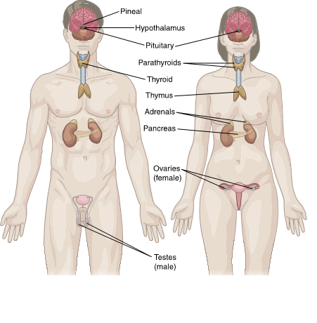
(ATI, 2019a)
Thymus
As shown in Figure 1, the thymus lies beneath the sternum and contains lymphoid tissue. It reaches its maximum size at puberty and then gradually atrophies. The thymus produces the hormones thymopoietin, thymulin, thymus humoral factor, and thymosin, which promote the growth of peripheral lymphoid tissues. The thymus's primary role is to produce T-lymphocytes (T-cells), which are crucial for cell-mediated immunity (Rogers & Brashers, 2023; Venes, 2025).
Hypothalamus
The hypothalamus lies in the lower central part of the brain, above the pituitary gland and the brainstem (refer to Figure 2). Its key role is to maintain the body's homeostasis. It is comparable in size to an almond and is fundamental for regulating body temperature, metabolism, and satiety. As mentioned, the hypothalamus controls the release of hormones from the pituitary gland and monitors hormone levels. The hypothalamus and the pituitary gland work together through a feedback mechanism known as the HPA axis. Through this axis (refer to Figure 3), signals and feedback from several areas of the central nervous system reach the hypothalamus regarding...
...purchase below to continue the course
evels, and the hypothalamus then sends a message to the pituitary gland to release or inhibit hormones in response (ATI, n.d.; Rogers & Brashers, 2023).
Figure 2
Hypothalamus, Pituitary, and Pineal Gland Anatomy

(ATI, 2019e)
Figure 3
Hypothalamic-pituitary axis

(ATI, 2017b)
The hypothalamus serves as a central coordinator for the entire endocrine system. Hypothalamic dysfunction can result from tumors, infections, head injuries, or iatrogenic factors such as surgery, medications, or radiation therapy. Dysfunction of this gland can lead to disturbances in appetite, sleep, thirst, mood, sexual function, growth, excretion, breastfeeding, and labor. Although the hypothalamus generates oxytocin and ADH, both are transported to the posterior pituitary for release. The hypothalamus also directly regulates appetite by secreting orexin and ghrelin—both appetite stimulants—and receives feedback from adipose tissue via the hormone leptin, an appetite suppressant (Shahid et al., 2023; Welt, 2025).
Pituitary Gland
As shown in Figure 2, the pituitary is about the size of a pea and lies beneath the hypothalamus. Based on input from the hypothalamus and continuous monitoring of circulating hormone levels, the pituitary secretes hormones that regulate the function of the other endocrine glands. Hypopituitarism occurs when the pituitary fails to produce a hormone or produces insufficient amounts. In contrast, hyperpituitarism occurs when the pituitary produces excess hormones (Rogers & Brashers, 2023). Examples of hormones regulated by the hypothalamic/pituitary system are listed in Table 1 and Figure 4.
The pituitary gland consists of two lobes, the anterior and posterior, which produce several hormones. The anterior lobe comprises several cell types: lactotrophs, somatotrophs, corticotrophs, gonadotrophs, thyrotrophs, and melanocytes. The posterior lobe of the pituitary is innervated by hypothalamic nerve cells and serves as a reservoir for oxytocin and ADH (Rogers & Brashers, 2023). Refer to Table 1 and Figure 4 for the hormones produced in each area and their functions.
Table 1
Hypothalamic/Pituitary System Hormones and Their Function
Releasing Hormone Produced by the Hypothalamus | Stimulating Hormone Produced by the Anterior Pituitary | Target Tissue/Organ | Target Hormone | Function of Target Hormone |
Thyrotropin-releasing hormone (TRH) | Thyroid-stimulating hormone (TSH) | Thyroid gland | Thyroid hormones (triiodothyronine [T3] and thyroxine [T4]) | T3 (triiodothyronine) and T4 (thyroxine) regulate metabolism, growth, development, and responsiveness to sympathetic nervous system stimulation. |
Corticotropin-releasing hormone (CRH) | Adrenocorticotropic hormone (ACTH) | Adrenal cortex | Cortisol | In response to stress, cortisol increases blood glucose (BG), cardiac output, and oxygen consumption. |
Gonadotropin-releasing hormone (GnRH) | Follicle-stimulating hormone (FSH), luteinizing hormone (LH) | Testes/ovaries | Estrogen, progesterone, testosterone | Estrogen, progesterone, and testosterone control sexual function and development. LH stimulates ovulation and formation of the corpus luteum in the ovary and promotes progesterone production. FSH stimulates the maturation of the ovarian follicles (females) or maintains spermatogenesis (males). |
Growth hormone-releasing hormone (GHRH) | Growth hormone (GH) | Liver (primary), bone, muscles, and other tissues | Insulin-like growth factor-1 (IGF-1) | IGF-1 mediates many of the growth-promoting effects of GH, including linear bone growth, cell proliferation, tissue growth, and protein synthesis. |
Dopamine (inhibitory) | Prolactin (PRL) | Mammary glands | Prolactin (PRL) | PRL stimulates mammary gland growth and development, as well as milk production following childbirth (refer to Figure 5) |
| Hormone Synthesized in the Hypothalamus and Released by the Posterior Pituitary | Target Tissue/Organ | | Function |
| Antidiuretic hormone (ADH) | Kidneys and blood vessels |
| ADH, or vasopressin, is stored in and released by the posterior pituitary. It regulates water reabsorption in the kidneys and contributes to vasoconstriction. |
| Oxytocin | Mammary glands and uterus |
| Oxytocin is also stored/released in the posterior pituitary. It contracts the uterus during childbirth and stimulates milk ejection (refer to Figure 5) |
(Carmichael, 2025; Kamel-ElSayed & Schwartz, 2023; Rogers & Brashers, 2023; Shahid et al., 2023; Welt, 2025)
Figure 4
Pituitary Hormones and Their Target Sites

(ATI, 2019f)
Figure 5
Hypothalamic-Pituitary Regulation of Prolactin and Oxytocin During Breastfeeding

(ATI, 2019c)
Pineal Gland
The pineal gland—sometimes called the pineal body—is located in the middle of the brain (refer to Figures 1 and 2). It is also known as the “spiritual third eye” or “third eye,” as research suggests that the pineal gland contains neurons that connect to the retina and that light affects its secretion. This gland helps maintain the circadian rhythm (sleep-wake cycle) and the timing and release of reproductive hormones through melatonin secretion. The production of melatonin is regulated by the hypothalamus and the pineal gland in response to signals from the retinal ganglion cells to regulate the sleep-wake cycle (Chaudhary et al., 2022; Rogers & Brashers, 2023).
Thyroid and Parathyroid Glands
The thyroid gland, shaped like a small butterfly, lies anterior to the trachea between the cricoid cartilage and the suprasternal notch (Figures 1 and 6). It produces hormones that regulate metabolism. The hypothalamic-pituitary-thyroid axis regulates thyroid hormone production (Rogers & Brashers, 2023; Young, 2025).
Figure 6
Thyroid and Parathyroid Glands

In children, thyroid hormones support bone, brain, and nervous system development. In adults, they affect blood pressure (BP), heart rate (HR), muscle tone, and reproductive functions. Thyroid hormones also regulate body temperature and metabolism and influence how tissues outside the thyroid function. The thyroid gland produces two primary hormones: thyroxine (T4) and triiodothyronine (T3). As shown in Figure 7, the release of T3 and T4 is regulated by the hypothalamic-pituitary-thyroid (HPT) axis. The process is initiated by the hypothalamus, which releases TRH. TRH stimulates the anterior pituitary gland to produce TSH. TSH binds to receptors on thyroid follicular cells, triggering the production and release of T3 and T4. The amount of TSH released into the bloodstream depends on the amount of T3 and T4 that the pituitary perceives, as it operates on a negative feedback loop (refer to Figure 7). The pituitary constantly monitors T3/T4 levels and responds to changes to maintain equilibrium. If the pituitary senses insufficient T4, it will boost TSH production, signaling the thyroid gland to produce more T4. Once T4 reaches an acceptable level in the blood, TSH production decreases. In circulation, T3 and T4 are bound to proteins such as thyroxine-binding globulin, albumin, and transthyretin (Rogers & Brashers, 2023; Young, 2025).
Figure 7
Hypothalamic-Pituitary-Thyroid Axis Feedback Mechanism

(ATI, 2019d)
Parathyroid Glands
The parathyroid glands are embedded in the thyroid gland's surface, as shown in Figures 1 and 6. The two pairs of parathyroid glands release parathyroid hormone (PTH), which regulates serum calcium and phosphorus levels and bone metabolism. When serum calcium levels are too low, PTH stimulates bone calcium release and reduces renal calcium excretion to raise them. It also increases calcitriol production by converting calcidiol to calcitriol in the kidneys, thereby enhancing calcium absorption in the digestive system (Lofrese et al., 2023; Rogers & Brashers, 2023).
Adrenal Glands
The adrenal glands lie superior to the kidneys and comprise two parts: the medulla and the cortex (refer to Figures 1 and 8). The adrenal cortex is the outer portion and consists of three distinct zones: the outer zona glomerulosa, the middle zona fasciculata, and the inner zona reticularis. Cortisol is produced in the zona fasciculata in response to ACTH. The inner zona produces androgens (Banasik, 2026; Rogers & Brashers, 2023).
Figure 8
Adrenal Gland

(ATI, 2017a)
The adrenal cortex produces corticosteroids (three types: mineralocorticoids, glucocorticoids, and androgens; refer to Table 2) that play a vital role in regulating metabolism, salt and water balance, the immune system, and sexual function (Grossman, 2026; Young, 2025). Steroids are lipid-soluble hormones produced on demand and released into the systemic circulation, where they bind to proteins such as albumin and transcortin (corticosteroid-binding globulin; Banasik, 2026). Cortisol (a glucocorticoid) and androgens bind to their specific proteins, whereas aldosterone (a mineralocorticoid) does not. The adrenal gland is regulated by a negative feedback system involving the hypothalamus, which secretes CRH, triggering the corticotrophs in the anterior pituitary to secrete ACTH. Rising levels of adrenal hormones in the circulation inhibit the secretion of both CRH and ACTH, completing the hypothalamus-pituitary-adrenal axis (Kamel-ElSayed & Schwartz, 2023). The medulla—the inner part of the adrenal gland—produces catecholamines, including epinephrine (adrenaline) and norepinephrine (fight-or-flight hormones). Catecholamines help the body respond to emotional or physical stress by increasing HR and BP (Grossman, 2026; Young, 2025).
When the adrenal glands do not produce enough hormones, adrenal insufficiency or Addison's disease can result. Understanding normal cortisol physiology is essential to understanding adrenal dysfunction. Cortisol helps control the body's use of fats, proteins, and carbohydrates; suppresses inflammation; regulates BP via vascular tone (a decrease in cortisol production is associated with a decrease in BP); increases BG; and may decrease bone formation. Cortisol influences the body’s response to stress and stressful situations (cortisol is a fight-or-flight hormone). Increased cortisol levels are needed to cope with acute stressors such as surgery. An inadequate release of cortisol in these situations can be fatal. Cortisol regulates the sleep/wake cycle and boosts energy during periods of increased stress. Cortisol levels vary throughout the day: the lowest occur at midnight, and the highest early in the morning (Grossman, 2026).
Table 2
Steroids of the Adrenal Gland and Their Function
Location | Type of Hormone | Actions |
Cortex | Mineralocorticoids | Aldosterone | Maintain sodium and water balance by retaining sodium and excreting potassium in the distal tubules of the kidneys in response to the renin-angiotensin system Regulates blood pH Regulates BP |
Glucocorticoids | Cortisol | Assists with the metabolism of glucose and opposes the effect of insulin (increases BG levels) Protein catabolism by releasing proteins stored in muscles to provide amino acids for the production of glucose in the liver (gluconeogenesis) Protection against stress Regulate the inflammatory and immune responses |
Adrenal Androgens(sex hormones) | Dehydroepiandrosterone (DHEAS) | Converts to form estrogen in the ovaries. Converts to androgens such as testosterone in the testes. Responsible for the development of secondary sex characteristics |
Medulla
| Catecholamines | Epinephrine (adrenaline) and Norepinephrine (“fight or flight”) | Sympathetic nervous system stimulation to assist with managing emotional/physical stress by increasing HR and BP |
(Banasik, 2026)
Testes and Ovaries
The gonads—testes and ovaries (refer to Figures 1 and 9)—are the reproductive glands and the primary source of sex hormones. They are regulated by the hypothalamus-pituitary-gonadal (HPG) axis through the secretion of GnRH, LH, and FSH. The hormones primarily produced by the testes are androgens; the most important is testosterone, which controls facial and pubic hair and sexual development. In male patients, testosterone helps regulate spermatogenesis and libido (sexual desire) and influences muscle and bone mass in both sexes. In female patients, the ovaries produce progesterone, estrogen, and testosterone. These hormones control the development of secondary sexual characteristics (e.g., breasts, pubic hair), menstruation, libido, and ovulation. Estrogen also affects libido, muscle and lipid mass, and fertility in males. These androgens and estrogen are also excreted in smaller quantities by the adrenal glands (Endocrine Society, 2022; Rogers & Brashers; Young, 2025). Testosterone must first be converted to estradiol by aromatase to function properly, affect the gonads, and promote hair growth; it must be converted to dihydrotestosterone by the enzyme 5-alpha-reductase to act on bone tissue (Snyder, 2026).
Figure 9
Hypothalamic-Pituitary-Gonadal (HPG) Axis

(ATI, 2019b)
The physiologic functioning of the reproductive system relies heavily on the appropriate secretion of numerous hormones. The HPG axis is central to this regulation, involving the hypothalamus's production of GnRH, followed by the anterior pituitary's secretion of LH and FSH (Figure 9). LH and FSH bind to G protein-coupled receptors and trigger the secretion of androgens by the testes and of estrogen and progesterone by the ovaries. LH also stimulates the secretion of progesterone and testosterone by the ovaries. In premenopausal adolescent and adult females, an LH surge also prompts ovulation. FSH stimulates the secretion of estrogen and the development of the ovum. Estrogen levels peak around ovulation and decline during menstruation (Endocrine Society, 2022; Kamel-ElSayed & Schwartz, 2023; Shahid et al., 2023).
In males, LH stimulates testicular testosterone secretion, and FSH supports spermatogenesis. Testosterone is crucial for developing secondary sexual characteristics during puberty, including penile and testicular enlargement, hair growth, voice deepening, increased bone and muscle mass, and increased skeletal height. Beyond puberty, testosterone supports spermatogenesis, libido, and the maintenance of bone and muscle mass (Endocrine Society, 2022; Kamel-ElSayed & Schwartz, 2023; Shahid et al., 2023).
Pancreas
The pancreas (refer to Figures 1 and 10) lies behind the stomach in the posterior abdomen. It has both digestive and hormonal functions, with endocrine (insulin) and exocrine (digestive enzymes) roles. Insulin and glucagon are the two hormones secreted by the pancreas that help regulate blood glucose (refer to Figure 10). Pancreatic dysfunction can lead to DM (Rogers & Brashers, 2023; Young, 2025).
Figure 10
Pancreas and Its Hormones

(ATI, 2020)
Hormonal Release Mechanisms
As mentioned previously, the endocrine system operates via negative feedback. The hypothalamus and pituitary glands continuously monitor target hormone levels. When these levels decline, the hypothalamus secretes releasing hormones, which stimulate the pituitary gland to secrete stimulating or pituitary hormones. As listed in Table 1, the pituitary hormones have numerous and varied effects on the human body. These trophic hormones stimulate their target glands to increase hormone production. As target gland hormone levels rise, the secretion of releasing and stimulating hormones decreases. This process is known as the HPA. This mechanism can be complex, as many of these hormones exhibit secondary effects. For example, PRL affects the hypothalamus-pituitary-gonadal axis: increased circulating PRL levels inhibit hypothalamic GnRH secretion, leading to reduced pituitary LH/FSH secretion and reduced secretion of sex hormones (estrogen, progesterone, and androgens) by the target glands. TRH, primarily responsible for triggering the secretion of TSH by the anterior pituitary, also appears to facilitate the production of PRL (Kamel-ElSayed & Schwartz, 2023; Shahid et al., 2023; Welt, 2025).
Chemical regulation occurs when endocrine glands not regulated by the pituitary gland are controlled by other substances that stimulate hormone secretion. Examples include BG levels that regulate the release of glucagon and insulin by the pancreas and serum calcium levels, which affect the release of PTH by the parathyroid glands. The third method of hormonal control involves the central nervous system, which may provide sensory input through the hypothalamus or directly to one of the glands of the endocrine system, such as the regulation of melatonin production by the pineal gland in response to ambient light or the release of catecholamines by the adrenal gland in response to stress (Kamel-ElSayed & Schwartz, 2023; Shahid et al., 2023; Welt, 2025).
Aging
Older adults usually experience changes in their endocrine system function as part of aging. Significant endocrine changes associated with aging may include the following:
- declining production of sex hormones (estrogen, progesterone, and testosterone)
- decreasing aldosterone levels, which can contribute to orthostatic hypotension
- increasing PTH, which may lead to osteoporosis
- rising FSH, LH, and norepinephrine levels
- alterations in glucose metabolism, such as increased BG when faced with physical or mental stress (Rogers & Brashers, 2023; Warde et al., 2023)
Endocrine System Dysfunction
Certain endocrine disorders can result from hyposecretion, in which insufficient amounts of a hormone are produced or released. This can result from problems with the trophic hormones (ACTH, TSH), which cause the target gland to secrete less of the hormone. Alternatively, disorders of the endocrine system can result from hypersecretion, in which excess amounts of a hormone are produced and released. Decreased responsiveness may also result from hormone resistance caused by receptor abnormalities, receptor deficiencies, or postreceptor signaling defects.
This resistance often presents with clinical manifestations similar to those of hyposecretion despite normal or elevated levels of circulating hormones (Banasik, 2026).
For learners who are eager to access additional content related to the endocrine system and endocrine disorders, please refer to the following NursingCE courses:
- Diabetes
- Osteoporosis
- Thyroid Dysfunction
- Sexual Dysfunction
References
Assessment
Technologies Institute. (n.d.). Engage adult medical surgical RN. Retrieved
July 15, 2026, from www.atitesting.com.
Assessment
Technologies Institute. (2017a). Adrenal gland [image].
https://cms.ascendlearning.com/share/page/site/digital-asset-librarytest/document-details?nodeRef=workspace://SpacesStore/10bd6be1-5bcb-4186-abc6-dcec37068e7b
Assessment
Technologies Institute. (2017b). Hypothalamic pituitary axis [image].
https://cms.ascendlearning.com/share/page/site/digital-asset-librarytest/document-details?nodeRef=workspace://SpacesStore/4b8521bf-c996-4ea5-a66e-5c94bde5ebcb
Assessment Technologies Institute. (2019a). The endocrine system [image]. https://cms.ascendlearning.com/share/page/site/digital-asset-librarytest/document-details?nodeRef=workspace://SpacesStore/39e2d9e3-e7cf-49d3-943f-efda9f22eab2
Assessment
Technologies Institute. (2019b). Hypothalamic-pituitary-gonadal (HPG) axis
[image]. https://cms.ascendlearning.com/share/page/site/digital-asset-librarytest/document-details?nodeRef=workspace://SpacesStore/97299daa-3f8c-47cf-8e98-b9f4b3ecd26a
Assessment Technologies Institute. (2019c). Hypothalamic-pituitary regulation of prolactin and oxytocin during breastfeeding [image]. https://cms.ascendlearning.com/share/page/site/digital-asset-librarytest/document-details?nodeRef=workspace://SpacesStore/448645c5-8f29-4e03-b9da-065c9ad1563d
Assessment Technologies Institute. (2019d). Hypothalamic-pituitary-thyroid axis feedback mechanism [image]. https://cms.ascendlearning.com/share/page/site/digital-asset-librarytest/document-details?nodeRef=workspace://SpacesStore/905fd39c-2cd1-4804-ac79-dc704a400a93
Assessment Technologies Institute. (2019e). Hypothalamus, pituitary, and pineal gland cross-section view of anatomy [image]. https://cms.ascendlearning.com/share/page/site/digital-asset-librarytest/document-details?nodeRef=workspace://SpacesStore/4d9ec632-b888-43ad-a05e-e0e69af8da6f
Assessment Technologies Institute. (2019f). Pituitary hormones and their target sites [image]. https://cms.ascendlearning.com/share/page/site/digital-asset-librarytest/document-details?nodeRef=workspace://SpacesStore/9dba5cc9-d935-4359-848d-59b066e13b9d
Assessment
Technologies Institute. (2020). Pancreas and its hormones [image]. https://cms.ascendlearning.com/share/page/site/outdoor-emergency-care-6th-edition/document-details?nodeRef=workspace://SpacesStore/cdefa5a1-8db4-42aa-86fb-ae15c9a54076
Banasik, J. L. (2026). Pathophysiology (8th ed.). Elsevier.
Carmichael, J. D. (2025). Gigantism and acromegaly. Merck
Manual Professional Version. Retrieved July 15, 2026, from https://www.merckmanuals.com/professional/endocrine-and-metabolic-disorders/pituitary-disorders/gigantism-and-acromegaly
Centers for Disease Control and Prevention. (2026). National diabetes statistics report. Retrieved
July 14, 2026, from https://usdss.cdc.gov/diabetes/report.html
Chaudhary, S., Rishi, K. B., Yadav, S., & Upadhayay, P. (2022). Pineal gland and the third eye anatomy history revisited – a systematic review of literature. Cardiometry, (25),
1363-1368. https://doi.org/10.18137/cardiometry.2022.25.13631368
Endocrine Society. (2022). Reproductive hormones. Retrieved July 15, 2026, from https://www.endocrine.org/patient-engagement/endocrine-library/hormones-and-endocrine-function/reproductive-hormones
Grossman, A. B. (2026). Overview of adrenal function. Merck Manual Professional Version. Retrieved July 15, 2026, from https://www.merckmanuals.com/professional/endocrine-and-metabolic-disorders/adrenal-disorders/overview-of-adrenal-function
Kamel-ElSayed, S. A., & Schwartz, J. (2023). Physiology, pituitary gland. StatPearls. Retrieved July 15, 2026, from https://www.ncbi.nlm.nih.gov/books/NBK459247
Lofrese, J. J., Basit, H., & Lappin, S. L. (2023). Physiology, parathyroid. StatPearls. Retrieved July 15, 2026, from https://www.ncbi.nlm.nih.gov/books/NBK482510
National Institute of Diabetes and Digestive and Kidney Disease. (2024). Endocrine diseases & metabolic diseases. Retrieved
July 15, 2026, from
https://www.niddk.nih.gov/about-niddk/research-areas/endocrine-metabolic-diseases
Rogers, J. L. & Brashers, V. L. (Eds.). (2023). McCance
and Huether's pathophysiology: The biologic basis for disease in adults
and children (9th ed.). Elsevier.
Santo, L., Peters, Z. J., Guluma, L., & Ashman, J. (2025). Visits to health centers, by selected characteristics: United States, 2023 (National Health Statistics Reports No. 216). National Center for
Health Statistics. https://www.cdc.gov/nchs/data/nhsr/nhsr216.pdf
Shahid, Z., Asuka, E., & Singh, G. (2023). Physiology, hypothalamus. StatPearls. Retrieved
July 15, 2026, from https://www.ncbi.nlm.nih.gov/books/NBK535380/
Snyder, P. J. (2026). Testosterone treatment of male hypogonadism. UpToDate. Retrieved
July 15, 2026, from https://www.uptodate.com/contents/testosterone-treatment-of-male-hypogonadism
Venes, D. (2025). Taber's cyclopedic medical dictionary (25th ed.). F. A. Davis Company.
Warde, K. M., Smith, L. J., & Basham, K. J. (2023). Age-related changes in the adrenal cortex: Insights and implications. Journal
of the Endocrine Society, 7(9).
https://doi.org/10.1210/jendso/bvad097
Welt, C. K. (2025). Hypothalamic-pituitary axis. UpToDate. Retrieved July 15, 2026, from https://www.uptodate.com/contents/hypothalamic-pituitary-axis
Young, W. F., Jr. (2025). Overview of endocrine system. Merck Manual Professional Version. Retrieved July
15, 2026, from https://www.merckmanuals.com/professional/endocrine-and-metabolic-disorders/principles-of-endocrinology/overview-of-the-endocrine-system
Powered by Froala Editor