About this course:
This course reviews the most common blood clotting and bleeding disorders, including each disease process’s clinical manifestations and implications, and the clinical considerations for prescribing therapy to enhance clinical practice and improve patient outcomes.
Course preview
Blood Clotting and Bleeding Disorders for Nurses
This course reviews the most common blood clotting and bleeding disorders, including each disease process’s clinical manifestations and implications, and the clinical considerations for prescribing therapy to enhance clinical practice and improve patient outcomes.
By the completion of this module, learners should be able to:
- review the physiology of normal bleeding and blood clotting, including the distinction between primary and secondary hemostasis and the clotting cascade and coagulation factors
- explore the pathologic mechanisms of blood clotting failure or impairment, delineating the most common domains of dysfunction
- discuss anticoagulation therapy, reviewing the most common classes of medications, their indications, monitoring parameters, and side effects
- describe the most common disorders of excessive blood clotting, including inheritance patterns, clinical manifestations, and recommended treatments
- describe the most common bleeding disorders, including inheritance patterns, clinical manifestations, and recommended treatments
When the body’s physiologic mechanisms of blood clotting fail, excessive blood clotting or bleeding can ensue. Blood clotting disorders occur when factors that promote blood clotting (procoagulants) and those that inhibit clotting (anticoagulants) are imbalanced. Blood clotting disorders fall into two main categories: bleeding disorders and hypercoagulable states. Bleeding disorders are characterized by an impaired ability to effectively form a blood clot (or thrombus) in response to an injury. Hypercoagulable states occur when there is an increased tendency to develop a thrombus in the arteries and veins. These conditions can be inherited (congenital) due to specific genetic defects present at birth or acquired (developed), resulting from an underlying medical condition, trauma, surgery, or medication. If not promptly identified and effectively treated, blood clotting disorders can lead to serious health consequences, including hemorrhage, myocardial infarction, stroke, organ failure, and death. To understand blood clotting disorders, it is first essential to understand the physiologic basis of blood cell production and the mechanisms of bleeding and blood clotting (Norris, 2024; Rodgers & Young, 2024).
Hematopoiesis
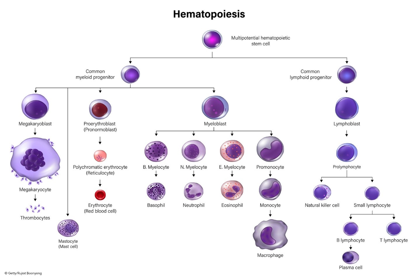
Hematopoiesis is the ongoing process of blood cell production in the human body that primarily occurs in the bone marrow. The process is regulated through a series of steps in which undifferentiated cells are biochemically stimulated to undergo mitotic cell division (i.e., proliferation) and cell maturation (i.e., differentiation). Hematopoiesis continues throughout the life span, maintaining homeostasis within the body in response to infection or injury. When there is an increase in the destruction of circulating cells, such as during an acute bleeding event, hematopoiesis accelerates to generate more cells and compensate for the loss. In long-term dysfunction, as in chronic illness, there is a more significant increase in hematopoiesis than in acute conditions such as hemorrhage. As demonstrated in Figure 1, every cell type in the body originates from hematopoietic stem cells. The stem cells grow, multiply, and differentiate under the control of cytokines and growth factors. During differentiation, the stem cells follow distinctive paths to maturity and travel down committed lines of blood cells, primed to perform a specific function. The average human body requires nearly 100 billion new blood cells per day, which is why hematopoietic stem cells are self-renewing or can proliferate by themselves so that a relatively constant population of stem cells is always readily available (Libretti & Puckett, 2023; Rodgers & Young, 2024; Rogers, 2022).
Figure 1
Hematopoiesis
Components of Blood
Blood is a v ital component of the body. It serves several important functions, including transporting oxygen and nutrients to tissues within the body, regulating temperature, and responding to blood vessel injuries to maintain homeostasis. The amount of blood within each individual depends on their size, but the average adult has approximately 5 L (1.3 gallons) of blood circulating. Blood consists of both solid (formed) elements (i.e., white blood cells [WBCs], red blood cells [RBCs], and platelets) and liquid components (i.e., plasma; Norris, 2024; Rodgers & Young, 2024).
WBCs
WBCs are components of the immune system that work to fight infection and other illnesses. WBCs comprise five specific subtypes (neutrophils, monocytes, macrophages, eosinophils, and basophils). Each WBC serves a particular function in mediating the inflammatory and immune response to infection. The most common type of WBC is the neutrophil, accounting for 55%–70% of the total WBC count. WBCs have variable life spans; some may live only 24 hours, but the average WBC life span is 13–20 days. When whole blood is separated into its components, the WBCs and platelets are contained within the middle layer (i.e., buffy coat) and constitute 1% of the total blood volume (American Society of Hematology [ASH], n.d.; Norris, 2024; Rodgers & Young, 2024).
RBCs
RBCs are mature red blood cells (erythrocytes) and carry hemoglobin, a protein that transports oxygen from the lungs to all the tissues within the body. The body relies on oxygen as a critical component for all cellular functioning and processes. Hemoglobin also carries waste products (mainly carbon dioxide) from the tissues to the lungs, where waste is expelled through breathing. Erythrocytes comprise about 45% of total blood, have an average life span of 120 days, and appear pink due to their high hemoglobin content. Hematocrit reflects the percentage of RBCs in a given blood volume (ASH, n.d.; Norris, 2024; Rodgers & Young, 2024).
Platelets
Platelets, also called thrombocytes, are critical in all aspects of bleeding and clotting. They are produced in the bone marrow from cells called megakaryocytes. The process of platelet differentiation is regulated under the control of thrombopoietin (TPO), a hormone produced by the liver and kidneys. TPO influences the growth and differentiation of myeloid stem cells into megakaryoblasts, the precursor cell to the promegakaryocyte, which, in turn, develops into a giant megakaryocyte. As demonstrated earlier in Figure 1, megakaryocytes are depicted as notably larger than all the other cells because each megakaryocyte is prepared to produce and release more than 1,000 platelets, accounting for their large size. Platelets are colorless blood cell fragments essential for blood clotting in response to an injury. They contain proteins on their surfaces, allowing them to stick to each other and blood vessel walls. Their primary function is to gather at the...
...purchase below to continue the course
Plasma
Plasma is an aqueous component of blood that carries nutrients, proteins, and hormones throughout the body and transports waste products to the kidneys and digestive tract for removal. Plasma accounts for about 55% of whole blood volume and comprises water (92%), with 8% of dissolved substances (solutes). Most of these dissolved solutes are plasma proteins, encompassing about 7% of the total plasma volume. These include albumin, immunoglobulins, and clotting factors. Electrolytes, nutrients, wastes, and other solutes make up the remaining 1%. The liver produces the majority of plasma proteins. Clotting factors function to stop bleeding from damaged vessels and promote coagulation (ASH, n.d.; Norris, 2024; Rodgers & Young, 2024; Rogers, 2022).
Physiologic Mechanisms of Blood Clotting (Coagulation)
Blood clotting, commonly called coagulation, is a vital mechanism that protects against blood loss due to bleeding. A complex interplay mediates the process between vascular mechanisms, enzymes, hormones, proteins, and cells. Hemostasis, or the cessation of bleeding, refers to the body’s physiologic response to stop bleeding from an injured blood vessel to prevent blood loss. The coagulation process is divided into two stages: primary hemostasis (which results in the formation of a weak platelet plug) and secondary hemostasis (which stabilizes the weak platelet plug, evolving into a secure and insoluble fibrin clot; Garmo et al., 2023; Norris, 2024). The defining components of each process are as follows:
- Primary hemostasis:
- vascular spasm or vasoconstriction of the blood vessel
- platelet adhesion
- platelet activation (or secretion)
- platelet aggregation
- Secondary hemostasis:
- activation of clotting factors
- conversion of prothrombin to thrombin
- conversion of fibrinogen to fibrin (Garmo et al., 2023)
Upon recognizing an injury, the blood vessel constricts (tightens) to reduce blood flow to the area to prevent further blood loss. The damaged endothelium synthesizes endothelin-1, a potent vasoconstrictor. Collagen and proteins, including von Willebrand factor (vWF), leak into the bloodstream. vWF is a protein and the primary mediating factor for platelet adhesion, acting like a glue that binds the platelets with enough strength to withstand stress and prevent detachment. Thromboxane A2, a potent vasoconstrictor, is formed during platelet activation; it functions as a stimulus for platelet aggregation and mediates the release of adenosine diphosphate (ADP). Platelets primarily bind to collagen and exposed vWF at the site of a damaged blood vessel. This binding triggers the first platelets to release signals that recruit additional platelets and activate them. One key signal is ADP, a platelet agonist that attracts and activates more platelets. ADP also enables platelets to clump together and change shape, increasing their surface area. Platelets circulating in the blood travel to the injury site and adhere to the vessel walls, forming a temporary platelet plug. Calcium is released and attaches to the phospholipids (a main component of cellular membranes), which appear secondary to the platelet activation, offering a surface for various coagulation factors. Thrombin, formed during the coagulation cascade (which will be reviewed in the next section), is the most potent activator of platelets and is considered the key coagulation enzyme. Fibrinogen and platelets create a weak platelet plug that temporarily protects against further bleeding. Secondary hemostasis subsequently kicks in, and the clotting factors stabilize the platelet plug and convert it into a hard, insoluble fibrin clot (Garmo et al., 2023; Kennelly et al., 2026; Norris, 2024).
Clotting Factors
There are 12 clotting factors (F) labeled in Roman numerals (FI through FXIII), as outlined in Table 1. Of note, there is no clotting FVI. Clotting factors are proteins secreted primarily by the platelets and liver; they depend on calcium ions (Ca2+) and vitamin K. Ca2+ is vital for accelerating blood clotting. Vitamin K is a naturally occurring fat-soluble vitamin synthesized by bacteria residing in the large intestine and is also consumed in the diet. Vitamin K assists with blood clotting, bone metabolism, and the regulation of blood calcium levels and is required to produce prothrombin (FII). Therefore, vitamin K deficiencies can lead to bleeding and hemorrhage, bone mineralization, and osteoporosis. Ca2+, otherwise referred to as FIV, is derived from the breakdown of bone and is also acquired through the diet (Norris, 2024; Rogers, 2022).
Table 1
Clotting Factors
Factor | Common Name | Function |
FI | Fibrinogen | Assists with fibrin clot formation |
FII | Prothrombin* | Assists FVIIIa, FIXa, and FVa to form thrombin |
FIII | Tissue factor (TF) | Assists FVII and FIV in activating FIX and FX |
FIV | Ca2+ | Activates multiple clotting factors |
FV | Proaccelerin (labile factor) | Assists FVIIIa, FIxa, FXa, and FII in the formation of thrombin |
FVII | Proconvertin (stable factor)* | Assists TF and FIV in activating FIX and FX |
FVIII | Antihemophilic factor (AHF) | Amplifies additional thrombin formation |
FIX | Plasma thromboplastin* component (PTC) or Christmas factor | Assists FVa, FVIIIa, FXa, and II to form thrombin |
FX | Stuart-Prower factor* (thrombokinase) | Assists FVIIIa, FIxa, FVa, and II to form thrombin |
FXI | Plasma thromboplastin antecedent (PTA) or antihemophilic factor C | Increases thrombin production inside fibrin clots through the intrinsic pathway; slows fibrinolysis |
FXII | Hageman factor (contact factor) | Contact activator of the kinin system |
FXIII | Fibrin stabilizing factor (FSF) | Forms bonds between fibrin strands during secondary hemostasis |
*Vitamin K required (Kennelly et al., 2026; Norris, 2024)
Coagulation Cascade
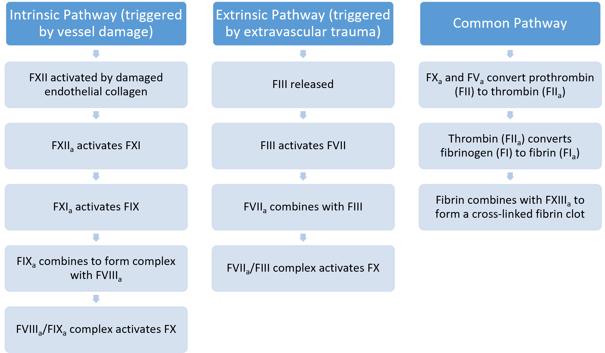
As Figure 2 demonstrates, the coagulation cascade is activated through two distinct yet interconnected pathways (intrinsic and extrinsic) that converge into the common pathway, leading to fibrin clot formation. All three pathways are mediated by hormones, proteins, and clotting factors through a series of complex events. The ultimate goal of the coagulation cascade is to convert fibrinogen (F1), a soluble plasma protein, into fibrin, a nonsoluble plasma protein (Kennelly et al., 2026; Norris, 2024). The following three essential steps can summarize the coagulation cascade:
- extrinsic and intrinsic pathways form the enzyme prothrombinase
- prothrombinase converts prothrombin to thrombin
- thrombin converts fibrinogen into fibrin (Kennelly et al., 2026; Norris, 2024)
Extrinsic Pathway
Clotting factors involved in the extrinsic pathway include FVII and FIII. The beginning of the process of fibrin clot formation is carried out by the extrinsic pathway, which is activated in response to external tissue trauma, in which blood escapes from the vascular system. Once the damage is identified, TF (FIII), a cell membrane protein, is released from the damaged cells. TF binds with procovertin (FVII or stable factor), activating it into FVIIa, thereby initiating the clotting cascade. The extrinsic pathway is quicker and less complex than the intrinsic pathway (Garmo et al., 2023; Kennelly et al., 2026).
Intrinsic Pathway
Clotting factors in the intrinsic pathway include FXII, FXI, FIX, and FVIII. The intrinsic pathway is activated by trauma that begins inside the vascular system, prompted by internal damage to the vessel wall. FXII is activated into FXIIa when it comes in contact with the damaged endothelial collagen, thereby igniting the intrinsic pathway. This pathway is slower than the extrinsic pathway but is considered more important (Garmo et al., 2023; Kennelly et al., 2026).
Common Pathway
Clotting factors involved in the common pathway include FX, FV, FII, FI, and FXIII. The extrinsic and intrinsic pathways converge into the common pathway with the activation of FX into FXa, which subsequently combines with FVa and Ca2+ on phospholipid surfaces. This process activates FII into thrombin, which then activates FXIIIa. FXIIIa interacts with fibrin to form the stabilized clot (Garmo et al., 2023; Kennelly et al., 2026).
Figure 2
The Steps of the Coagulation Cascade
Antithrombotic Mechanisms
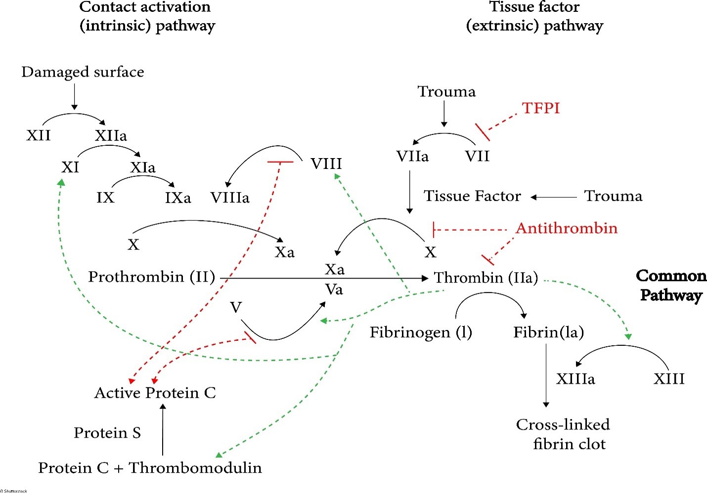
Parallel to the coagulation cascade, the body has an innate, highly regulated process for controlling and preventing abnormal blood clot development. Antithrombotic mechanisms are naturally occurring anticoagulants in the body. An anticoagulant refers to any substance that opposes coagulation. Coagulation is regulated by several antithrombotic mechanisms to preserve the fluidity of the blood and limit blood clotting to vascular injury sites, so blood clots do not form under normal conditions. The endothelial cells provide several antithrombotic effects. They produce substances that inhibit platelet binding, secretion, and aggregation and generate anticoagulant factors such as tissue factor pathway inhibitor (TFPI) and antithrombin (AT). Various circulating plasma anticoagulants instinctively function to limit the coagulation process to the area of injury and to reestablish and maintain the patency of blood vessels. Endothelial cells also activate the fibrinolytic mechanisms by producing tissue plasminogen activator 1 (tPA) and plasminogen activator inhibitor. Further, basophils of the immune system also play a fundamental role in preventing blood clotting by releasing heparin, a short-acting anticoagulant that opposes prothrombin. Heparin is additionally found on the surfaces of cells lining the blood vessels and promotes blood fluidity (Garmo et al., 2023; Kennelly et al., 2026). The major physiologic antithrombotic pathways and their impact on the coagulation cascade are illustrated in Figure 3 (written in red) and described in what follows.
Figure 3
Coagulation Cascade with Antithrombotic Mechanisms
AT
Previously referred to as antithrombin III, AT is the major inhibitor of thrombin and other clotting factors in the coagulation cascade. AT is activated by binding to heparin on endothelial cell surfaces. It binds to and inactivates FIXa, FXa, FXIa, and FXIIa and opposes the conversion of prothrombin (FII) to thrombin in the common pathway. AT neutralizes thrombin and other activated clotting factors on the vascular surface (Garmo et al., 2023; Rodgers & Young, 2024).
Protein C and Protein S
Proteins C and S (Protein C/S) are plasma proteins that regulate blood clot formation. Protein C inactivates clotting factors involved in the intrinsic pathway, which is a natural anticoagulant when activated by thrombin. Thrombin-induced activated protein C (APC) occurs on thrombomodulin, a transmembrane binding site for thrombin on the endothelial cell surface. APC subsequently inactivates FVa and FVIIIa, slowing down blood clot formation. Protein S exists in two forms: free and bound to another protein. As demonstrated in Figure 3, free protein S is a cofactor to protein C, accelerating the inactivation of FVa and FVIIIa (Garmo et al., 2023; Rodgers & Young, 2024).
TFPI
TFPI is generated primarily by endothelial cells and inhibits the TF/FVIIa/FXa complex, turning off the initiation of the coagulation cascade’s extrinsic pathway. TFPI is bound to lipoprotein and can be released by heparin. It also circulates in small quantities in plasma in a free form, acting as a physiologically active anticoagulant (Garmo et al., 2023; Rodgers & Young, 2024).
Fibrinolytic System
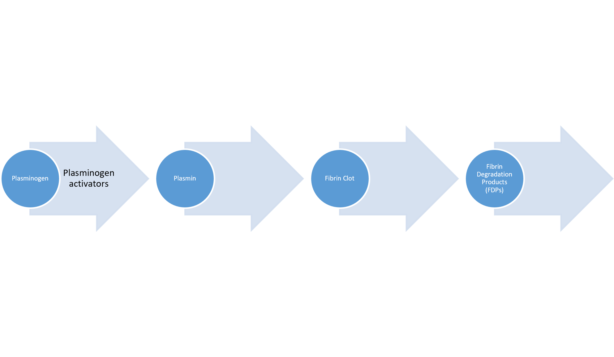
Any thrombin that evades the inhibitory effects of the body’s natural anticoagulant system converts fibrinogen to fibrin. However, once the injured blood vessel heals, the clot must eventually be removed to reestablish normal blood flow and maintain the patency of the blood vessel. Therefore, the fibrinolytic system and fibrinolysis process are activated to facilitate the breakdown and removal of these unwanted fibrin deposits. In addition to fibrin degradation, fibrinolysis regulates the abnormal development of blood clots and manipulates the extent of blood clot formation. Plasmin is the primary enzyme of the fibrinolytic system, functioning to digest fibrin into fibrin degradation products (FDPs). Plasmin is formed through a series of reactions, as demonstrated in Figure 4. tPA is released from endothelial cells and binds to the fibrin clot. Subsequently, inactivated plasminogen (an enzyme that degrades fibrin clots) is converted into active plasmin. The process of fibrinolysis can be primary or secondary. Primary fibrinolysis is the body’s natural process of maintaining homeostasis, as described previously. In contrast, secondary fibrinolysis refers to the dysfunctional breakdown of blood clots secondary to medical disorders, infections, or medications and can induce severe bleeding (Garmo et al., 2023; Leung, 2025; Rodgers & Young, 2024).
Figure 4
Fibrinolytic System
Thrombosis
Thrombosis is a pathologic process with an increased tendency to develop abnormal blood clots (thrombi) within uninjured blood vessels. A thrombus is a stationary clot attached to the vessel wall, composed primarily of fibrin and other blood cells. The thrombus can grow large enough to reduce or obstruct blood flow within the vessel, depriving tissues and organs of vital nutrients and inducing tissue ischemia and hypoxia (Hinkle et al., 2025; Rogers, 2022).
Virchow’s Triad
Rudolf Virchow, often referred to as the “father of modern pathology,” is a 19th-century physician credited with identifying three significant predisposing abnormalities that can lead to the development of thrombosis. Branded “Virchow’s triad,” the three pathologic mechanisms influencing blood clot formation are as follows:
- damage to the blood vessel wall
- blood flow abnormalities
- hypercoagulability of the blood (Hinkle et al., 2025; Rogers, 2022)
Endothelial Injury
As described earlier, vascular endothelial injury under physiologic conditions leads to platelet activation and blood clot formation to prevent blood loss and restore the integrity of the vascular wall. However, when these mechanisms are impaired or there is an underlying defect, pathologic consequences can ensue. Endothelial injury can be precipitated by numerous insults that extend beyond external tissue trauma, such as smoking, surgery, atherosclerotic disease, inflammation, or chronic elevations in blood pressure. Regardless of the precipitating event, normal blood flow is altered when the endothelium is injured, inducing “turbulence” within the vessel. Turbulence is the increased blood flow rate over the injured surface, generating disordered currents and increased friction within the vessel. Atherosclerotic plaques from hyperlipidemia commonly cause turbulent environments within blood vessels (Bauer & Lip, 2026; Hinkle et al., 2025; Rogers, 2022).
Venous Stasis
Venous stasis refers to the loss of blood flow within the vessel. Blood pooling is a significant risk factor for thrombus formation, as during venous stasis, platelets remain in contact with the endothelium for a prolonged period. Clotting factors, usually diluted by blood flow, become concentrated and potentially activated. The most common conditions predisposing patients to venous stasis include immobility, paralysis, venous insufficiency, varicose veins, elevated body mass index (BMI), heart failure, and low cardiac output (Bauer & Lip, 2026; Hinkle et al., 2025; Rogers, 2022).
Hypercoagulability
Abnormalities within the coagulation cascade, fibrinolytic pathways, or platelet function contribute to hypercoagulability and subsequent thrombus formation. Hypercoagulability may result from genetic or acquired etiologies and is usually multifactorial, with lifestyle and environmental factors playing key roles. Hypercoagulability resulting from inherited defects includes Factor V Leiden (FVL), prothrombin mutation (G20210A), or deficiencies of the natural proteins that prevent clotting (described in the next section of this module). Some individuals may present with increased levels of certain clotting factors without any identifiable genetic mutation. The most common causes of acquired hypercoagulability include malignancy, inflammation, immobility from paralysis or prolonged bed rest, surgery (especially the immediate postoperative period), pregnancy, and postpartum. Certain medications are known to increase the coagulability of the blood, such as oral contraceptives and hormone replacement therapy (Bauer & Lip, 2026; Hinkle et al., 2025; Rogers, 2022). Additional risk factors for thrombus formation that extend beyond those identified within Virchow’s triad include the following:
- personal history of blood clots
- family history of blood clots (even without an identifiable inherited disorder)
- hypertension
- chronic inflammatory diseases
- diabetes
- elevated BMI
- age (increased risk in people over the age of 60)
- presence of multiple concurrent risk factors (i.e., smoking cigarettes while taking oral contraceptive medications; Ashorobi et al., 2024; Bauer & Lip, 2026; Hinkle et al., 2025; Rodgers & Young, 2024)
Thrombosis can be provoked or unprovoked, which is essential for determining the duration of therapy. “Unprovoked” implies that no identifiable risk factor or underlying cause is evident; a provoked thrombus is caused by a known event, such as surgery or prolonged immobility. Unprovoked events are considered more serious and require specific diagnostic workup and treatment, usually under the care of a hematologist (Lip & Stevens, 2025).
Arterial Versus Venous Thrombosis
A thrombus can develop anywhere within the cardiovascular system, with the majority classified as venous (within a vein) or arterial (within an artery). Arteries are the blood vessels responsible for carrying oxygen-rich blood away from the heart to the body tissues, whereas veins are the blood vessels that carry blood back to the heart. Venous thrombosis is also called venous thromboembolism (VTE) and includes two major types: deep vein thrombosis (DVT) and superficial vein thrombosis (SVT). DVT is the most common type, arising in the large veins of the lower extremity. Arterial thrombi almost always arise from the heart (i.e., aorta, coronary arteries) but may also occur in the brain’s cerebral arteries. Arterial thrombosis is commonly caused by arteriosclerosis, a condition in which a fatty substance called plaque builds up along the walls of arteries. This plaque formation hardens and narrows arteries, and the plaque can crack or rupture, attracting platelets and eventually leading to blood clots. While thrombi vary in shape and size, they all commonly retain an area of attachment to an underlying vessel or heart wall. The distinguishing features and characteristics of venous and arterial thrombi are outlined in Table 2 (Ashorobi et al., 2024; National Heart, Lung, and Blood Institute [NHLBI], 2022c; Streiff, 2025d).
Table 2
Comparison of Venous and Arterial Thrombi
Venous Thrombosis | Arterial Thrombosis |
More common than arterial thrombosis | Less common than venous thrombosis |
Occurs in sites of venous stasis (slow-moving blood) | Begins at sites of endothelial injury or turbulence (rapid blood flow) |
Tend to extend in the antegrade direction (direction of blood flow toward the heart) | Tend to grow in the retrograde direction (away from the heart) from the point of attachment |
Mixed or red color Composed mostly of fibrin and more RBCs due to sluggish blood flow | Pale/white color Composed of mostly platelets with some fibrin, RBCs, and WBCs |
Not firmly attached and prone to fragment (easily detach and embolize) | Firmly adhere to the injured endothelial wall; however, an embolus is possible |
(Ashorobi et al., 2024; Hinkle et al., 2025; Rodgers & Young, 2024)
Complications of Venous Thrombosis
Since thromboses can block the blood flow in both veins and arteries, complications depend on the clot’s location. The most serious and dangerous type of VTE is a pulmonary embolism (PE) or a blood clot within the lung. A thrombus detaches from the vessel wall and circulates within the bloodstream, causing a sudden blockage of a vessel called an embolism. As demonstrated in Figure 5, PE is most commonly caused by DVT, as a piece of the blood clot within the extremity breaks off, travels through the veins, and becomes lodged in the lung’s blood vessels. A PE can completely obstruct blood flow and induce sudden death in some patients. It can also cause hypoxia or low oxygen levels in the blood, leading to permanent lung damage or damage to other organs due to insufficient oxygen (Ashorbi et al., 2024; Hinkle et al., 2025; Rogers, 2022).
Figure 5
Deep Vein Thrombosis with Embolus Development
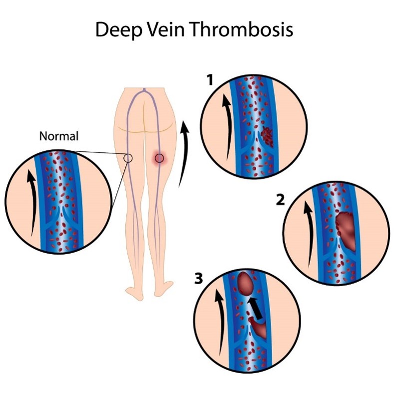
VTE can also lead to serious long-term complications, including postthrombotic syndrome (PTS), which is the most common complication of a DVT. PTS causes chronic pain and swelling in the affected extremity and, in severe cases, can lead to venous ulcers. Less commonly, patients can develop chronic thromboembolic pulmonary hypertension (CTEPH), in which abnormally high blood pressure in the arteries of the lungs causes the right side of the heart to work harder than normal, leading to heart failure (Ashorbi et al., 2024; Hinkle et al., 2025; Rogers, 2022).
Complications of Arterial Thrombosis
Arterial thrombosis and emboli can cause tissues to become starved of blood and oxygen, leading to necrosis and infarction of vital tissue and organs (i.e., heart, brain, kidney, spleen). As shown in Figure 6, coronary thrombosis is a blockage within an artery of the heart and causes a myocardial infarction (MI) or heart attack (Ashorbi et al., 2024; Hinkle et al., 2025; Rogers, 2022).
Figure 6
Coronary Thrombosis Inducing an MI
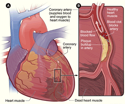
(NHLBI, 2022b)
Arterial thrombosis can also cause an ischemic stroke, which occurs when cerebral blood flow is reduced to critical levels or stopped altogether, as demonstrated in Figure 7. An ischemic stroke can be caused by a thrombus that originates within an artery that supplies blood to the brain or an embolus that becomes lodged in and blocks an artery that supplies blood to the brain. In both types of ischemic stroke, the plaque or clot keeps oxygen-rich blood from getting to a portion of the brain, and neurons deprived of oxygen will die within minutes (NHLBI, 2023).
Figure 7
Embolic Stroke
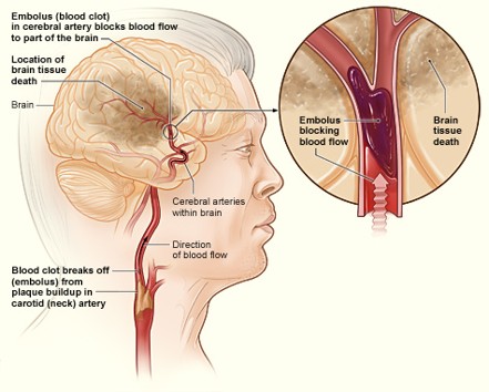
(NHLBI, 2023)
Ischemic stroke is most commonly caused by atherosclerosis but can also be due to atrial fibrillation (Afib), a type of cardiac arrhythmia. Afib is characterized by an irregular rhythm in which blood is not appropriately pumped out of the heart, leading to periods of blood pooling within the heart’s atrium. Afib increases the risk of embolic stroke if a blood clot forms there, is expelled, and travels up to the brain (NHLBI, 2023; Rogers, 2022).
Arterial emboli can also occur in the periphery, such as the legs and feet, reducing blood flow or causing acute ischemia of the lower limbs. Arterial emboli can occur in one of two ways: from an arterial embolism or an arterial thrombus induced by an atherosclerotic artery (atheroembolism) within the extremity. When emboli originate in the heart, over 70% will obstruct the lower limbs. The femoral artery at the femoral bifurcation is the most common site affected, accounting for 35%–50% of all cases. Other lower extremity arteries commonly affected include the popliteal and iliac. Complete obstruction of blood flow can lead to gangrene, in which the extremity becomes cold and painful and eventually turns black from necrosis or cell death. In these cases, patients usually require amputation (Braun, 2026; Casas, 2019).
Signs and Symptoms of Thrombosis
Since thrombosis can lead to significant morbidity and mortality, resulting in serious and life-threatening consequences, early identification and intervention are essential. The nurse should be well-versed in the most common signs and symptoms, which can vary depending on the location within the body affected. The most common signs and symptoms according to the affected body part are outlined in Table 3.
Table 3
Common Signs and Symptoms of Thrombosis
Diagnosis | Affected Body Part | Symptoms |
DVT | Extremity (arm or leg) |
|
PE | Lung |
|
Coronary thrombosis | Heart |
|
Stroke | Brain |
|
Arterial Embolism of the Lower Limb | Lower extremity |
|
(Casas, 2019; Centers for Disease Control and Prevention [CDC], 2024d; Rodgers & Young, 2024; Rogers, 2022)
Inheritance Patterns of Disease
It is essential to establish a basic understanding of the inheritance patterns of bleeding and clotting disorders that are attributed to genetic defects. Heterozygous is the term used to describe patients with a mutation in only one copy of the gene. In contrast, homozygous describes a patient who has inherited a mutation in both gene copies (National Institute of Health [NIH], 2021; Padiath, 2025).
Autosomal Dominant (AD)
In AD conditions, the abnormal gene is dominant and located on one of the chromosomes. The term “dominant” means that the patient only needs one mutated gene copy to be affected by the disorder. In some cases, an affected person inherits the condition from an affected parent. In other cases, the condition may develop from a new gene mutation, which occurs randomly in people with no history of the disorder in their family. As demonstrated in Figure 8 (top), a person with an AD disorder has a 50% chance of having an affected child with one abnormal gene and a 50% chance of having an unaffected child with two normal genes (NIH, 2021; Padiath, 2025).
Autosomal Recessive (AR)
In conditions that are AR, one abnormal gene must be inherited from each parent, as the patient requires two copies of the mutation to be affected by the disease. A carrier of the condition has one abnormal gene (recessive) and one normal gene (dominant) but is unaffected by the disorder. Therefore, as demonstrated in Figure 8 (bottom), two carriers have a 25% chance of having an unaffected child with two normal genes, a 50% chance of having a child who is an unaffected carrier, and a 25% chance of having a child with two recessive genes affected by the disorder. AR disorders are not usually seen in every generation of an affected family (NIH, 2021; Padiath, 2025).
Figure 8
Autosomal Dominant (top) vs. Autosomal Recessive (bottom) Disease Inheritance


X-linked Dominant
X-linked dominant disorders are caused by mutations in genes on the X chromosome, one of the two sex chromosomes in each cell. Females have two X chromosomes, but a mutation in one of the two copies of the gene in each cell is sufficient to cause the disorder. Since males only have one X chromosome, a mutation in that copy of the gene causes the condition. While a single copy of the mutation causes the disease in males and females, males generally experience more severe symptoms than females, and some are lethal in male embryos. A characteristic trait of X-linked inheritance is that there is no male-to-male transmission; male parents cannot pass the X-linked traits to their male offspring since male parents only pass on their Y chromosome to their male offspring and their X chromosome to their female offspring. As demonstrated in Figure 9 (top), the male offspring of a male with an X-linked dominant disorder will not be affected, whereas 100% of their female offspring will inherit the condition. The female parent passes one of their X chromosomes to each child. Therefore, a female with an X-linked dominant disorder has a 50% chance of having an affected offspring with each pregnancy, as demonstrated in Figure 9 (bottom; NIH, 2021; Padiath, 2025).
Figure 9
X-linked Dominant Disease Inheritance


X-linked Recessive
Genetic mutations on the X chromosome also cause X-linked recessive disorders. In males, one altered gene copy in each cell is sufficient to cause the condition; in females, a mutation would have to occur in both copies of the gene to cause the disorder. Since females are unlikely to acquire two altered copies of the gene, males are much more likely to be affected by X-linked recessive disorders than females. As demonstrated in Figure 10, the male offspring of a male with an X-linked recessive disorder will not be affected, while all of their female offspring will carry one copy of the mutated gene. A female with an X-linked recessive disorder has a 50% chance of having male offspring affected by the condition and a 50% chance of having female offspring who carry one copy of the mutated gene (NIH, 2021; Padiath, 2025).
Figure 10
X-linked Recessive Disorder Inheritance


Clinical Approach to the Patient with Bleeding and Clotting Disorders
When a patient presents with symptoms suggestive of a thrombus, a detailed personal and family history is essential in determining the chronicity of symptoms and the likelihood of the inherited disorder. A complete history can also provide important clues to underlying conditions contributing to thrombus formation. One of the important factors related to thrombosis diagnosis is determining whether the event is idiopathic or has an identifiable precipitating event. Family history can help determine whether there is a potential genetic predisposition and how strong that may be. Since patients with thrombophilic conditions are already at heightened risk of blood clotting, mitigating this risk by controlling modifiable risk factors, such as oral contraceptive use, hormone replacement therapy, and smoking, is essential. Other factors that increase blood clot risk include increasing age, elevated BMI, trauma, surgery, and pregnancy (Bauer & Stevens, 2025; NIH, 2023; Rodgers & Young, 2024).
Diagnostic Approach to the Patient with Thrombosis
The diagnostic approach for patients with suspected thrombosis is based on their presenting signs and symptoms. If an arterial thrombosis or PE is suspected, the diagnostic workup is emergent, critical, and potentially lifesaving. It should be geared toward appropriately managing the suspected condition (i.e., stroke or MI) to reduce morbidity and mortality and improve clinical outcomes. The diagnostic approach to the patient presenting with clinical suspicion of thrombosis is beyond this module’s scope or intent (Streiff, 2025d). However, for information regarding the diagnostic workup for these conditions, please refer to the following NursingCE.com courses:
- The Diagnosis, Management, and Rehabilitation of the Acute Stroke Patient
- Venous Thromboembolism
- Acute Myocardial Infarction
Nurses should consider predisposing factors to thrombosis. Some of these factors may be clinically obvious (e.g., prolonged immobilization, recent surgery or trauma, generalized atherosclerosis, cancer), while others require further evaluation (Streiff, 2025d). A more in-depth evaluation is required for patients with no readily apparent predisposing factors who meet the following criteria:
- more than one episode of venous thrombosis
- unusual sites of venous thrombosis (i.e., mesenteric veins, cavernous sinus)
- family history of venous thrombosis
- venous or arterial thrombosis before age 50 (Streiff, 2025d)
Since 50% of spontaneous DVTs have a genetic predisposition, further laboratory testing should be conducted, including the following:
- clotting assay for resistance to activated protein C
- clotting assay for lupus anticoagulant
- genetic test for prothrombin gene mutation (G20210A)
- genetic test for FVL
- measurement of plasma homocysteine levels
- antigenic assays of total and free proteins S
- immunoassays for antiphospholipid antibodies
- functional assay for proteins C and S
- functional assay for AT (Streiff, 2025d)
D-dimer is a protein fragment produced when the body forms or breaks down blood clots. Under physiologic conditions, the D-dimer level is usually undetectable or very low; the levels can increase when there is significant formation and breakdown of fibrin clots in the body. Therefore, the D-dimer test measures the amount of the substance released into the bloodstream when fibrin proteins in a blood clot dissolve. The D-dimer test is commonly used as a screening test for VTEs, as a negative (low or undetectable) D-dimer test indicates that it is highly unlikely that a thrombus is present. However, the test is not specific, as a positive D-dimer test (elevated levels) cannot definitively predict whether or not a clot is present. While a positive D-dimer can indicate an abnormally high level of fibrin degradation products, raising clinical suspicion for a thrombus formation or breakdown in the body, it does not tell the location or etiology (Killeen & Kok, 2025).
The ASH 2018 guidelines for diagnosing VTE were reviewed in 2022, and no changes were made. The authors of the guidelines caution about the high rate of false-positive results with D-dimer testing, especially in certain patient populations such as post-surgical or pregnant patients. The guidelines recommend starting with a D-dimer to exclude a PE for patients with a low or intermediate pretest probability (PTP), followed by a ventilation-perfusion (VQ) scan or computed tomography pulmonary angiography (CTPA) for patients requiring additional testing. A positive D-dimer alone should not be used to diagnose a PE; additional VQ scan or CTPA testing should be done. For patients with a high PTP, a D-dimer alone should not be used to diagnose PE. Similarly, the ASH guidelines recommend against using a D-dimer alone to diagnose a DVT in patients with low, intermediate, or high PTP. A proximal or whole-leg ultrasound is recommended for these patients to confirm a DVT diagnosis (Lim et al., 2018).
Pharmacologic Therapy for Thrombosis
Once a thrombosis has been diagnosed, anticoagulation is the mainstay of treatment for managing and preventing blood clots. Anticoagulants, commonly referred to as “blood thinners,” function to reduce the risk of morbidity and mortality associated with thrombotic events by preventing or dissolving existing blood clots, as well as slowing down the body’s process of generating new blood clots (Witt et al., 2018).
Type and Duration of Anticoagulation Therapy
The choice of anticoagulant therapy is based on several factors, including the severity of thrombosis or thrombophilia disorder, patient and clinician preference, adherence to therapy, and potential drug and dietary interactions. The 2018 ASH guidelines on the optimal management of anticoagulants do not specify which medication is preferred for VTE treatment but, instead, guide the use of the different groups of medications and their clinical considerations. The reversal of each anticoagulant and management of bleeding are also specific to each type of therapy. The duration of anticoagulant therapy depends on the etiology of the condition, whether the thrombotic event was provoked or unprovoked, specific patient clinical risk factors, and the presence of any comorbid conditions. However, interruptions in anticoagulation therapy in the first 3 months should be minimized because of the high recurrence rate (Garcia & Vazquez, 2026; Witt et al., 2018).
Anticoagulation therapy is complex, with many benefits and associated risks, including life-threatening bleeding complications. Benefits of treatment include prevention of clot extension, fatal PE in the setting of an acute illness, recurrent VTE, hemodynamic collapse, and even death. Surgical patients who develop a postoperative VTE are considered low risk (less than 1% after 1 year, 3% after 5 years) for recurrence. Patients with VTE not related to surgery but instead associated with pregnancy, prolonged immobility, or exogenous estrogen therapy are considered at intermediate risk for recurrence (5% after 1 year, 15% after 5 years). Anticoagulation is generally recommended for at least 3 months in patients with VTE, including low- and intermediate-risk patients. Patients who have endured their first provoked DVT will likely be treated with anticoagulation for at least 3 months or until the risk has resolved. The risk for recurrence is higher for high-risk patients (those with unprovoked or cancer-related VTE). Patients with cancer have a 15% recurrence rate and should be treated until cured and for at least 6 months. Patients with unprovoked VTE have a 10% risk of recurrence after 1 year and 30% after 5 years. They should be treated indefinitely if their bleeding risk is low to intermediate, or for 3–6 months if they are at high risk for bleeding. Male patients are at twice the risk of recurrence compared to female patients. Most experts and clinical guidelines support continuing chronic anticoagulation therapy for patients with two or more VTE episodes or if a risk factor for clotting persists (Garcia & Vazquez, 2026; Witt et al., 2018).
In patients on anticoagulation therapy for arterial clots, the duration of therapy is more complex and depends on the clinical circumstances and the patient’s underlying risk factors. For example, patients with an acute arterial embolism to the lower extremities should have systemic anticoagulation initiated as soon as the clinical diagnosis is made. Most anticoagulation therapies for arterial thrombosis are continued for significantly longer durations, such as 2–5 years or indefinitely. Some patients may be prescribed chronic oral anticoagulation, followed by lifelong aspirin (acetylsalicylic acid [ASA]) therapy. However, these guidelines are more fluid and less finite than those available for VTEs (Ashorobi et al., 2024; Braun, 2026).
Treatment of inherited thrombophilia disorders varies widely and is usually based on the patient’s medical history and clinical manifestations. Those with inherited thrombophilia (i.e., FVL) who have never developed an abnormal blood clot are not routinely or prophylactically treated with anticoagulant therapy across most of these conditions. However, those with a prior blood clot are usually prescribed anticoagulant therapy, with the average treatment lasting 3–6 months. The decision to continue anticoagulation therapy beyond this time frame or indefinitely depends on whether the thrombosis was provoked or unprovoked and other clinical risk factors. For most patients, lifelong treatment with anticoagulant therapy is generally not advised unless there are other critical risk factors. For example, the risk of recurrence is much higher in patients with VTE that is unprovoked, life-threatening, at an unusual site, or following more than one episode of VTE (Middeldorp, 2025b; Streiff, 2025c).
Vitamin K Antagonists (VKA). VKAs are the oldest class of oral anticoagulants and have a variety of medical indications beyond the treatment and prevention of VTEs, such as Afib, ischemic stroke, acute MI, patients with mechanical prosthetic cardiac valves, and patients undergoing orthopedic surgery. VKAs are also used as prophylaxis for VTE in high-risk patients, such as those with embolic peripheral and arterial disease. VKAs prevent coagulation by suppressing the synthesis of vitamin K-dependent factors. They act as antagonists to vitamin K, impairing the liver’s ability to process vitamin K into clotting factors, thereby curbing blood clotting. The goal of VKA therapy is to decrease the clotting tendency of blood but not to prevent clotting entirely. The use of VKAs is challenging because their therapeutic range is narrow, and dosing can be affected by many factors, including medications and dietary intake. There are several types of VKAs, such as warfarin (Coumadin), acenocoumarol (Ascumar), phenprocoumon (Marcoumar), and coumatetralyl (Racumin). However, warfarin (Coumadin) is the most well-known and widely used oral VKA anticoagulant in the USA and will be the focus of discussion within this section (Douketis, 2026; Garcia & Vazquez, 2026; Patel et al., 2024).
Patients prescribed warfarin (Coumadin) require ongoing monitoring for therapeutic levels of the medication, as vitamin K is found in many foods, such as green leafy vegetables and certain oils. If the consumption of vitamin K–rich foods increases, the patient will require a higher warfarin (Coumadin) dose to maintain therapeutic levels. Similarly, the contrary is also true; with a reduced intake of foods rich in vitamin K, the warfarin (Coumadin) dose may need to be reduced to prevent bleeding. Therefore, patients should be advised to maintain a balanced diet with consistent vitamin K intake. Similarly, if patients skip a dose (or take an extra dose) of warfarin (Coumadin), the warfarin (Coumadin) level may be altered for several days. In addition, there are several other special considerations regarding dietary and medication interactions while on warfarin (Coumadin) therapy. Various medications, dietary supplements, and herbal preparations can interfere with the metabolism of warfarin (Coumadin), leading to an increased risk for bleeding or reduced efficacy of the medication, thereby increasing the risk for blood clot formation. Therefore, patients must be counseled on the importance of reporting new medications or dietary preparations before starting them to ensure no potential interactions (Douketis, 2026; Garcia & Vazquez, 2026; Patel et al., 2024). Some of the most common interactions include the following:
- common drug interactions:
- acetylsalicylic acid (Aspirin)
- nonsteroidal anti-inflammatories (NSAIDs) such as ibuprofen (Motrin)
- acetaminophen (Tylenol)
- antacids
- laxatives
- numerous types of antibiotics
- antifungal medications such as fluconazole (Diflucan)
- antiarrhythmic medications such as amiodarone (Pacerone)
- common herbal preparations and dietary supplement interactions:
- coenzyme Q10 (ubiquinone)
- garlic
- Ginkgo biloba
- ginseng
- green tea
- St. John’s wort
- vitamin E
- Aside from vitamin K, other foods and drinks that can potentially interact with warfarin (Coumadin) include the following:
- cranberries or cranberry juice
- grapefruit or grapefruit juice
- alcohol
- black licorice (Douketis, 2026; Garcia & Vazquez, 2026; Patel et al., 2024)
Before initiating warfarin (Coumadin), baseline testing should be completed, including prothrombin time (PT), international normalized ratio (INR), and activated partial thromboplastin time (aPTT). In addition, a complete blood count (CBC) should be done to identify thrombocytopenia, serum creatinine to estimate glomerular filtration rate, and liver function tests to identify any alterations in metabolism. The frequency of INR monitoring is fluid and depends on the patient, their clinical risk factors, the reason for VKA therapy, and the patient’s comorbid conditions. The target INR for patients with VTE is usually between 2 and 3; however, in patients who develop recurrent VTE while on VKA or direct oral anticoagulant (DOAC), the target INR is 3.5. In patients receiving warfarin (Coumadin) for VTE treatment, the ASH recommends following an INR monitoring interval of 4 weeks or less. In patients receiving maintenance VKA for the treatment of VTE, a longer interval of 6–12 weeks is recommended once stable INR control has been obtained. In patients receiving maintenance VKA therapy for the treatment of VTE, the ASH recommends using patient self-testing of INR with home point-of-care monitoring in patients who have demonstrated competency to perform and afford this (Douketis, 2026; Garcia & Vazquez, 2026; Patel et al., 2024; Witt et al., 2018).
VKAs are contraindicated in patients with a history of hemorrhagic stroke or clinically significant bleeding. Use should also be avoided within 72 hours of major surgery due to the risk of severe bleeding. As with most medications and medical treatments, complications may arise with anticoagulant medications, and the primary potential complication is bleeding. In patients on VKA, this may present as an unsafe/highly elevated INR (above 4.5), leading to dangerous bleeding. The ASH suggests discontinuing the medication for patients with an INR of 4.5–10 without clinically relevant bleeding. However, they do not suggest administering vitamin K. In the case of life-threatening bleeding, 4-factor prothrombin complex concentrate (PCCS; Kcentra) with phytonadione (vitamin K1) should be administered by slow intravenous (IV) injection (Douketis, 2026; Garcia & Vazquez, 2026; Patel et al., 2024; Witt et al., 2018). PCCS (Kcentra) is approved by the U.S. Food & Drug Administration (FDA, 2023) for the urgent reversal of acquired coagulation factor deficiency induced by VKA therapy in adult patients with major bleeding. Available as a single-use injection, dosing is based on the INR value and the patient’s body weight. Vitamin K should be administered concurrently with PCCS (Kcentra) to maintain therapeutic factor levels once the effects of PCCS (Kcentra) have diminished. Since PCCS (Kcentra) is generated from human blood, it carries a risk of transmitting infectious agents, particularly human viruses, and hypersensitivity reactions. The most common adverse reactions include headache, nausea, vomiting, arthralgia, hypotension, and, rarely, thromboembolic events, including stroke, PE, and DVT (FDA, 2023).
DOACs. DOACs are a relatively novel anticoagulation therapy group that is as safe and effective as VKAs across randomized clinical trials for managing VTE. They are also approved for patients at risk for thromboembolic complications, such as ischemic stroke, and prophylaxis and treatment of DVT and PE. DOACs work by directly inhibiting specific proteins within the clotting cascade. Compared to warfarin (Coumadin), DOACs have fewer drug interactions and produce a more predictable, less labile anticoagulant effect. However, DOACs differ in their reliance on the kidneys for elimination and frequency of administration. Oral agents are typically preferred compared to parenteral agents. However, limited data are available to guide clinical decision-making about the best approach to reverse the anticoagulant effects of DOACs. DOACs are unsuitable for patients with severe renal insufficiency, active malignancy, hemodynamically unstable PE, or pregnant individuals (American Society of Health-System Pharmacists [ASHP], n.d.; Douketis, 2026; Garcia & Vazquez, 2026; Leung, 2026a).
DOACs are classified into two basic categories: direct thrombin inhibitors and factor Xa inhibitors. Dabigatran (Pradaxa) is a direct thrombin inhibitor that exerts its anticoagulant effects by binding directly to thrombin, thereby inhibiting soluble and fibrin-bound thrombin. Since thrombin enables the conversion of fibrinogen into fibrin during the coagulation cascade, its inhibition prevents thrombus development. Rivaroxaban (Xarelto), apixaban (Eliquis), and edoxaban (Savaysa) are direct factor Xa inhibitors, which work by selectively and reversibly blocking the activity of FXa, thereby preventing clot formation. They affect FXa presence within the bloodstream and a preexisting clot, but do not affect platelet aggregation. The American College of Chest Physicians (ACCP) guidelines and the European Society of Cardiology (ESC) guidelines recommend using DOACs over other medications as they are noninferior in terms of efficacy with an improved safety profile based on a reduced risk of major bleeding compared to warfarin (Coumadin). They also carry the added advantage of a rapid onset of action and a predictable pharmacokinetic profile, which negates the need for monitoring and dose adjustments. If using dabigatran (Pradaxa), they recommend using low molecular weight heparin (LMWH) for at least 5 days first, whereas rivaroxaban (Xarelto) and apixaban (Eliquis) do not require antecedent treatment with LMWH (Konstantinides & Meyer, 2019; Leung, 2026a; Stevens et al., 2024).
With direct factor Xa inhibitors, no monitoring is required to ensure therapeutic drug levels. However, routine creatinine clearance (CrCl) monitoring is recommended. Other common adverse effects of direct factor Xa inhibitors include nausea, bruising, and anemia. Less commonly, patients can experience hypotension, thrombocytopenia, or skin rash. With dabigatran (Pradaxa), clinical data are limited in patients with CrCl 30-50 mL/min, and there is also limited data for its use in patients with a body weight of less than 50 kg or greater than 100 kg. Similarly to direct factor Xa inhibitors, no monitoring is required to ensure therapeutic levels. However, regular monitoring of CrCl is recommended. The most common adverse effects include nausea, dyspepsia, diarrhea, abdominal pain, anemia, and hemorrhage. Less commonly, patients can develop gastrointestinal ulcers, gastroesophageal reflux disease, esophagitis, and thrombocytopenia. DOACs reach peak efficacy within 1–4 hours of ingestion, so bridging therapy is not required when switching from initial treatment therapies (Garcia & Vazquez, 2026; Leung, 2026a; Witt et al., 2018).
Currently, idarucizumab (Praxbind) is the only antidote approved by the FDA in 2015 to reverse the anticoagulant effects of dabigatran (Pradaxa); however, it does not reverse the anticoagulant effects of other DOACs. Idarucizumab (Praxbind) is a fully-humanized monoclonal antibody fragment that binds specifically to dabigatran (Pradaxa) to inhibit its anticoagulant effects. It does not independently cause hemostasis and is incapable of activating clotting; despite having structural features similar to thrombin, it is ineffective in reversing other DOACs. Idarucizumab (Praxbind) is administered as two consecutive IV infusions (ASHP, n.d.; Garcia & Crowther, 2026).
For patients on a DOAC other than dabigatran (Pradaxa) who endure acute, severe, and potentially life-threatening bleeding complications, the ASH recommends stopping the anticoagulant medication and administering 4-factor PCCS (Kcentra) or coagulation factor Xa (recombinant), inactivated-zhzo (andexanet alpha or Andexxa) if on rivaroxaban (Xarelto), edoxaban (Savaysa), or apixaban (Eliquis). In contrast, the ACCP/ESC guidelines state that andexanet alpha (Andexxa) should be used to reverse apixaban (Eliquis) or rivaroxaban (Xarelto) in cases of life-threatening bleeding but do not mention its use for patients taking edoxaban (Savaysa). For most mild-to-moderate bleeding cases that are not life-threatening, the ACCP/ESC guidelines recommend simply stopping the medication and providing supportive care due to their short half-life. In most cases of mild-to-moderate bleeding that is not life-threatening, clinicians are advised to stop the DOAC and monitor patients closely. The guidelines suggest restarting oral anticoagulants within 90 days of major bleeding for patients with a moderate to high risk of recurrent VTE and a low to moderate risk of recurrent bleeding (Garcia & Crowther, 2026; Konstantinides & Meyer, 2019; Witt et al., 2018).
Parenteral Direct Thrombin Inhibitors. In addition to the oral agent dabigatran (Pradaxa), there are a few IV direct thrombin inhibitors that have primarily been developed and evaluated for the treatment of heparin-induced thrombocytopenia (HIT), acute coronary syndrome, and the management of VTE. These agents include argatroban (Acova), bivalirudin (Angiomax), and lepirudin (Refludan). However, lepirudin (Refludan) was discontinued by the manufacturer in 2012. Argatroban (Acova) is administered via continuous IV infusion because it has limited bioavailability (terminal elimination half-life of 40–50 minutes). It is hepatically cleared and should be avoided in patients with underlying hepatic dysfunction and impairment (or with dosing adjustments). Bivalirudin (Angiomax) is administered as an IV bolus or by continuous infusion (half-life is 25 minutes), and dosing does not require adjustment in patients with hepatic impairment. The continuous infusion dose may need to be adjusted for moderate to severe renal impairment (Leung, 2026a).
UFH. UFH is a rapidly acting anticoagulant agent that collaborates with AT to block clot formation. UFH binds to AT and enhances its ability to inhibit FXa and FIIa quickly. While UFH does not break down clots, it functions to prevent new ones from forming, allowing the body to dissolve any existing thrombi gradually. UFH is administered via subcutaneous injection or as a continuous IV infusion. Dosing is determined by body weight, and patients require frequent monitoring while on treatment with UFH to ensure appropriate dosing and safety. Since UFH does not rely heavily on the kidneys for its excretion, it is considered the treatment of choice for patients with underlying renal dysfunction and those with poor subcutaneous absorption (i.e., BMI above 30 or below 18.5 or patients with edema). The primary advantages of UFH include its rapid onset and relatively rapid termination following discontinuation of the medication. It is the preferred treatment for patients at heightened risk for bleeding due to its short half-life and reversibility. While the definitive half-life of UFH depends on the dose, the average half-life is about 30 to 90 minutes in most healthy adults (Garcia & Vazquez, 2026; Lip & Stevens, 2026; Rodgers & Young, 2024).
Protamine sulfate (Prosulf) is commonly used to reverse the anticoagulation effect of UFH. In patients taking UFH who develop life-threatening bleeding, the ASH recommends stopping the anticoagulant and administering protamine (Prosulf). Aside from uncontrolled bleeding events, additional potential side effects of UFH include redness and irritation at the injection site, elevations in liver enzymes, reduced bone strength, and HIT. HIT is a potentially life-threatening disorder that occurs in response to the administration of UFH and will be described in greater detail within the section on blood clotting disorders (Garcia & Burnett, 2026; Kantorovich, n.d.; Witt et al., 2018).
LMWH. LMWHs are subcutaneous injections that work by inhibiting thrombin and factor Xa. Some of the most common LMWH agents include dalteparin (Fragmin), enoxaparin (Lovenox), nadroparin (Fraxiparin), and tinzaparin (Innohep). LMWHs are generally preferred over UFH or DOACs for the initial treatment of acute VTE because of their once-daily dosing regimen and reduced rate of complications. LMWHs are dosed based on the patient’s body weight. ASH guidelines recommend using the patient’s actual body weight when calculating the dose, including those with a BMI greater than 25. Further, the doses of LMWH should be adjusted for renal function in patients with CrCl greater than 30 mL/min, as advised by the medication’s manufacturing label. Alternatively, patients with severe renal dysfunction can be changed to an alternative anticoagulant such as UFH. For patients on LMWH who develop life-threatening bleeding, the ASH recommends stopping the anticoagulant and administering protamine (Prosulf; Schünemann et al., 2018; Witt et al., 2018).
Dosing requirements may need adjusting for patients with a BMI over 25. Clinicians should check the platelet count regularly (i.e., on days 5 and 9) for all patients receiving LMWH to detect potential HIT. In patients with a CrCl of less than 30 mL/min, tinzaparin (Innohep), dalteparin (Fragmin), or a reduced dose of enoxaparin (Lovenox) should be used. If a patient develops severe renal insufficiency during hospitalization, LMWH should be discontinued, and UFH should be initiated (Douketis & Mithoowani, 2026).
Fondaparinux (Arixtra) is a factor Xa inhibitor chemically related to LMWHs; it is more effective than LMWH in most studies but has an increased risk of bleeding. It works as a synthetic selective factor Xa inhibitor and is most commonly used to manage superficial thrombophlebitis, which can progress to a VTE if left untreated. Many clinicians believe they should be treated to avoid progression. For patients who refuse or cannot take parenteral anticoagulation, rivaroxaban (Xarelto) 10 mg daily is recommended (Scovell, 2026; Stevens et al., 2024).
Patient Education
Due to the risk of adverse bleeding events while on anticoagulation therapy, all patients on anticoagulants should receive supplementary education that begins before prescribing anticoagulation therapy and continues on an ongoing basis throughout treatment. Clinicians are responsible for ensuring that patients are extensively counseled about the potential risk of bleeding events while taking these high-risk medications. Patients should be provided with information regarding the major complications of anticoagulation therapy and the importance of monitoring for and reporting any adverse effects. The major complications include blood clotting due to underdosing (or subtherapeutic dosing) or bleeding due to excessive anticoagulation. The most serious bleeding includes gastrointestinal and intracerebral bleeds, although excessive bleeding can occur in any area (Patel et al., 2024; Witt et al., 2018). Patients taking warfarin (Coumadin) should be advised to monitor for signs of abnormal bleeding while on anticoagulation therapy and to seek medical attention for any of the following:
- fall or head injury
- headache that is more severe than usual, confusion, weakness, or numbness, which can be signs of intracerebral bleeding
- hemoptysis
- hematemesis
- bleeding that will not stop (Patel et al., 2024; Witt et al., 2018)
Transitioning Between Anticoagulants
The need to transition patients between anticoagulation therapies is a common occurrence that clinicians routinely face. There are specific considerations when transitioning between anticoagulant agents, and the guidelines on this topic are constantly changing. It is recommended that interruptions in anticoagulation therapy be limited in the first 3 months because switching medications can increase the risk of bleeding or recurrent clots (Lip & Stevens, 2026; Witt et al., 2018). However, there are specific indications when switching agents is reasonable, including:
- development of renal insufficiency
- poor adherence or perceived burden to INR testing for warfarin (Coumadin)
- pain or inflammation at injection sites
- costs associated with the treatment
- resolution of active cancer
- need for invasive procedures
- VTE recurrence despite therapeutic anticoagulation (Lip & Stevens, 2026)
When transitioning from a DOAC to a VKA, the ASH guidelines recommend overlapping therapies until the INR is within the therapeutic range. Previously, bridging therapy using LMWH or UFH was advised, but this is no longer considered standard practice (Lip & Stevens, 2026; Witt et al., 2018).
Monitoring and Safeguarding Patients from Adverse Events
While anticoagulation therapy can be lifesaving for many patients, it can pose serious risks for life-threatening bleeding events. The ASH developed a guideline in 2018 (Witt et al., reaffirmed in 2022) for the optimal management of anticoagulation therapy, which advises clinicians to understand anticoagulation therapy principles, including the clinical implications of routine laboratory monitoring, to ensure an optimal balance between its therapeutic properties and risks of serious bleeding. The most common indicator for evaluating a patient’s risk of bleeding is through a series of laboratory tests, which may be referred to as the coagulation profile, as shown in Table 4 (Witt et al., 2018).
Table 4
Coagulation Profile
Test | Reference Range |
PT | 11–13 seconds |
INR | 0.8–1.1 |
aPTT | 25–35 seconds |
Platelets | 150,000–450,000/μL |
Fibrinogen, plasma | 200–400 mg/dL (2.0–4.0 g/L) |
D-dimer | ˂0.5 μg/mL |
(American Board of Internal Medicine [ABIM], 2026)
One of the most common reasons for performing PT/INR testing is to monitor warfarin (Coumadin) levels. Higher than normal PT/INR levels indicate a higher risk of bleeding due to the body’s impaired ability to clot. While on warfarin (Coumadin), the PT/INR must remain under the therapeutic thresholds; otherwise, the risk of bleeding increases. The aPTT test is commonly performed alongside the PT/INR when evaluating patients with suspected bleeding or blood clotting disorders. While the PT test assesses how well all coagulation factors in the extrinsic and common pathways of the coagulation cascade are functioning collectively, the aPTT evaluates the clotting factors within the intrinsic and common pathways. Therefore, the functioning of the extrinsic pathway is best monitored using the PT/INR test, whereas the intrinsic and common pathways are represented by the partial thromboplastin time (PTT)/aPTT test (Zaidi & Rout, 2025).
Fibrinogen can be measured in two ways (Devkota, 2025):
- A fibrinogen antigen test measures the amount of fibrinogen in the blood, as shown in Table 4, or
- A fibrinogen activity test, done less commonly, evaluates how well fibrinogen functions in helping to form a blood clot.
Disorders of Excessive Blood Clotting
Excessive blood clotting disorders (i.e., hypercoagulable conditions or thrombophilia) are characterized by an increased propensity or predisposition to develop blood clots. These conditions can affect the venous and arterial circulation systems and may be inherited (genetic) or acquired. Most inherited blood clotting disorders affect the venous system, increasing the risk of VTEs (Senst et al., 2023; Streiff, 2025g).
FVL Thrombophilia
A mutation in the FV gene causes FVL. FVL gene mutations cause Factor V to be inactivated more slowly than normal, allowing the clotting process to remain active longer. As a result, this increases the patient’s risk of VTE. Considered the most common inherited form of thrombophilia, FVL affects approximately 3%–8% of people with European ancestry. Although the condition increases the risk of VTE, only about 10% of affected individuals ever develop abnormal clots, with DVT and PE among the most common. FVL is inherited in an AD manner. The chance of developing abnormal blood clots depends on whether the individual has inherited one or two copies of the FVL mutation in each cell. Those who inherit two copies of the mutation are at a higher risk of developing a thrombus than those who inherit just one copy. Most individuals affected by the condition have one “normal” F5 gene and one with the FVL gene mutation. In the general population, 1 in 1,000 people per year (PPY) develop an abnormal blood clot. In patients with one copy of the FVL mutation, the risk of thrombus formation is 3–8 in 1,000 PPY, whereas two copies of the mutation heighten the risk to 80 in 1,000 PPY (Albagoush et al., 2023; Middeldorp, 2025b).
Diagnosis of FVL can be suspected based on notable personal or family history of DVTs or PEs, particularly if occurring at a young age or with no identifiable risk factors. Testing for FVL is generally performed with the APC resistance assay, a screening test that evaluates the anticoagulant response to APC, or with targeted mutation analysis, a genetic test evaluating the F5 gene for the Leiden mutation. The targeted mutation analysis is considered the definitive test for diagnosis. It is generally recommended that patients who test positive by another means should subsequently undergo this test for confirmation and to distinguish heterozygotes from homozygotes (individuals with mutations in both copies of the gene). Treatment of FVL depends on the patient’s underlying medical history and clinical findings. Patients with a prior DVT or PE are usually treated with anticoagulation therapy, such as DOACs. Warfarin (Coumadin) or LMWHs are used in some instances, as described previously. Anticoagulation therapy is generally given for a finite period, ranging from 3 to 6 months. Lifelong anticoagulation is not recommended for most patients with FVL unless additional risk factors warrant continued therapy. Patients who have never had a blood clot are not usually treated prophylactically with anticoagulation therapy. Instead, prevention is centered on limiting other factors that will heighten the risk of blood clots. Temporary treatment with anticoagulation therapy may be advised for patients undergoing major surgeries (Albagoush et al., 2023; Middeldorp, 2025b).
Prothrombin G20210A Mutation (Prothrombin Thrombophilia)
The prothrombin G20210A gene mutation is also referred to as prothrombin thrombophilia. Patients with this mutation produce excess amounts of prothrombin (FII), which leads to an abundance of thrombin in the circulation, thereby increasing the tendency to form VTE. A G20210A mutation is present in about 1 in 50 non-Hispanic White people and is more common in those of European ancestry. It is considered the second most common inherited form of thrombophilia. Prothrombin thrombophilia follows AD inheritance, and the risk of developing a VTE is linked to the inheritance pattern of the condition. Heterozygous people have a risk of 2–3 in 1,000 PPY, whereas homozygous people have a risk of up to 20 in 1,000 PPY. Testing for the prothrombin G20210A mutation is performed using a genetic test on a blood sample to see if there is a mutation in the prothrombin gene (Genetic and Rare Diseases [GARD] Information Center, 2026; Middeldorp, 2025c; Streiff, 2025g).
Treatment for this condition depends on whether a blood clot has occurred and whether there are additional risk factors. Asymptomatic patients who have never had a VTE are not usually treated with routine anticoagulation therapy. Treatment is reserved for patients who have a VTE or are considered to be at risk of developing another VTE. Patients with a current VTE should be treated with anticoagulation therapy for at least 3–6 months. Continuing therapy beyond this time frame depends on the circumstances surrounding thrombosis, whether it was provoked or unprovoked, and the patient’s risk factors. Indefinite anticoagulation is generally advised for many patients with an unprovoked thromboembolic event, regardless of whether an inherited thrombophilia is identified (GARD, 2026; Middeldorp, 2025c; Moake, 2025c).
Deficiencies of Natural Proteins that Prevent Clotting
AT Deficiency. AT deficiency is a rare condition affecting 1 in 3,000 to 1 in 5,000 individuals within the USA, affecting males and females equally. Only about 1% of patients who develop a VTE or PE have congenital AT deficiency. Caused by mutations in the SERPINC1 gene, patients with inherited AT deficiencies have reduced levels of AT circulating within their blood. As previously mentioned, AT limits blood clotting, serving as a natural anticoagulant. As the primary thrombin inhibitor, those affected by AT deficiency have a lifelong predisposition to developing VTEs. Most patients affected by inherited AT develop their first VTE before age 40 and usually manifest as a DVT or SVT, as arterial thrombosis is rare in AT deficiency. About 40% of patients with AT deficiency develop a PE, which makes the condition particularly dangerous. AT deficiency follows an AD inheritance pattern, but unlike previously discussed conditions, this one has variable clinical penetrance in heterozygotes. More specifically, not all patients who inherit one copy of the altered genetic mutation will be affected. Some may never experience a VTE event. Unfortunately, homozygotes with two copies of the altered gene are seen primarily in newborns who do not survive. Of note, AT deficiency is not always inherited. It can be acquired infrequently as a complication of another medical condition, such as surgery, trauma, pregnancy, childbirth, metastatic cancer, and liver failure. This is important because while inherited AT deficiency increases the risk of blood clots, acquired AT deficiency usually does not (Middeldorp, 2025a; Streiff, 2025a).
AT deficiency is diagnosed by a blood test that measures the level of AT within the circulation, with lower-than-normal levels suggesting AT deficiency. The normal AT level is about 80%–120%, whereas patients with inherited AT deficiency usually have lower levels (40%–60%). Clinicians should be aware that warfarin (Coumadin) can increase AT levels. Therefore, a normal level in the setting of warfarin (Coumadin) therapy does not definitively rule out the presence of an AT deficiency. According to the National Organization for Rare Disorders (NORD), there are limited clinical trial data regarding the treatment of AT deficiency. Opinions differ among clinicians regarding the treatment of the condition. While most agree that an individual with a definitive AT deficiency who has experienced a prior blood clot should be on indefinite anticoagulant therapy, managing asymptomatic patients with AT deficiency is less clear. Clinical decisions are made based on the individual’s coexisting risk factors and patient–provider shared decision-making (Middeldorp, 2025a; NORD, 2018; Streiff, 2025a).
Deficiency in Protein C/S. Since protein C and protein S function as part of the body’s innate anticoagulation mechanisms, if there is a deficiency in one or both of these proteins or if either protein’s functioning is impaired, clot formation can occur unregulated, leading to hypercoagulable states. Patients with deficiencies in either or both proteins are at increased risk for VTE events. These conditions are usually inherited but can also be acquired (Garmo et al., 2023; NIH, 2013, 2024; Rodgers & Young, 2024; Streiff, 2025c, 2025f).
Protein C Deficiency. A genetic mutation in the protein C gene causes inherited protein C deficiency. Protein C deficiency is primarily inherited in an AD pattern, meaning the degree of the condition can be mild to severe. The patient may never develop a blood clot in milder forms of the condition. The number of protein C gene mutations determines the severity of the deficiency. In the rare instance that a patient inherits two mutated copies of the protein C gene (one from each parent), the disease can be very severe, heightening the risk for intracranial thromboembolism in infants and VTE during childhood. The type of mutation present may also vary, leading to two subtypes of the condition. Type 1 deficiency is more common and is characterized by an insufficient quantity of protein C. Type 2 deficiency results in reduced (or altered) protein function. Acquired protein C deficiency is usually due to an underlying condition such as infection, cancer, liver disease, or kidney disease and resolves with treatment of the underlying disorder. It can also be caused by disseminated intravascular coagulation (DIC), sepsis, vitamin K deficiency, use of warfarin (Coumadin), or certain types of chemotherapy (Garmo et al., 2023; NIH, 2013; Rodgers & Young, 2024; Streiff, 2025f).
Protein S Deficiency. Inherited protein S deficiency is caused by a genetic mutation in the PROC1 gene and is inherited primarily in an AD manner. Patients who inherit two abnormal copies are at higher risk for developing a more severe form of thrombosis called purpura fulminans, a life-threatening condition involving severe clotting throughout the body. Purpura fulminans is characterized by blood spots, bruising, and discoloration of the skin from coagulation in the small blood vessels within the skin, which can lead to tissue necrosis. There are three types of inherited protein S deficiency: type one is characterized by insufficient quantity, type two results in abnormal functioning of protein S, and type three involves decreased free protein S levels. Acquired protein S deficiency is rare but can be due to liver disease or vitamin K deficiency (Garmo et al., 2023; NIH, 2024; Rodgers & Young, 2024; Streiff, 2025e).
Testing for protein C/S deficiencies is performed as two tests routinely done together. The tests measure the amount of each protein and evaluate whether they are properly performing their designated functions. Treatment for protein C/S deficiency depends on the severity of the condition. However, most patients never develop an abnormal blood clot or require treatment. Patients who have endured a VTE are usually treated with anticoagulation therapy to prevent another blood clot from developing in the future. There is only one FDA-approved treatment for preventing and treating VTE and purpura fulminans secondary to severe congenital protein C deficiency. Protein C concentrate [human] (Ceprotin) is an IV injection generated from human plasma that was first approved for use in 2007. Since it is human plasma, it carries a risk of transmitting infectious agents, particularly viruses and bleeding events. The dose, administration frequency, and treatment duration depend on the severity of the protein C deficiency and the patient’s age and plasma level of protein C. High doses of IV protein C concentrate (Ceprotin) help thin the blood and protect it from blood clots. It can also be used as a preventive treatment against blood clots during surgery, pregnancy/delivery, prolonged immobility, or sepsis. However, it is not routinely used in clinical practice. According to the FDA’s prescribing guidelines, treatment must be initiated under the supervision of a hematologist experienced in replacement therapy with coagulation factors and inhibitors and in a setting where monitoring of protein C activity is possible (Chan & Bhatt, 2025; FDA, 2021; Streiff, 2025e).
Hyperhomocysteinemia
Homocysteine is an amino acid, a chemical the body uses to generate proteins. Under physiologic conditions, vitamins B6, B12, and B9 (folic acid) break down homocysteine in the blood and transform it into substances the body needs, leaving minimal homocysteine circulating within the bloodstream. Hyperhomocysteinemia is characterized by increased levels of homocysteine in the blood, associated with a marked increase in the risk for VTE. Elevated homocysteine levels carry prothrombotic properties, causing vascular injury, platelet accumulation, and increased risk of forming occlusive thrombosis. Approximately 10% of primary episodes of VTE are due to elevated homocysteine levels. Hyperhomocysteinemia can be inherited or acquired. It is generally ignited by homocystinuria, a rare inherited disorder that prevents the body from breaking down certain proteins (methionine), leading to an abnormal accumulation of homocysteine and its metabolites in blood and urine. Homocystinuria is inherited in an AR pattern. The accumulation of homocysteine and its metabolites is caused by disruption of any of the three interrelated pathways of methionine metabolism—deficiency in the cystathionine B-synthase (CBS) enzyme, defective methylcobalamin synthesis, or abnormality in methylenetetrahydrofolate reductase (MTHFR). Patients who inherit the condition usually have a mutation in MTHFR, causing increased homocysteine production (Mandava, 2024; NIH, 2025).
Acquired causes of elevated homocysteine blood levels include vitamin deficiency in B6, B12, or folate; chronic kidney disease; cigarette smoking; or certain medications. These factors can also worsen the condition and provoke symptoms in patients who have inherited the condition. In the USA, approximately 30% of the population is heterozygous for the thermolabile variant of MTHFR, and 10% is homozygous. Homozygosity is responsible for the increased plasma level of homocysteine and is generally accompanied by a low folate level. Clinical evidence recommends not testing for or treating hyperhomocysteinemia unless homocystinuria is suspected or confirmed. The condition is evaluated by measuring the level of homocysteine in the blood. Patients with marked elevations can then be sent for genetic testing to evaluate for inheritance of the genetic mutation. The normal homocysteine levels range between 5 and 15 µmol/L (Rosenson et al., 2026). Diagnostically, hyperhomocysteinemia has been classified as follows:
- moderate (15–30 µmol /L)
- intermediate (30–100 µmol /L)
- severe (greater than 100 µmol /L; Rosenson et al., 2026)
No definitive clinical consensus exists on the best treatment for this condition. Anticoagulation therapy is used for the management of VTEs when indicated. Since plasma homocysteine levels are reduced by folic acid supplementation and correcting vitamin B12 deficiency, treatment can include supplementation of both. Initiation of vitamin B12, folic acid, and B6 therapy tends to normalize homocysteine in 4–8 weeks. During and after surgery, aggressive hydration and prophylaxis for DVT are strongly recommended (Rosenson et al., 2026).
Antiphospholipid Antibody Syndrome (APS)
APS is an acquired autoimmune condition in which the body’s own antibodies attack and damage phospholipids. As previously described, phospholipids are present on the lining of blood vessels and play an important role in maintaining the integrity of cell wall membranes. When the antibodies attack phospholipids, the blood cells become damaged and degraded, leading to thrombosis in the veins and arteries. Thrombosis is the hallmark sign of the condition, with VTEs more common than arterial thromboses. In APS, excessive and unregulated blood clot formation can block the blood flow to organs, impacting multiple body systems and leading to stroke, MI, renal damage, and venous complications, including PE and DVT. The condition can be fatal in severe cases. To further add to the complexity of the condition, patients who have APS are at higher risk for thrombocytopenia, as either the antibodies destroy the platelets or the platelets are used up during the clotting process; this heightens the risk for mild to serious bleeding events (Erkan & Ortel, 2025; NHLBI, 2022a; Rout et al., 2024).
While the definitive etiology of this acquired condition is not completely understood, it is usually associated with infection, cancer, certain medications, and some rheumatologic conditions such as systemic lupus erythematosus (SLE). Approximately 10% of patients who have SLE also have APS, and approximately 50% of all patients with APS also have another autoimmune or rheumatic disorder. Although rare, some patients affected by APS develop catastrophic antiphospholipid syndrome (CAPS), a condition characterized by excessive blood clots within weeks or months. CAPS should be suspected in patients who develop one or more unprovoked venous or arterial thrombotic events, especially young patients. It should also be considered in patients who endure specific adverse outcomes related to pregnancy, such as fetal death after 10 weeks of gestation, premature birth due to severe preeclampsia or placental insufficiency, or multiple embryonic losses under 10 weeks of gestation. The diagnosis is based primarily on antiphospholipid antibody testing of immunoassays for IgG and IgM antibodies to cardiolipin and beta2-glycoprotein I. Antibody titers must be repeated at least 12 weeks apart, demonstrating positive antibodies in both to confirm APS. Sometimes, a functional assay for the lupus anticoagulant is also performed. Not all patients with APS antibodies will develop clinical signs and symptoms of the disorder, as some remain asymptomatic carriers. There is no cure for APS, and patients usually require long-term care under the management of a hematologist. The primary treatment focuses on anticoagulation therapy to prevent thrombi from forming and existing clots from enlarging. In patients affected by thrombocytopenia, the bleeding risk associated with anticoagulation therapy is complex, and patients need to be monitored closely (Erkan & Ortel, 2025; NHLBI, 2022a; Rout et al., 2024).
Paroxysmal Nocturnal Hemoglobinuria (PNH)
PNH is a rare acquired and potentially life-threatening blood disorder characterized by hemolytic anemia (destruction of RBCs), bone marrow failure, and abnormal thrombosis formation (especially in the hepatic, portal, mesenteric, splenic, and renal veins), and in the brain (cerebral veins). Clinical manifestations include weakness, fatigue, pallor, shortness of breath, thrombosis, and, less commonly, hemorrhage. Patients typically display hemoglobinuria (abnormally high hemoglobin levels in the urine) manifested by dark or blood-colored urine. This finding is most prominent in the morning as the urine concentrates overnight during sleep. PNH affects between 1 and 5 per million people and is most common in adults aged 35–40. Up to 30% of newly diagnosed cases evolve from aplastic anemia, and up to 30% develop following immunosuppressive therapy for aplastic anemia. Further, up to 30% of those affected by PNH develop thrombosis, especially VTEs, although arterial blood clots can occur. While the median survival for the condition is about 10 years, some patients can survive for decades with only minor symptoms (Brodsky, 2025; Brodsky & DeZern, 2024; NORD, 2024).
PNH is caused by a somatic mutation in the phosphatidylinositol glycan anchor biosynthesis class A (PIGA) gene. This is not an inherited condition and cannot be passed down to the children of affected individuals. Instead, this is an acquired condition in which the gene changes occur throughout a patient’s lifetime and are present only in certain cells. In PNH, somatic mutations of the PIGA gene occur within the hematopoietic stem cells, leading to the development of abnormal blood cells, which subsequently multiply. The proportion of abnormal blood cells is directly correlated with the severity of the condition. Chronic hemolysis contributes to the development of blood clots, which are most common in the abdominal veins, which can lead to Budd-Chiari syndrome, a rare condition caused by occlusion of the hepatic veins that drain the liver. Patients develop severe abdominal pain, ascites, jaundice, and hepatomegaly (Brodsky, 2025; Brodsky & DeZern, 2024; NORD, 2024).
The diagnosis of PNH is usually suspected in patients who present with intravascular hemolysis, such as hemoglobinuria or abnormally high serum lactate dehydrogenase concentration, with no other identifiable causes. The diagnostic test for patients with PNH is flow cytometry, a blood test that can identify PNH cells. Treatment for PNH is directed largely at the specific symptoms of the condition and is multifactorial. Concerning the thrombosis component of the condition, anticoagulation therapy may be prescribed. Some individuals may be placed on long-term anticoagulant therapy. Still, anticoagulation therapy requires strict monitoring in these patients due to the risk of excessive bleeding due to low platelet numbers in some patients. Patients affected by Budd-Chiari syndrome may be treated with thrombolytic therapy to break down or dissolve blood clots. The only cure for individuals with PNH is bone marrow transplantation, usually reserved for patients with serious complications such as severe bone marrow failure or repeated, life-threatening thrombosis events (Brodsky, 2025; Brodsky & DeZern, 2024; NORD, 2024).
HIT
HIT is a potentially fatal acquired disorder that occurs in response to the administration of UFH. Affected patients form antibodies against heparin and the platelet factor-4 (PF4) complex. Immune complexes of HIT antibodies and PF4/heparin bind to the surface of platelets and activate them. Activated platelets adhere to the blood vessel lining, promote clotting activity, and clump together, causing overuse of platelets, thereby inducing thrombocytopenia. Damage to the blood vessel wall and platelet clumping associated with HIT can lead to blood clots forming despite UFH. UFH administration is the most common cause of the condition. However, LMWH agents and fondaparinux (Arixtra) each also carry a reduced risk, respectively. The prevalence of HIT can occur in up to 5% of patients receiving some form of heparin therapy, and if left untreated, 50% to 89% of affected patients will develop a thrombosis (Crowther & Pishko, 2025; Kuter, 2024; Nicolas et al., 2023). Although it remains unclear why some patients develop HIT while others do not, some of the strongest risk factors include the following:
- longer duration of heparin therapy (greater than 5 days)
- higher doses of heparin
- indication for treatment, with surgical (orthopedic) and trauma patients at the highest risk, followed by patients with cardiovascular disease and cardiovascular interventions
- sex, with female patients affected more commonly than males (Crowther & Pishko, 2025; Kuter, 2024; Nicolas et al., 2023)
The ASH defines the following patients as being at low risk for developing HIT: medical or obstetric patients or patients following minor surgeries or trauma on LMWH or fondaparinux (Arixtra). Patients on LMWH or fondaparinux (Arixtra) following major surgery, trauma, or medical/obstetric patients on UFH are considered to be at intermediate risk for HIT. Patients on UFH after major surgery or trauma are defined as being at high risk for the development of HIT. Usually, UFH is administered 7–10 days before the onset of HIT symptoms, and the clinical presentation can be variable. The most common clinical signs include bleeding, bruising, ecchymosis, new thrombosis formation, or an increase in the size of a current thrombosis despite being on anticoagulation therapy. For most patients, the first sign is thrombocytopenia identified on a routine CBC test, in which the platelet count declines by at least 50% from baseline. Therefore, even if the patient’s platelet count remains within normal limits, a 50% reduction while receiving UFH or an LMWH should still warrant clinical suspicion for the condition (Crowther & Pishko, 2025; Cuker et al., 2018; Kuter, 2024; Nicolas et al., 2023). Many clinicians follow the “4T’s” score for the diagnosis of HIT, evaluating the following:
- the degree of thrombocytopenia
- the timing of platelet decline following heparin administration (days of heparin therapy)
- the presence of thrombus
- the probability of other causes of thrombocytopenia (Crowther & Pishko, 2025; Kuter, 2024; Nicolas et al., 2023)
Further, coagulation testing usually demonstrates a normal PT and PTT time since those coagulation pathways are not affected by HIT. Patients suspected of having the condition should undergo HIT antibody testing to confirm the presence of HIT antibodies in the blood. The serotonin release assay, a functional evaluation of platelet activation, is considered the gold standard for diagnosing HIT (Crowther & Pishko, 2025; Kuter, 2024; Nicolas et al., 2023).
Management of HIT requires the discontinuation of all forms of heparin immediately, including heparin-coated catheters, as well as any LMWH agents. The platelet count may take up to 30 days to recover, and due to the increased risk of thrombosis formation, most patients will still require anticoagulation therapy. Recommendations for oral anticoagulation options include nonheparin anticoagulant agents such as direct thrombin inhibitors (lepirudin [Refludan], argatroban [Acova], bivalirudin [Angiomax]), or danaparoid (Orgaran), an AT-dependent inhibitor of FXa. Since these drugs do not necessitate AT-III for their desired anticoagulation effects, they are considered the drugs of choice for patients with HIT. Warfarin (Coumadin) should not be used in patients with HIT due to the increased risk of developing warfarin (Coumadin)-induced skin necrosis and venous limb gangrene. In patients in whom warfarin (Coumadin) may have already been initiated, the recommendation is to reverse the effect with vitamin K and replete proteins C/S, so as to help prevent gangrene. Platelet transfusions are not routinely advised for HIT management; however, they can be considered in patients who are actively bleeding or undergoing invasive procedures that pose an additional bleeding risk. Patients with HIT are treated with anticoagulation for at least 4 weeks, which extends to 3–6 months for patients affected by a thrombosis (Crowther & Pishko, 2025; Kuter, 2024; Nicolas et al., 2023).
DIC
DIC is a complex condition characterized by the systemic activation of coagulation, leading to widespread intravascular fibrin formation, organ failure, and/or profuse (internal and external) bleeding. DIC is always a secondary disorder that occurs in response to an underlying condition, causing the activation of the coagulation cascade, the release of cytokines, and overuse and increased consumption of the body’s supply of platelets and clotting factors. In DIC, small blood clots form throughout the body’s small and midsized vessels, obstructing blood supply to organs and contributing to organ failure. While clotting and bleeding can characterize DIC, one or the other usually predominates. The condition is highly convoluted and can be attributed to a multitude of diseases or disorders but most commonly develops as a result of a severe infection such as sepsis, systemic inflammatory conditions, trauma, or as a by-product of exposure to procoagulant materials within the bloodstream from malignancy or obstetric complications. The presentation of the condition is variable but is generally characterized by severe bleeding that occurs internally and externally. There are two forms of DIC: acute and chronic. Acute DIC develops over a few hours or days and causes serious bleeding. Chronic DIC develops over weeks to months and generally does not lead to excessive bleeding but is more closely associated with increased thrombosis. However, in acute and chronic DIC, blood clots often form or travel to the lungs, resulting in PE (Costello et al., 2024; Leung, 2026b; Streiff, 2025b).
There is not one single laboratory test that can definitively and quickly diagnose DIC. Instead, DIC is diagnosed through a series of clinical findings in conjunction with laboratory abnormalities of coagulation or thrombocytopenia. In 2001, the International Society on Thrombosis and Haemostasis (ISTH) published guidance on specific criteria for DIC based on a simple scoring system widely utilized throughout health care settings. Early identification and treatment of DIC can reduce mortality (Iba et al., 2025).
DIC treatment focuses primarily on identifying, managing, or eliminating the underlying condition. Therapeutic interventions to treat hemodynamic instability are primarily supportive, as patients with severe DIC require strict control of hemodynamic parameters, respiratory support, and surgical interventions if indicated. However, most therapeutic options for DIC are not based on high levels of evidence. Usually, treatment is based on an algorithm-based approach to managing the underlying condition and clinical manifestations. Some patients will need adjunctive pharmacologic therapy aimed at the coagulation system, to control either the bleeding or thrombosis (Costello et al., 2024; Leung, 2026b; Streiff, 2025b).
If bleeding is present, it should first be controlled, followed by the consideration of anticoagulant treatment to control ongoing thrombosis formation. Treatment for bleeding is generally required in patients with marked thrombocytopenia (platelet count of 10,000–20,000/μL). For symptomatic thrombocytopenia, blood product support should be initiated with platelet transfusions; platelet concentrates at a dose of 1–2 units/10 kg of body weight are considered sufficient for most patients with DIC. Other therapies include the administration of fresh frozen plasma (FFP), a blood product derived from the liquid portion of whole blood, or platelets. Generally, 1 unit of FFP increases coagulation factors by 30% in adults without DIC. Cryoprecipitate (plasma fraction enriched with FVIII and vWF) may be used to manage low fibrinogen levels in some settings. In contrast, patients with DIC experiencing excess thrombosis formation will require pharmacologic agents to control the coagulation. In the event of vitamin K deficiency, patients may need supplementation. Antifibrinolytic therapy may be necessary in severe cases of excessive hyperfibrinolysis (Costello et al., 2024; Leung, 2026b; Streiff, 2025b).
Anticoagulation is the first-line treatment when bleeding is not part of the initial presentation. There is a consensus that LMWH agents should be considered for prophylactic anticoagulation therapy if there is laboratory evidence of DIC, the patient is not bleeding, and there is no evidence of thrombosis. If thromboembolism is obvious, or organ failure related to occlusive thrombosis, therapeutic (not prophylactic) anticoagulation is recommended. The most common therapeutic anticoagulant used in acutely ill hospitalized patients with known thrombosis is UFH, administered as a continuous infusion of 5–10 units/kg/hour. This management technique has variable outcomes and is not generally associated with enhanced survival. Some studies report using AT supplementation or thrombomodulin; however, their clinical efficacy and safety are still being established (Costello et al., 2024; Leung, 2026b; Streiff, 2025b).
Bleeding Disorders
When the physiologic mechanisms of blood clotting fail, are deficient, or insufficient, bleeding can ensue. Bleeding disorders are a group of conditions that are characterized by impaired hemostasis or the inability to form a blood clot properly. Parallel to excessive blood clotting disorders, bleeding disorders can also be inherited or acquired. However, acquired disorders are more common. Patients with genetic deficiencies experience lifelong recurrent external and internal bleeding episodes that may occur spontaneously or in response to an injury. External bleeding can occur from dental procedures, oral or nasal cavities, or superficial skin cuts that do not clot or stop bleeding. Internal bleeding can occur in the joints, muscles, soft tissues, and closed spaces, including intracranial hemorrhage. In addition, females affected by bleeding disorders most commonly experience menorrhagia, or heavy menstrual bleeding and cramping (Doherty & Kelley, 2023; Ma, 2025; National Bleeding Disorders Foundation [NBDF], n.d.; Rodgers & Young, 2024).
Hemophilia
Hemophilia is a factor deficiency disorder, inherited in an X-linked recessive manner. It comprises several types, but the following two are the most common: hemophilia A (FVIII deficiency) and hemophilia B (FIX deficiency). According to the CDC (2024b), hemophilia A is four times more common than hemophilia B, affecting 1 in 5,000 male births and about 400 babies annually. Within the USA, most patients with hemophilia are diagnosed during infancy, between 1 month and 36 months, depending on the severity of the condition. Two-thirds of the babies born with hemophilia have a known family history of the condition; about one-third have no known family history. Patients with hemophilia bleed longer than others, as they cannot properly form a blood clot. There is no cure for this lifelong condition, as patients will be subject to periods of acute bleeding, prolonged bleeding, and bleeding crises. Since FVIII and FIX serve more important roles in deep tissues, hemophilia tends to cause more bleeding within the joints and muscles. There are federally funded hemophilia treatment centers throughout the USA, providing comprehensive medical care to patients suffering from this condition. Given the complexity of the condition and its life-threatening nature, specialized medical teams led by skilled hematologists are vital (CDC, 2024b; Mehta & Reddivari, 2023; NBDF, n.d.).
Hemophilia A, also called classic hemophilia, is caused by a genetic mutation in FVIII, causing it to be defective or missing entirely. The condition is most commonly passed from parent to child. Hemophilia B is caused by a deficiency or absence of FIX. As outlined in Table 5, the severity of hemophilia (for either A or B) exists on a scale ranging from mild to severe. The severity of symptoms is directly correlated with the stage (Doherty & Kelley, 2023; Ma, 2025; Mehta & Reddivari, 2023; NBDF, n.d.).
Table 5
Hemophilia Disease Severity and Associated Symptoms
Severity | FVIII Levels in the Blood | Symptoms |
Normal levels of FVIII range from 50% to 100% | ||
Mild | 6%–49% |
|
Moderate | 1%–5% |
|
Severe | Less than 1% |
|
(Doherty & Kelley, 2023; Ma, 2025; Mehta & Reddivari, 2023; NBDF, n.d.)
Diagnosis of hemophilia is made through a series of blood tests, including a coagulation panel, to see if the blood is clotting properly. If not, then testing usually includes clotting factor tests or factor assays to determine the levels of clotting factors present in the blood. As outlined in Table 5, the percentage of clotting factors in the blood determines the severity of the condition. Treatment is focused on replenishing or replacing the missing blood-clotting factor. In hemophilia A, the primary medication is concentrated FVIII product, the clotting factor. Recombinant factor products are created synthetically instead of through human-derived plasma products, which are safer. Around 75% of patients with hemophilia take a recombinant FVIII product. The treatment for hemophilia B is essentially the same, except that the primary medication is concentrated FIX product, which is also available as a recombinant factor product. These factor therapies are infused intravenously. Patients with severe hemophilia may receive treatments regularly as prophylaxis to prevent significant bleeding (CDC, 2024b; Doherty & Kelley, 2023; Ma, 2025; NBDF, n.d.).
Aminocaproic acid (Amicar) is an antifibrinolytic agent that may help prevent the breakdown of blood clots. It is formulated as an oral preparation (as a liquid or tablet) and is often recommended before dental procedures and to treat nasal and oral bleeds. It is recommended that a dose of clotting factor be given first to form a clot, followed by aminocaproic acid (Amicar) to preserve the clot and prevent premature breakdown. Desmopressin acetate (DDAVP) is a synthetic form of vasopressin, the body’s natural antidiuretic hormone that helps stop bleeding. It is dispensed as both an injectable and a nasal spray. It is used in patients with mild hemophilia to manage joint and muscle bleeds, bleeding in the mucous membranes of the mouth or nose, or before and after surgery (Doherty & Kelley, 2023; Ma, 2025; NBDF, n.d.).
About 15%–20% of patients with hemophilia (1 in 5 with hemophilia A; 3 in 100 with hemophilia B) will develop an antibody or inhibitor. An inhibitor prevents the clotting factors from being able to clot the blood and stop the bleeding. Inhibitors make controlling a bleeding episode significantly more challenging because they counteract and destroy the treatment. Managing bleeding episodes becomes increasingly complex when this occurs, as the patient generally requires more and/or different types of clotting factors. The reasons why some patients develop inhibitors are not well understood. They most commonly develop during the first 50 infusions of clotting factor concentrates; however, they can potentially develop at any time. Patients who develop inhibitors tend to endure more joint disease and other complications from bleeding (CDC, 2024e).
VWD
von Willebrand disease (VWD) is the most common bleeding disorder. It affects up to 1% of the US population; about 3.2 million (or 1 in every 100) people in the USA are living with the disease. VWD is caused by defective or absent vWF, impairing platelet plug formation during the coagulation cascade and clotting process. It is carried on chromosome 12 and occurs equally in male and female patients; however, female patients are more likely to experience symptoms of increased bleeding during their menstrual cycle, pregnancy, and childbirth. The three types of VWD have different inheritance patterns, as outlined in Table 6. Most cases are type 1 and type 2, which are inherited primarily in an AD pattern, whereas type 3 is inherited predominantly in an AR pattern (CDC, 2024a; James, 2025; Sabih & Babiker, 2023).
Table 6
Three Types of VWD
Type | Primary Inheritance | Description |
Type 1 | AD |
|
Type 2 | AD |
|
Type 3 | AR |
|
(CDC, 2024a; James, 2025; Sabih & Babiker, 2023)
VWF targets the skin and mucous membranes, including the lining of the nose, mouth, intestines, uterus, and vagina, so VWD tends to cause more bleeding. The main signs and symptoms of VWD include bruising easily, blood in stools, extended gum bleeding following dental procedures, and frequent episodes of bleeding that are difficult to stop. Epistaxis is the most common form of spontaneous bleeding in VWD, occurring more than five times per year and lasting for at least 10 minutes, requiring packing or cautery to stop the bleeding. Females commonly experience heavy menstrual bleeding with blood clots larger than the size of a quarter, usually soaking a pad every 2 hours. Diagnosis of VWD usually starts with routine blood work such as CBC, aPTT, PT, and fibrinogen; however, these test results will be normal in most patients with VWD. Several specific tests are needed to diagnose VWD and distinguish it from other bleeding disorders. Diagnostic workup is usually performed under the care of a hematologist. The tests often need to be repeated several times due to the fluctuation of clotting factors in the blood (CDC, 2024a, 2024c; James, 2025; Sabih & Babiker, 2023). The following tests are core components of the diagnostic workup for VWD:
- FVIII clotting activity measures the amount of FVIII in the blood
- VWF antigen measures the amount of vWF in the blood
- VWF multimers, which measure the makeup or structure of vWF
- Platelet aggregation tests measure how well the platelets are working
- Ristocetin cofactor measures how well the vWF is functioning (CDC, 2024c)
Given the complexity of the condition, the NHLBI (2008) created a 112-page guideline with evidence tables and treatment algorithms to assist clinicians with diagnosing, monitoring, and treating VWD. Treatment is based on the severity of the condition, but since most patients have mild cases, treatment may be required only if the patient is undergoing surgery, a dental procedure, or after trauma from an accident or injury. The focus of pharmacologic treatment is to stop bleeding and prevent the breakdown of blood clots by increasing the release of vWF and FVIII into the bloodstream. This can be accomplished by administering vWF replacement therapy or synthetic recombinant FVIII agents. As with hemophilia, desmopressin acetate (DDAVP) is often administered to patients with Type 1 and 2 VWD to facilitate the release of more vWF or FVIII. Antifibrinolytic therapy, such as Aminocaproic acid (Amicar), may also be used in conjunction with DDAVP or replacement therapy to help prevent the breakdown of blood clots. Patients with VWD Type 3 may also develop an inhibitor, which follows the same complex treatment trajectory as when it occurs in patients with hemophilia (CDC, 2024c; NHLBI, 2008).
Immune Thrombocytopenic Purpura (ITP)
ITP is an autoimmune bleeding disorder in which the immune system produces antibodies against platelets, destroying them and preventing blood from clotting normally. ITP affects both children and adults but is more common in children. In the USA, ITP affects approximately 5.3 per 100,000 children per year and about 3.3 per 100,000 adults per year. According to NORD (2022), among adults, the incidence of ITP increases with age and is more common in adults over the age of 60. Children often develop ITP after a viral infection and recover fully without treatment. Conversely, the disorder is more commonly chronic in adults and requires treatment. In adults, ITP can develop following viral infection, during pregnancy, from specific medications, or may present as part of an immune disorder. Patients often develop purpura or purple bruises on the skin or mucous membranes (including the mouth and nose) caused by bleeding from small blood vessels under the skin. They may also develop petechiae, pinpoint-sized red or purple dots on the skin that resemble a skin rash and most commonly occur around the shin. Other signs or symptoms of ITP include excessive bruising, abnormally heavy menstruation, hematuria, and minor cuts and lacerations that can take to stop bleeding. In addition, ITP is commonly associated with debilitating fatigue and impaired quality of life for many affected patients (Arnold & Cuker, 2026; NORD, 2022).
ITP is considered a diagnosis of clinical exclusion, in which other causes of thrombocytopenia are disqualified. The CBC demonstrates thrombocytopenia, and inspection of a peripheral blood smear under a microscope will demonstrate that the platelets are reduced in number and exclude any confounding etiologies. Some patients may undergo a bone marrow biopsy to exclude other causes of thrombocytopenia, such as malignancy. There is no established cure for ITP. Treatment depends on the severity of symptoms, the degree of thrombocytopenia, and patient-specific factors such as age, lifestyle, comorbid conditions, and response to therapy (Arnold & Cuker, 2026; NORD, 2022).
In adults, first-line therapy is corticosteroids, such as prednisone (Deltasone), which suppresses the clearance of antibody-coated platelets and may also decrease the risk of bleeding by improving blood vessel lining cell function. Corticosteroid therapy effectively increases the platelet count in about 50% of patients. In cases where the platelet count does not improve following corticosteroid therapy or if the patient develops severe bleeding, many patients will receive an IV immunoglobulin (IVIG) infusion in an outpatient setting every 2–4 weeks. IVIG is a blood product that comes from the human plasma of healthy donors. When patients with ITP receive IVIG, it is thought to manipulate parts of the immune system that attack the platelets, thereby reducing the severity of the condition. The most common adverse reactions to IVIG therapy are infusion-related reactions, which can include fever, shaking, chills, hypotension, hives, rash, and fatigue. After the plasma is acquired from donors, it undergoes an extensive purification process to mitigate the patient’s risk of infection. Still, IVIG therapy carries a small risk of viral infection. Generally, platelet transfusions are reserved for emergent situations because the autoantibodies will likely destroy them quickly. Surgical intervention with splenectomy is generally reserved for when medication therapies fail. Patients with ITP should not take acetylsalicylic acid (Aspirin), nonsteroidal anti-inflammatories such as ibuprofen (Motrin), warfarin (Coumadin), or antiplatelet medications such as clopidogrel (Plavix), as these drugs interfere with platelet function and blood clotting and can increase the risk for bleeding (Arnold & Cuker, 2026; NORD, 2022).
References
Albagoush, S. A., Koya, S., Chakraborty, R. K., & Schmidt, A. E. (2023). Factor V Leiden mutation. In StatPearls [Internet]. StatPearls Publishing. https://www.ncbi.nlm.nih.gov/books/NBK534802
American Board of Internal Medicine. (2026). ABIM laboratory test reference ranges—January 2026. https://www.abim.org/Media/bfijryql/laboratory-reference-ranges.pdf
American Society of Health-System Pharmacists. (n.d.). Managing and reversing direct oral anticoagulants: A discussion guide. Retrieved May 20, 2026, from https://ashpadvantagemedia.com/doacresources/files/doacresources-discussion-guide.pdf
American Society of Hematology. (n.d.). Blood basics. Retrieved May 14, 2026, from https://www.hematology.org/education/patients/blood-basics
Arnold, D. M., & Cuker, A. (2026). Immune thrombocytopenia (ITP) in adults: Clinical manifestations and diagnosis. UpToDate. Retrieved May 26, 2026, from https://www.uptodate.com/contents/immune-thrombocytopenia-itp-in-adults-clinical-manifestations-and-diagnosis
Ashorobi, D., Ameer, M. A., & Fernandez, R. (2024). Thrombosis. In StatPearls [Internet]. StatPearls Publishing. https://www.ncbi.nlm.nih.gov/books/NBK538430
Bauer, K. A., & Lip, G. Y. H. (2026). Overview of the causes of venous thrombosis in adults. UpToDate. Retrieved May 16, 2026, from https://www.uptodate.com/contents/overview-of-the-causes-of-venous-thrombosis-in-adults
Bauer, K. A., & Stevens, S. M. (2025). Evaluation for thrombophilia and occult malignancy in adults with venous thromboembolism. UpToDate. Retrieved May 23, 2026, from https://www.uptodate.com/contents/evaluation-for-thrombophilia-and-occult-malignancy-in-adults-with-venous-thromboembolism
Braun, J. D. (2026). Embolism to the lower extremities. UpToDate. Retrieved May 16, 2026, from https://www.uptodate.com/contents/embolism-to-the-lower-extremities
Brodsky, R. A. (2025). Paroxysmal nocturnal hemoglobinuria: Treatment and prognosis. UpToDate. Retrieved May 26, 2026, from https://www.uptodate.com/contents/treatment-and-prognosis-of-paroxysmal-nocturnal-hemoglobinuria
Brodsky, R. A., & DeZern, A. E. (2024). Clinical manifestations and diagnosis of paroxysmal nocturnal hemoglobinuria. UpToDate. Retrieved May 26, 2026, from https://www.uptodate.com/contents/clinical-manifestations-and-diagnosis-of-paroxysmal-nocturnal-hemoglobinuria
Casas, A. L. F. (2019). Acute arterial embolism of the lower limb. https://www.intechopen.com/books/embolic-diseases-evolving-diagnostic-and-management-approaches/acute-arterial-embolism-of-the-lower-limb
Centers for Disease Control and Prevention. (2024a). About von Willebrand disease. https://www.cdc.gov/ncbddd/vwd/facts.html
Centers for Disease Control and Prevention. (2024b). Data & statistics on hemophilia. https://www.cdc.gov/ncbddd/hemophilia/data.html
Centers for Disease Control and Prevention. (2024c). Diagnosing von Willebrand disease. https://www.cdc.gov/ncbddd/vwd/diagnosis.html
Centers for Disease Control and Prevention. (2024d). Know the risks, signs & symptoms of blood clots infographic. https://www.cdc.gov/blood-clots/toolkit/know-the-risks.html
Centers for Disease Control and Prevention. (2024e). Testing for inhibitors and hemophilia. https://www.cdc.gov/ncbddd/hemophilia/inhibitors.html
Chan, A. K. C., & Bhatt, M. D. (2025). Neonatal thrombosis: Management and outcome. UpToDate. Retrieved May 23, 2026, from https://www.uptodate.com/contents/neonatal-thrombosis-management-and-outcome
Costello, R. A., Leslie, S. W., & Nehring, S. M. (2024). Disseminated intravascular coagulation. In StatPearls [Internet]. StatPearls Publishing. https://www.ncbi.nlm.nih.gov/books/NBK441834
Crowther, M., & Pishko, A. (2025). Management of heparin-induced thrombocytopenia. UpToDate. Retrieved May 26, 2026, from https://www.uptodate.com/contents/management-of-heparin-induced-thrombocytopenia
Cuker, A., Arepally, G. M., Chong, B. H., Cines, D. B., Greinacher, A., Gruel, Y., Linkins, L. A., Rodner, S. B., Selleng, S., Warkentin, T. E., Wex, A., Mustafa, R. A., Morgan, R. L., & Santesso, N. (2018). American Society of Hematology 2018 guidelines for management of venous thromboembolism: heparin-induced thrombocytopenia. Blood Advances, 2(22), 3360–3392. https://doi.org/10.1182/bloodadvances.2018024489
Devkota, B. P. (2025). Fibrinogen. https://emedicine.medscape.com/article/2085501-overview#a1
Doherty, T. M., & Kelley, A. (2023). Bleeding disorders. In StatPearls [Internet]. StatPearls Publishing. https://www.ncbi.nlm.nih.gov/books/NBK541050
Douketis, J. D. (2026). Medications for deep venous thrombosis. https://www.merckmanuals.com/professional/cardiovascular-disorders/peripheral-venous-disorders/medications-for-deep-venous-thrombosis
Douketis, J. D., & Mithoowani, S. (2026). Prevention of venous thromboembolic disease in acutely ill hospitalized medical adults. UpToDate. Retrieved May 21, 2026, from https://www.uptodate.com/contents/prevention-of-venous-thromboembolic-disease-in-acutely-ill-hospitalized-medical-adults
Erkan, D., & Ortel, T. L. (2025). Antiphospholipid syndrome: Diagnosis. UpToDate. Retrieved May 26, 2026, from https://www.uptodate.com/contents/antiphospholipid-syndrome-diagnosis
Garcia, D. A., & Burnett, A. E. (2026). Heparin and LMW heparin: Dosing and adverse effects. UpToDate. Retrieved May 21, 2026, from https://www.uptodate.com/contents/heparin-and-lmw-heparin-dosing-and-adverse-effects
Garcia, D. A., & Crowther, M. (2026). Management of bleeding in patients receiving direct oral anticoagulants. UpToDate. Retrieved from May 20, 2026, from https://www.uptodate.com/contents/management-of-bleeding-in-patients-receiving-direct-oral-anticoagulants
Garcia, D. A., & Vazquez, S. R. (2026). Warfarin and other VKAs: Dosing and adverse effects. UpToDate. Retrieved May 19, 2026, from https://www.uptodate.com/contents/warfarin-and-other-vkas-dosing-and-adverse-effects
Garmo, C., Bajwa, T., & Burns, B. (2023). Physiology, clotting mechanism. In StatPearls [Internet]. StatPearls Publishing. https://www.ncbi.nlm.nih.gov/books/NBK507795
Genetic and Rare Diseases Information Center. (2026). Thrombophilia due to thrombin defect. https://rarediseases.info.nih.gov/diseases/10815/prothrombin-related-thrombophilia
Hinkle, J. L., Cheever, K. H., Overbaugh, K., & Bradley, C. E. (2025). Brunner & Suddarth’s textbook of medical-surgical nursing (16th ed.). Wolters Kluwer.
Iba, T., Levy, J. H., Maier, C. L., Helms, J., Umemura, Y., Moore, H., Othman, M., Thachil, J., Connors, J. M., Levi, M., & Scarlatescu, E. (2025). Updated definition and scoring of disseminated intravascular coagulation in 2025: Communication from the ISTH SSC subcommittee on disseminated intravascular coagulation. Journal of Thrombosis and Haemostasis, 23(7), 2356–2362. https://doi.org/10.1016/j.jtha.2025.03.038
James, P. (2025). Clinical presentation and diagnosis of von Willebrand disease. UpToDate. Retrieved May 27, 2026, from https://www.uptodate.com/contents/clinical-presentation-and-diagnosis-of-von-willebrand-disease
Kantorovich, A. (n.d.). Protamine reversal calculator: Protamine dosing for neutralizing heparin and LMWH. Retrieved May 21, 2026, from https://clincalc.com/Protamine
Kennelly, P. J., Botham, K. M., McGuinness, O., Rodwell, V. W., & Weil, A. (2026). Harper’s illustrated biochemistry (33rd ed.). McGraw-Hill.
Killeen, R. B., & Kok, S. J. (2025). D-dimer test. In StatPearls [Internet]. StatPearls Publishing. https://www.ncbi.nlm.nih.gov/books/NBK431064
Konstantinides, S. V., & Meyer, G. (2019). The 2019 ESC guidelines on the diagnosis and management of acute pulmonary embolism. European Heart Journal, 40(42), 3453–3455. https://doi.org/10.1093/eurheartj/ehz726
Kuter, D. J. (2024). Thrombocytopenia: Other causes. https://www.merckmanuals.com/professional/hematology-and-oncology/thrombocytopenia-and-platelet-dysfunction/thrombocytopenia-other-causes
Leung, L. L. K. (2025). Overview of hemostasis. UpToDate. Retrieved May 16, 2026, from https://www.uptodate.com/contents/overview-of-hemostasis
Leung, L. L. K. (2026a). Direct oral anticoagulants (DOACs) and parenteral direct-acting anticoagulants: Dosing and adverse effects. UpToDate. Retrieved May 20, 2026, from https://www.uptodate.com/contents/direct-oral-anticoagulants-doacs-and-parenteral-direct-acting-anticoagulants-dosing-and-adverse-effects
Leung, L. L. K. (2026b). Evaluation and management of disseminated intravascular coagulation (DIC) in adults. UpToDate. Retrieved May 26, 2026, from https://www.uptodate.com/contents/evaluation-and-management-of-disseminated-intravascular-coagulation-dic-in-adults
Libretti, S., & Puckett, Y. (2023). Physiology, homeostasis. In StatPearls [Internet]. StatPearls Publishing. https://www.ncbi.nlm.nih.gov/books/NBK559138
Lim, W., Gal, G. L., Bates, S. M., Righini, M., Haramati, L. B., Lang, E., Kline, J. A., Chasteen, S., Snyder, M., Patel, P., Bhatt, M., Patel, P., Braun, C., Begum, H., Wiercioch, W., Schünemann, H. K., & Mustafa, R. A. (2018). American Society of Hematology 2018 guidelines for management of venous thromboembolism: Diagnosis of venous thromboembolism. Blood Advances, 2(22), 3226–3256. https://doi.org/10.1182/bloodadvances.2018024828
Lip, G., & Stevens, S. M. (2025). Overview of the treatment of lower extremity deep vein thrombosis (DVT). UpToDate. Retrieved May 16, 2026, from https://www.uptodate.com/contents/overview-of-the-treatment-of-lower-extremity-deep-vein-thrombosis-dvt
Lip, G., & Stevens, S. M. (2026). Venous thromboembolism: Anticoagulation after initial management. UpToDate. Retrieved May 21, 2026, from https://www.uptodate.com/contents/venous-thromboembolism-anticoagulation-after-initial-management
Ma, A. (2025). Approach to the adult with a suspected bleeding disorder. UpToDate. Retrieved May 27, 2026, from https://www.uptodate.com/contents/approach-to-the-adult-with-a-suspected-bleeding-disorder
Mandava, P. (2024). Homocystinuria/homocysteinemia. https://emedicine.medscape.com/article/1952251-overview#a1
Mehta, P., & Reddivari, A. K. R. (2023). Hemophilia. In StatPearls [Internet]. StatPearls Publishing. https://www.ncbi.nlm.nih.gov/books/NBK551607
Middeldorp, S. (2025a). Antithrombin deficiency. UpToDate. Retrieved May 23, 2026, from https://www.uptodate.com/contents/antithrombin-deficiency
Middeldorp, S. (2025b). Factor V Leiden and activated protein C resistance. UpToDate. Retrieved May 19, 2026, from https://www.uptodate.com/contents/factor-v-leiden-and-activated-protein-c-resistance
Middeldorp, S. (2025c). Prothrombin G20210A. UpToDate. Retrieved May 23, 2026, from https://www.uptodate.com/contents/prothrombin-g20210a
National Bleeding Disorders Foundation. (n.d.). What is a bleeding disorder? Retrieved May 27, 2026, from https://www.hemophilia.org/bleeding-disorders-a-z/overview/what-is-a-bleeding-disorder
National Heart, Lung, and Blood Institute. (2008). The diagnosis, evaluation, and management of von Willebrand disease. https://www.nhlbi.nih.gov/health-topics/diagnosis-evaluation-and-management-of-von-willebrand-disease
National Heart, Lung, and Blood Institute. (2022a). Blood clotting disorders: Antiphospholipid syndrome (APS). https://www.nhlbi.nih.gov/health/antiphospholipid-syndrome
National Heart, Lung, and Blood Institute. (2022b). Heart attack: Causes and risk factors. https://www.nhlbi.nih.gov/health/heart-attack/causes
National Heart, Lung, and Blood Institute. (2022c). What is venous thromboembolism? https://www.nhlbi.nih.gov/health/venous-thromboembolism
National Heart, Lung, and Blood Institute. (2023). Stroke: Causes and risk factors. https://www.nhlbi.nih.gov/health/stroke/causes
National Institute of Health. (2013). Protein C deficiency. National Library of Medicine. https://medlineplus.gov/genetics/condition/protein-c-deficiency/#diagnosis
National Institute of Health. (2021). What are the different ways a genetic condition can be inherited? National Library of Medicine. https://medlineplus.gov/genetics/understanding/inheritance/inheritancepatterns
National Institute of Health. (2023). Factor V Leiden thrombophilia. National Library of Medicine. https://ghr.nlm.nih.gov/condition/factor-v-leiden-thrombophilia#definition
National Institute of Health. (2024). Protein S deficiency. National Library of Medicine. https://medlineplus.gov/genetics/condition/protein-s-deficiency
National Institute of Health. (2025). Homocysteine test. National Library of Medicine. https://medlineplus.gov/lab-tests/homocysteine-test
National Organization for Rare Disorders. (2018). Antithrombin deficiency. https://rarediseases.org/rare-diseases/antithrombin-deficiency
National Organization for Rare Disorders. (2022). Immune thrombocytopenia. https://rarediseases.org/rare-diseases/immune-thrombocytopenia
National Organization for Rare Disorders. (2024). Paroxysmal nocturnal hemoglobinuria. https://rarediseases.org/rare-diseases/paroxysmal-nocturnal-hemoglobinuria
Nicolas, D., Nicolas, S., Hodgens, A., & Reed, M. (2023). Heparin-induced thrombocytopenia. In StatPearls [Internet]. StatPearls Publishing. https://www.ncbi.nlm.nih.gov/books/NBK482330
Norris, T. L. (2024). Porth’s pathophysiology: Concepts of altered health states (11th ed.). Wolters Kluwer.
Padiath, Q. S. (2025). Overview of genetics. https://www.merckmanuals.com/professional/special-subjects/general-principles-of-medical-genetics/overview-of-genetics
Patel, S., Singh, R., Preuss, C. V., & Patel, N. (2024). Warfarin. In StatPearls [Internet]. StatPearls Publishing. https://www.ncbi.nlm.nih.gov/books/NBK470313
Rodgers, G. P., & Young, N. S. (2024). The Bethesda handbook of clinical hematology (5th ed.). Wolters Kluwer.
Rogers, J. (2022). McCance & Huether’s pathophysiology: The biologic basis for disease in adults and children. (9th ed.). Elsevier.
Rosenson, R. S., Smith, C. C., & Bauer, K. A. (2026). Overview of homocysteine. UpToDate. Retrieved May 25, 2026, from https://www.uptodate.com/contents/overview-of-homocysteine
Rout, P., Goyal, A., & Singhal, M. (2024). Antiphospholipid syndrome. In StatPearls [Internet]. StatPearls Publishing. https://www.ncbi.nlm.nih.gov/books/NBK430980
Sabih, A., & Babiker, H. M. (2023). Von Willebrand disease. In StatPearls [Internet]. StatPearls Publishing. https://www.ncbi.nlm.nih.gov/books/NBK459222
Schünemann, H. J., Cushman, M., Burnett, A. E., Kahn, S. R., Beyer-Westendorf, J., Spencer, F. A., Rezende, S. M., Zakai, N. A., Bauer, K. A., Dentali, F., Lansing, J., Balduzzi, S., Darzi, A., Morgano, G. P., Neumann, I., Nieuwlaat, R., Yepes-Nuñez, J. J., Zhang, Y., & Wiercioch, W. (2018). American Society of Hematology 2018 guidelines for management of venous thromboembolism: Prophylaxis for hospitalized and nonhospitalized medical patients. Blood Advances, 2(22), 3198–3225. https://doi.org/10.1182/bloodadvances.2018022954
Scovell, S. (2026). Superficial vein thrombosis and phlebitis of the lower extremity veins. UpToDate. Retrieved May 21, 2026, from https://www.uptodate.com/contents/superficial-vein-thrombosis-and-phlebitis-of-the-lower-extremity-veins
Senst, B., Tadi, P., Basit, H., & Jan, A. (2023). Hypercoagulability. In StatPearls [Internet]. StatPearls Publishing. https://www.ncbi.nlm.nih.gov/books/NBK538251
Stevens, S. M., Woller, S. C., Kreuziger, L. B., Doerschug, K., Geersing, G.-J., Klok, F. A., King, C. S., Murin, S., Vintch, J. R. E., Wells, P. S., Wasan, S., & Moores, L. K. (2024). Antithrombotic therapy for VTE disease: Compendium and review of CHEST guidelines 2012-2021. Chest, 166(2), 388–404. https://doi.org/10.1016/j.chest.2024.03.003
Streiff, M. B. (2025a). Antithrombin deficiency. https://www.merckmanuals.com/professional/hematology-and-oncology/thrombotic-disorders/antithrombin-deficiency
Streiff, M. B. (2025b). Disseminated intravascular coagulation (DIC). https://www.merckmanuals.com/professional/hematology-and-oncology/coagulation-disorders/disseminated-intravascular-coagulation-dic
Streiff, M. B. (2025c). Factor V resistance to activated protein C (APC). https://www.merckmanuals.com/professional/hematology-and-oncology/thrombotic-disorders/factor-v-resistance-to-activated-protein-c-apc
Streiff, M. B. (2025d). Overview of thrombotic disorders. https://www.merckmanuals.com/professional/hematology-and-oncology/thrombotic-disorders/overview-of-thrombotic-disorders
Streiff, M. B. (2025e). Protein C deficiency. https://www.merckmanuals.com/professional/hematology-and-oncology/thrombotic-disorders/protein-c-deficiency
Streiff, M. B. (2025f). Protein S deficiency. https://www.merckmanuals.com/professional/hematology-and-oncology/thrombotic-disorders/protein-s-deficiency
Streiff, M. B. (2025g). Prothrombin (Factor II) 20210 gene mutation. https://www.merckmanuals.com/professional/hematology-and-oncology/thrombotic-disorders/prothrombin-factor-ii-20210-gene-mutation
US Food & Drug Administration. (2021). Ceprotin. https://www.fda.gov/vaccines-blood-biologics/approved-blood-products/ceprotin
US Food & Drug Administration. (2023). Kcentra. https://www.fda.gov/vaccines-blood-biologics/approved-blood-products/kcentra-prothrombin-complex-concentrate-human
Witt, D. M., Nieuwlaat, R. Clark, N. P., Ansell, J., Holbrook, A., Skov, J., Shehab, N., Mock, J., Myers, T., Dentali, F., Crowther, M. A., Agarwal, A., Bhatt, M., Khatib, R., Riva, J. J., Zhang, Y., & Guyatt, G. (2018). American Society of Hematology 2018 guidelines for management of venous thromboembolism: Optimal management of anticoagulation therapy. Blood Advances, 2(22), 3257–3291. https://doi.org/10.1182/bloodadvances.2018024893
Zaidi, S. R. H., & Rout, P. (2025). Interpretation of blood clotting studies and values (PT, PTT, aPTT, INR, anti-factor Xa, D-Dimer). In StatPearls [Internet]. StatPearls Publishing. https://www.ncbi.nlm.nih.gov/books/NBK604215
Powered by Froala Editor