About this course:
This module will review cervical precancer and cancer, risk factors, signs, symptoms, diagnosis, management, and side effects of common treatments. It will also discuss the core components of cervical cancer prevention and early detection to inform APRN practice and improve patient outcomes.
Course preview
Cervical Cancer for APRNs
This module will review cervical precancer and cancer, risk factors, signs, symptoms, diagnosis, management, and side effects of common treatments. It will also discuss the core components of cervical cancer prevention and early detection to inform APRN practice and improve patient outcomes.
By the completion of this learning activity, the APRN should be able to:
- discuss the epidemiology of cervical cancer in the United States and risk factors for the development of the disease
- review the anatomy of the cervix and the pathophysiology leading to the development of cervical precancer and cancer
- summarize cervical cancer screening and early detection guidelines, including the human papillomavirus (HPV) vaccine and immunization schedules
- identify the signs and symptoms of cervical cancer, components of the diagnostic workup, and clinical features as critical components of cervical cancer staging
- describe the management of cervical precancer and invasive cervical cancer, including an overview of treatment risks, side effects, and patient education
Cervical cancer is the fifth most common cause of cancer incidence and mortality in females worldwide, with 600,000 new cases reported in 2024, resulting in 280,000 deaths (World Health Organization [WHO], 2026). In the United States, the American Cancer Society (ACS, 2026a) estimates that approximately 13,490 females will be diagnosed with invasive cervical cancer in 2026, resulting in an estimated 4,200 deaths from the disease. Previously cited as one of the most common causes of cancer-related deaths, cervical cancer incidence and mortality rates have declined dramatically since the 1970s and have stabilized over the past 20 years. This is related primarily to the widespread utilization of the Papanicolaou (Pap) smear, followed by the introduction of the human papillomavirus (HPV) co-test. The advent of the HPV vaccination ignited an era in cervical cancer prevention in the United States, substantially reducing the incidence of cervical cancer in females 21 to 31 years of age by 27% during 2016–2021 (Jiang et al., 2026). When diagnosed early and managed effectively, cervical cancer is highly treatable and potentially curable. APRNs practicing in primary care and women’s health settings must remain informed on evidence-based guidelines regarding the prevention, early detection, and management of precancerous lesions and invasive cervical cancers to facilitate an early diagnosis and reduce the morbidity and mortality associated with the disease (ACS, 2025j, 2026b; Bruni et al., 2023).
Epidemiology in the United States
Based on 2023 data, the National Cancer Institute (NCI, 2026a) reports that approximately 0.6% of females will be diagnosed with cervical cancer during their lifetime, with the annual age-adjusted incidence rate of 7.7 per 100,000 females. Racial disparities in cervical cancer are large. Hispanic females have the highest incidence (10.1 per 100,000), followed by non-Hispanic American Indian/Alaska Native females (AI/AN; 9.8 per 100,000) and non-Hispanic Black femaies (NHB; 8.2 per 100,000). Lower incidence rates are seen in non-Hispanic White females (NHW; 6.8 per 100,000) and non-Hispanic Asian/Pacific Islanders (API, 5.9 per 100,000). NHB females are 30% more likely to develop and 60% more likely to die from the disease than non-Hispanic White females (ACS, 2022; Spencer et al., 2023). The median age at diagnosis is 50, and the disease occurs most frequently in females aged 35 to 44 (25.4%), followed by those aged 45 to 54 (21.6%). While 20% of cases occur in females over 65, cervical cancer rarely develops in those who have maintained compliance with recommended screenings (ACS, 2026a, 2026b; NCI, 2026a).
The overall 5-year survival rate of cervical cancer is 68%. Localized disease with no evidence that cancer is present outside of the cervix or uterus has a survival rate of 91%. This declines to 20% for females with metastatic cervical cancer. NHB females are more likely to be diagnosed at advanced stages of the disease, reducing their 5-year survival rate to 59% compared to 68% in NHW females (ACS, 2026a, 2026d). Worldwide, disparities resulting in inadequate access to prevention, screening, and treatment make cervical cancer the leading cause of death from cancer in 26 countries (WHO, 2026).
Risk Factors
Risk factors for cervical cancer include HPV infection, tobacco use, early onset of sexual activity, multiple sexual partners, and HIV infection. Predominantly transmitted through sexual contact, HPV is the chief risk factor for cervical cancer, leading to nearly 99% of diagnoses. Tobacco use is the only nonsexual behavior associated with cervical dysplasia (i.e., abnormal cell growth on the cervix) and cancer. Smokers are at least twice as likely as nonsmokers to be diagnosed with cervical cancer. Research has demonstrated that among females with HPV infections, those who smoke have a significantly higher viral load on the cervix, heightening the risk of cancer development (Ray, 2025; Wei et al., 2024). Early sexual activity and multiple sexual partners (especially those with numerous partners) increase the risk of HPV exposure. Immunocompromised people—such as those with HIV, transplant recipients, cancer patients, and those receiving immunosuppressive medications—are at higher risk for acquiring HPV (ACS, 2026a).
Pathophysiology
The cervix is the fibromuscular lower portion of the uterus connecting the uterine cavity to the vagina (Figure 1). It measures 1.6 in (4 cm) in length and 1.2 in (3 cm) in diameter in an adult female of childbearing age and undergoes progressive involution following menopause. The cervix os changes from a small, round appearance in nulliparous females (i.e., those who have never carried a pregnancy to term) to a transverse slit appearance in patients who have experienced a vaginal delivery (Laufer, 2026; Rogers & Brashers, 2023).
Figure 1
Female Reproductive System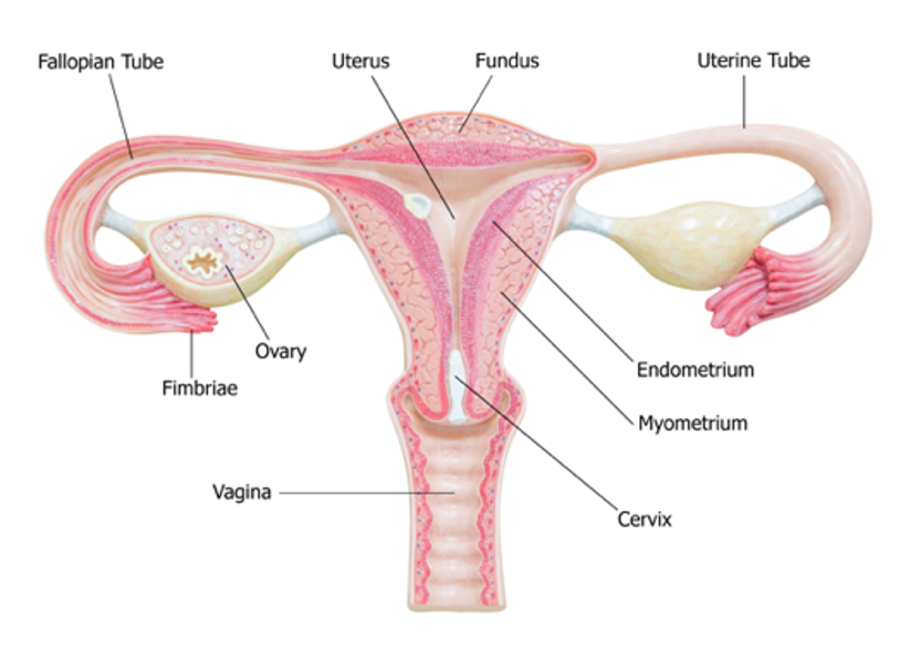
(iStock Photo ID: 538949875)
Two sections comprise the cervix: the ectocervix and the endocervix. The ectocervix extends into the vagina and is lined with stratified squamous nonkeratinized epithelium. This squamous epithelium contains four layers: basal, parabasal, intermediate, and superficial. The cervix opens into the vagina through the external os (i.e., external orifice), the readily visible portion of the cervix. The external os marks the transition point to the endocervix (i.e., endocervical canal), which is the inner part of the cervix. The endocervix is rarely visible on an internal pelvic examination, but it is lined with mucus-secreting columnar epithelium or glandular cells. It forms a passageway between the external...
...purchase below to continue the course
The area between the endocervix and the ectocervix is called the squamocolumnar junction (SCJ), where cervical dysplasia occurs most often. At birth, the SCJ lies close to the external os, as described previously, but moves internally with advancing age, parity, and menopause. The SCJ is no longer visible in a postmenopausal female as it has receded into the endocervix. The columnar epithelium of the endocervix expands outward onto the ectocervix, exposing the tissue to estrogen and irritants from the vagina’s acidic environment. Over time, the columnar epithelium is replaced with metaplastic squamous epithelium, forming the new SCJ. Metaplasia is a process by which one cell type transforms into another cell type not naturally present in that location. The area between the original and new SCJs is called the transformation zone (TZ). The TZ may be wide or narrow depending on individual factors such as age, parity, hormone exposure, and prior infections. More than 90% of cervical cancers originate in the TZ (ACS, 2025n; Laufer, 2026; Rogers & Brashers, 2023).
Human Papillomavirus
HPV is a ubiquitous group of tiny viruses that carry DNA, can adapt to their host, and evade the immune system. According to the Centers for Disease Control and Prevention [CDC, 2025], more than 9 out of 10 cervical cancers are caused by HPV, and approximately 11,000 new cases of HPV-associated cervical cancers are diagnosed in the United States annually. A growing body of evidence links HPV to other anogenital cancers (e.g., anal, vulvar, vaginal, penile) and head and neck cancers. Of the 200 identified strains of HPV, approximately 40 have a particular affinity for the genital region. While most of these strains are low-risk and nononcogenic (i.e., wart-causing), about 13 are high-risk and oncogenic (i.e., cancer-causing). High-risk HPV subtypes can cause several types of cancer. There are 7 HPV types associated with progression to cervical intraepithelial neoplasia (CIN) 3 or higher: HPV 16, 18, 31, 33, 35, 52, and 58. HPV-16 and HPV-18 are responsible for at least 70% of cervical cancers. While nearly all sexually active people will be infected with HPV during their lifetime, most of these infections remain clinically silent and dormant. The immune system typically mediates the spontaneous clearance of HPV from the cervix within two years of infection. However, about 10% of females infected with a high-risk HPV subtype will develop a persistent infection (Bowden et al., 2023; Bruni et al., 2023; CDC, 2025; Kusakabe et al., 2023; NCI, 2025a; Palefsky, 2024; WHO, 2026).
The Role of HPV in Cervical Carcinogenesis
Chronic high-risk HPV infections can lead to cervical precancers and cervical intraepithelial neoplasia (CIN). CIN is a term used to describe abnormal changes in the epithelial cells lining the surface of the cervix. CIN is a noninvasive, precancerous condition in which the abnormal cells are confined to the epithelium and relate directly to the integration of an HPV infection. Left untreated, CIN can invade local tissues and develop into invasive cancer. It can take 10 or more years for HPV-infected cells to develop into cervical cancer. HPV infection leads to CIN and invasive cancer by altering oncogenes and tumor suppressor genes. An oncogene is a mutation associated with cancer development; it can be inherited or caused by exposure to environmental carcinogenic substances, such as ultraviolet (UV) radiation and free radicals. In their normal, nonmutated state, oncogenes are called proto-oncogenes, which regulate healthy cell growth and division. When a proto-oncogene mutates into an oncogene, it becomes permanently activated. Oncogenes permit unregulated cell growth, leading to cancer growth. In their healthy state, tumor suppressor genes regulate normal cell growth and division, repair DNA errors, and induce apoptosis (i.e., programmed cell death). When tumor suppressor genes are mutated or inactivated, these processes become unregulated, allowing cancer to grow (ACS, 2026c; Bowden et al., 2023; Kusakabe et al., 2023; Palefsky, 2024).
Pap Smear and HPV Testing
Since CIN does not cause symptoms, cellular abnormalities are identified on a Pap smear. A Pap smear is a noninvasive test for cervical abnormalities, precancerous lesions, and invasive cancer. It can also diagnose benign conditions such as infections and inflammation, but it cannot diagnose HPV. HPV can be detected about a year after the cervix is infected, and HPV testing is a critical component of cervical cancer screening. United States cervical cancer screening guidelines support primary HPV testing alone or HPV co-testing. Primary HPV testing is a relatively novel screening modality performed alone, whereas HPV co-testing is done simultaneously with a Pap smear. The ACS (2025i) recommends the primary HPV test for cervical cancer screening in females aged 25 to 65 as the preferred screening option. The American Society for Colposcopy and Cervical Pathology (ASCCP) also endorses HPV–based testing as the basis for risk estimation (Perkins et al., 2020). The US Preventive Services Task Force (USPSTF) last issued final recommendations on cervical cancer screening in 2018. An update for this topic is currently in progress as of July 2026, with draft recommendations released in 2024 (USPSTF, 2024). Table 1 compares the ACS (2025i) guidelines and the USPSTF (2024) draft guidelines for the prevention and early detection of cervical cancer. The primary difference between the 2018 USPSTF final recommendation and the 2024 USPSTF draft recommendations is that HPV primary screening is the preferred screening method every 5 years starting at age 30, with an option for patient-collected specimens (USPSTF, 2024). The American College of Obstetricians and Gynecologists (ACOG) has also updated cervical cancer screening guidelines as of July 2026, with recommendations that align with the 2024 draft USPSTF recommendations on the age of onset for cytology alone and HPV testing. The ACOG recommendations advise that cytology testing alone after age 30 should be performed only if HPV testing is unavailable. If the HPV specimen is patient-collected, it should be repeated every 3 years (ACOG, 2026).
Table 1
ACS and USPSTF Cervical Cancer Screening Guidelines
ACS 2025 | USPSTF 2024 Draft |
|
|
|
|
|
|
|
|
(ACS, 2025i, 2025j; USPSTF, 2024)
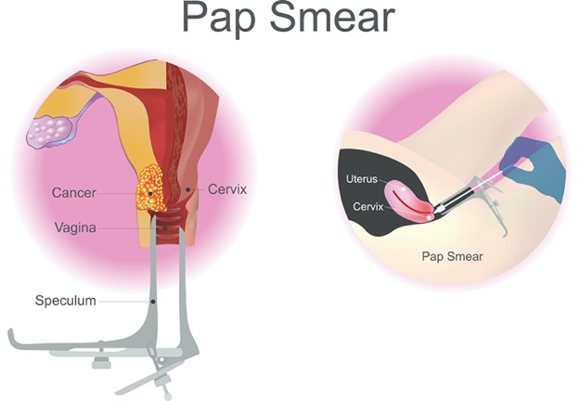
Currently, the US Food and Drug Administration (FDA) has approved three specific primary HPV tests and six co-testing options. The co-testing products offer the option of reflex testing when cytology results show atypical squamous cells of undetermined significance (ASC-US) or CIN. Regardless of the modality, all HPV tests assess the cells’ intracellular environment for signs of infection with high-risk viral subtypes. Viral nucleic acid (e.g., DNA or messenger ribonucleic acid [mRNA]) must be detected within the cervical cells to form a definitive diagnosis. Cervical HPV testing is performed by the healthcare provider (HCP) using the same procedure as a Pap smear. If the patient is self-collecting the HPV sample, they obtain a sample from the vagina utilizing the instrument provided by the manufacturer. The patient should utilize only FDA-approved self-collection kits. To perform a Pap smear, the clinician positions a vaginal speculum to visualize the cervix (Figure 2). Then, the cervix is scraped using a small, cone-shaped brush and a plastic spatula to collect cells. Next, the brush and spatula are rinsed in a liquid-filled vial and sent to the laboratory for processing. Most patients tolerate the test without complications or pain. Slight vaginal spotting may occur immediately following the Pap smear (Feldman & Mito, 2026).
Figure 2
Pap Smear
(iStock illustration ID:698717224)
Pap Smear Results
The Pap smear results indicate whether cervical cells are normal or abnormal, but some samples may return as unsatisfactory (see below). All Pap smears must include cells within the SCJ to produce a satisfactory result (Feldman & Mito, 2026).
Normal Results
Normal (negative) findings indicate that the sample was adequate and that no abnormal cells were identified, and patients do not require further workup or management. These results will be reported as negative intraepithelial lesions for malignancy (NILM). According to evidence-based cervical cancer screening guidelines, patients are advised to undergo routine screening every 3 to 5 years, as outlined in Table 1 (ACS, 2025d, 2025h; Huh et al., 2025; NCI, 2024; USPSTF, 2024).
Unsatisfactory Results
Unsatisfactory Pap smear results can indicate insufficient cells for evaluation or that the cells were obscured by blood or mucus. Patients with unsatisfactory results typically require repeat testing in 2 to 4 months (Huh et al., 2025; NCI, 2024; Perkins et al., 2020).
Abnormal Results
Abnormal (positive) results indicate cellular changes were seen in the sample. A pathologist classifies the severity of the abnormalities based on their risk of progressing to invasive cancer. Any grade of dysplasia warrants further workup. There are several abnormal Pap results; the most common are outlined in Table 2 (ACS, 2025d; Huh et al., 2025; NCI, 2024).
Table 2
Abnormal Pap Smear Results
Result | Description |
Atypical squamous cells of undetermined significance (ASC-US) | ASC-US is the most common abnormal Pap result and confirms the presence of abnormal cells. ASCUS is an inconclusive finding and does not determine the origin of the abnormality or whether the changes are secondary to an HPV infection. Several benign causes of atypical-appearing cells include yeast infection, irritation, polyps, cysts, and hormonal changes during pregnancy or menopause. ASC-US results do not indicate a malignancy, but HPV testing should be performed. |
Atypical squamous cells cannot exclude high-grade squamous intraepithelial lesion (ASC-H) | ASC-H is rare, occurring in less than 1% of cervical cytology specimens. ASC-H confirms the presence of abnormal squamous cells that may be suggestive, although not certain to be, of high-grade squamous intraepithelial lesions (HISL). Additional testing, such as colposcopy and biopsy, as well as HPV testing, is indicated. |
Atypical glandular cells (AGC) | AGC is rare, occurring in less than 1% of cervical cytology specimens. AGC signifies that some glandular cells from an endocervical or endometrial source have abnormal cytologic features that are too pronounced to be called inflammatory or reactive yet cannot be classified as malignant. AGC can indicate inflammatory conditions, but it can also indicate dysplasia or malignancy in up to 10% of cases. Management depends on the type of AGC but generally requires additional testing, such as colposcopy and/or an endometrial biopsy. About 50% of patients with AGC will have negative colposcopy and endometrial sampling results. |
Squamous intraepithelial lesion (SIL) | SIL refers to dysplastic changes in the cervix and is categorized into low-grade (LSIL) or high-grade (HSIL). LSIL denotes early cellular changes (mild dysplasia), meaning cells have only a few abnormal characteristics and still resemble healthy cells. LSIL is also classified as CIN 1. HSIL indicates moderate or severe dysplasia; cells appear highly abnormal under the microscope but are confined to the cervix’s surface. While HSIL does not indicate invasion into deeper parts of the cervix, it can progress to cervical cancer if not treated. HSIL can be classified as CIN 2 or CIN 3. Additional testing, such as colposcopy and HPV testing, is indicated. |
Adenocarcinoma in situ (AIS) | Less commonly, Pap smears can detect malignant cells (squamous or adenocarcinoma). However, invasive cervical cancer is rarely identified on a Pap smear in females who have undergone screening at regular intervals, as cervical abnormalities typically manifest as precancerous lesions. |
Invasive cervical cancer |
(ACS, 2025d; Goodman et al., 2025; Huh et al., 2025; NCI, 2024; Perkins et al., 2020)
HPV Test Results
HPV tests are easier to interpret than Pap smear results, as the result is either positive (high-risk HPV was detected) or negative (high-risk HPV was not detected). Results will also indicate which HPV strain is present, thereby helping to determine the potential risk for future cervical disease. As noted previously, HPV 16 and 18 are considered high-risk strains for progression to cervical disease (ACS, 2025k; NCI, 2025a; Perkins et al., 2020).
Follow-Up of Abnormal Results
Colposcopy
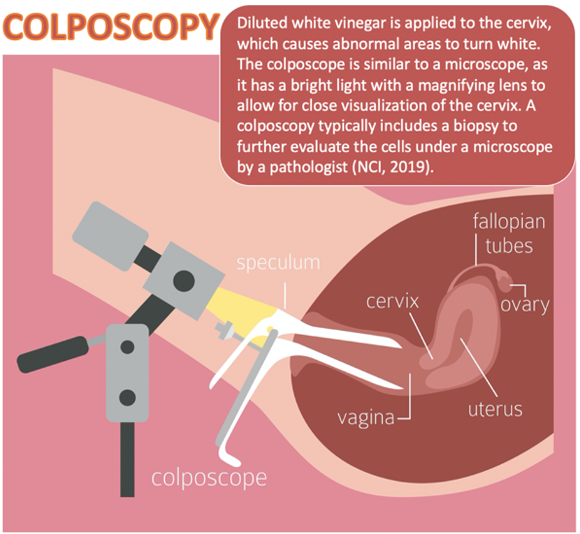
As shown in Figure 3, colposcopy is a noninvasive procedure typically performed in follow-up after abnormalities are detected on Pap or HPV testing. A colposcopy can detect precancerous changes and benign conditions, such as polyps, cervicitis (i.e., inflammation of the cervix), or genital warts. During the procedure, a 3% to 5% acetic acid solution is applied to the cervix. This solution turns abnormal areas white, allowing for direct visualization of cervical changes under a stereomicroscope (ACS, 2025i; Feltmate & Feldman, 2026).
Figure 3
Colposcopy
(iStock illustration ID: 1226029410)
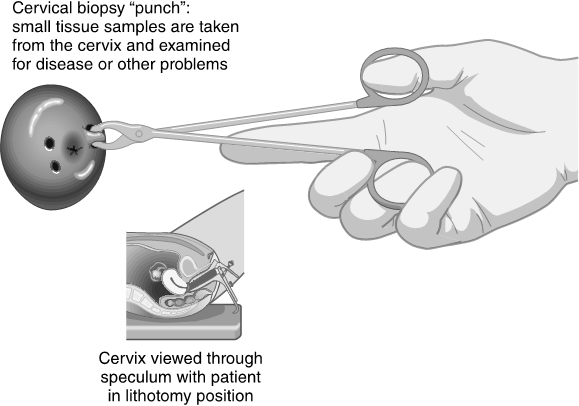
A biopsy of the abnormal area is performed, as tissue sampling and histologic analysis are the only ways to diagnose precancer or cancer definitively. There are three major types of cervical biopsies, as described in Table 3. A colposcopy is a safe procedure that carries minimal risks. The potential harms include pain, psychological distress, infection, and bleeding (ACS, 2025i; Feltmate & Feldman, 2026).
There is a lack of consensus regarding the need for analgesia before colposcopy and biopsy procedures, as well as on the optimal analgesic management option if used. Prior studies on the use of oral analgesics before the procedure found no decrease in patients’ pain scores. Some more recent studies have demonstrated mild improvement in the patient-reported pain levels with the use of topical lidocaine administered to the cervix prior to the procedure. In contrast, other studies found no difference in reported pain levels with or without anesthesia. One study using a local lidocaine injection to the cervix prior to the procedure demonstrated decreased perceived pain scores. There is reported pain with the use of analgesic injection administration, but it was reported as less painful than the discomfort experienced with a biopsy. Additional high‐quality RCTs are needed to determine the optimal route of administration and dose of local anesthetics (Akel et al., 2024; Feltmate & Feldman, 2026).
Table 3
Cervical Biopsy
Biopsy | Description | Risks/Benefits |
Punch biopsy (Figure 4) |
|
|
Endocervical curettage (ECC) |
|
|
Cold knife cone (CKC, conization; Figure 5) |
|
|
(ACS, 2025h; Feltmate & Feldman, 2026; Hoffman, 2025; Jakobsson & Norwitz, 2025)
Figure 4
Cervical Punch Biopsy
 (Assessment Technologies Institute [ATI], 2019a)
Figure 5
Cold Cone Biopsy

(ATI, 2019b)
CIN
CIN is classified into three grades based on the extent of dysplasia and the depth of abnormal cell invasion. CIN grade is determined by biopsy based on the appearance of cells under the microscope. As the CIN grade advances, the risk for progression to invasive cancer heightens, and natural regression is uncommon. The classification of CIN is further outlined in Table 4 (Wright, 2026b).
Table 4
CIN Classification
Grade | Thickness of Abnormal Epithelium | Classification |
CIN-1 |
|
|
CIN-2 |
|
|
CIN-3 |
|
|
(Bowden et al., 2023; Wright, 2026b)
CIN Management
CIN treatment aims to eradicate the precancerous lesions to prevent them from progressing to invasive cancer. Treatment selection depends on the extent and severity of CIN, HPV presence, the patient’s age, medical history, cost-effectiveness, availability, and each method’s potential risks and benefits. Procedures to treat CIN can affect future childbearing potential, so shared decision-making is crucial to ensure patients are fully informed of the potential risks before deciding on a therapy option. Most low-grade CINs resolve without intervention; only about 1% of cases will progress to cervical cancer. According to the ASCCP, most patients with CIN-1 can be adequately managed with close observation using Pap smears, HPV testing, and regular colposcopies. In contrast, patients with high-grade CIN typically require intervention. Treatment options include ablative and excisional techniques. Ablative techniques such as cryotherapy destroy abnormal tissues by freezing them, whereas excisional methods (e.g., loop electrosurgical excision procedure [LEEP] and CKC) remove abnormal tissue surgically. ASCCP strongly recommends excisional treatment over cryotherapy in all settings where a LEEP procedure is available and accessible. LEEP and CKC are the two most common and effective excisional procedures for CIN-2 and CIN-3 (Abu-Rustum et al., 2025; ACS, 2025g; Goodman et al., 2025; Perkins et al., 2020; Wright, 2026a).
LEEP
LEEP is a common technique for high-grade cervical dysplasia following colposcopy. During the procedure, a thin wire loop, which serves as a scalpel, is used to excise abnormal cells. An electric current is passed through the device to remove a thin layer of the cervix. Patients may feel mild discomfort during the procedure, so local anesthesia is typically used for pain control. The procedure is commonly performed in an office or outpatient setting and takes about 15 min. At the end of the procedure, a reddish-brown paste called ferric subsulfate (Monsel’s solution) may be applied to the cervix to control bleeding; alternatively, the blood vessels may be cauterized (heated with a device to control bleeding). The most common side effects include mild discomfort and slight bleeding for a few weeks after the procedure. To allow the cervix to heal fully, patients should avoid placing anything inside the vagina (including tampons and douching) and abstain from sexual intercourse for 2 to 4 weeks. Patients may shower as usual but should also avoid baths and swimming for 2 to 4 weeks. A LEEP is associated with a small increased risk for future pregnancy complications, including premature birth and low birth weight. In rare cases, the cervix may narrow after the procedure, causing menstrual irregularities (ACOG, 2022; ACS, 2025l; Hoffman, 2025; Pflugner, 2024).
Cryotherapy
Cryotherapy, called cryosurgery or laser ablation, involves applying a cooled metal disc (i.e., cryoprobe) to the cervix to freeze precancerous cells. The cryoprobe is cooled using compressed carbon dioxide (CO2) or nitrous oxide (NO) gas. Cryotherapy is a simple, fast outpatient procedure performed without anesthesia. It typically takes about 15 min and may cause mild discomfort. Following cryotherapy, the frozen area regenerates with normal epithelium within about 1 month, and some patients may experience watery vaginal discharge during this time. Patients should avoid sexual intercourse or placing anything in the vagina during the healing process for 4 to 6 weeks (Mello & Sundstrom, 2022; Perkins et al., 2020).
According to the ASCCP guidelines, cryotherapy should not be used in any of the following clinical circumstances:
- for lesions that extend into the canal
- for lesions covering more than 75% of the surface area of the ectocervix
- if the SCJ or upper limit of any lesion is not fully visualized
- if the CIN cannot be graded or is CIN-2 or higher
- after prior treatment for CIN-2 or higher, or
- in the setting of inadequate cervical biopsies to confirm a histologic diagnosis (Perkins et al., 2020, p. 118)
Surveillance Following CIN Treatment
The ASCCP supports continued surveillance with HPV testing or co-testing at 3-year intervals for at least 25 years after treatment for HSIL, CIN-2, CIN-3, or adenocarcinoma in situ (AIS). Extending surveillance beyond 25 years is acceptable if the patient’s life expectancy and screening capabilities are not compromised by concomitant health conditions (Perkins et al., 2020).
AIS Management
AIS occurs prior to cervical adenocarcinoma. It is less common than CIN 3 and requires higher management. The incidence of AIS has increased in recent decades among females aged 30 to 40. The average interval between the diagnosis of AIS and progression to early invasive cancer is about 5 years. While AIS may be detected on a Pap smear, a definitive diagnosis requires tissue sampling. AIS typically develops in the TZ and extends proximally to the endocervical canal. When diagnosed with a cervical biopsy, about 15% of patients have an invasive component (del Carmen & Schorge, 2026; Linder, 2024; Teoh et al., 2020). The gold standard treatment for AIS is hysterectomy (uterus removal) unless future fertility is desired. If fertility is desired, excisional procedures (e.g., LEEP or CKC) are acceptable if the patient is willing to adhere to recommendations for increased surveillance with HPV co-testing and endocervical sampling every 6 months for at least 3 years, then annually for at least 2 years or until hysterectomy. After 5 years of consistently negative co-testing results, surveillance can be extended to every 3 years until hysterectomy or indefinitely without hysterectomy. A hysterectomy is recommended after childbearing is complete (Linder, 2024; Perkins et al., 2020; Teoh et al., 2020).
Cervical Cancer Subtypes
When precancerous cells invade the basement membrane, invasive cervical cancer is diagnosed. Cervical cancer has two major subtypes: squamous cell carcinoma (SCC) and adenocarcinoma. Over 80% of cervical cancers are SCC, originating in the squamous epithelium in the TZ, and closely correlate with chronic HPV infection, as described earlier. Cervical adenocarcinoma begins in the glandular cells of the endocervix or the TZ and causes about 20% of cervical cancers. Adenocarcinomas differ from SCCs in that HPV does not uniformly cause them (Bruni et al., 2023; Li et al., 2025). Neuroendocrine carcinoma of the cervix (NECC) is a third, rare, and more aggressive type that accounts for only 1.5% of all cervical cancers. There are four subtypes of NECC tumors: small cell neuroendocrine carcinoma (SCNEC), large cell neuroendocrine carcinoma (LCNEC), typical carcinoid tumor, and atypical carcinoid tumor. SCNEC is the most common and aggressive type and carries the poorest prognosis. In contrast, carcinoid tumors typically have a more indolent disease trajectory and a more favorable prognosis. The role of HPV in mediating the development of NECC tumors is not clearly understood. Unlike SCC and adenocarcinoma, NECC is not universally preceded by preinvasive disease. Therefore, early detection via Pap smear screening is less common. The management of NECC is more complex and less precise due to the condition’s rarity, but it typically includes multiple chemotherapy agents (Leitao & Zivanovic, 2025; Li et al., 2025; Verma et al., 2023).
Signs and Symptoms
Females with early-stage cervical cancers typically do not have symptoms, as symptoms usually do not develop until the cancer advances and invades nearby tissue. The most common symptoms of advanced disease include the following:
- abnormal vaginal bleeding, such as spotting between periods, bleeding after intercourse, menstrual periods that are longer or heavier than usual, and postmenopausal bleeding
- dyspareunia (pain during or immediately following intercourse) or pain in the pelvis not related to intercourse
- abnormal vaginal discharge
- swelling of the legs due to impaired lymphatics caused by tumor compression
- urinary symptoms such as frequency, dysuria, inability to void, or hematuria
- constipation (ACS, 2025f)
Diagnostic Workup
The diagnosis and management of cervical cancer are multifactorial and involve combined modalities. Most institutions accredited by the NCI rely on evidence-based guidelines from the National Comprehensive Cancer Network (NCCN) to outline cancer diagnosis, staging, and treatment planning (Abu-Rustum et al., 2025). The NCCN is an alliance of leading cancer centers and world-renowned experts devoted to cancer care, research, and education. The NCCN provides evidence-based treatment guidelines for cancer based on subtype, pathology, genetics, staging, inheritance patterns, and other specific features, supported by rigorous clinical trial research, data compiled across institutions, and annual expert panel reviews. The guidelines are widely utilized in cancer care and guide medical decision-making throughout the patient’s disease trajectory. Once cervical cancer is diagnosed through biopsy and histologic analysis, the NCCN guidelines endorse various components of further diagnostic workup outlined in Table 5 (Abu-Rustum et al., 2025).
Table 5
Components of Diagnostic Workup
NCCN Cervical Cancer Diagnostic Workup |
Complete history and physical |
Laboratory tests (complete blood count [CBC], liver function tests [LFTs], and renal function studies) |
Smoking cessation and counseling intervention, if indicated |
Imaging studies to evaluate for other sites of disease as follows:
|
Possible HIV testing (particularly for younger patients)
|
Referral to a reproductive endocrinologist for fertility concerns if indicated |
(Abu-Rustum et al., 2025; Cibula et al., 2023)
Principles of Pathologic Review
The biopsy sample undergoes a series of tests to determine the cancer’s pathologic features, evaluate its behavior, and select the best treatment options. Tumor grade assesses how different the cancer cells look from healthy cells under the microscope. The grading determines the likelihood of cancer growth and spread and is defined as follows:
- Grade 1 is well-differentiated, appears similar to healthy cells, and is the least aggressive.
- Grade 2 is moderately differentiated, appears less like healthy cells, and is an intermediate grade (i.e., more aggressive than grade 1).
- Grade 3 is poorly differentiated or undifferentiated, does not resemble healthy cells, is high-grade, and is the most aggressive. Grade 3 cancer grows and spreads more quickly
- Grade 4 is undifferentiated, does not resemble healthy cells, is high-grade, grows rapidly, and spreads more quickly (Abu-Rustum et al., 2025; NCI, 2022).
Gene and Molecular Biomarker Analyses
Tumor protein p53 (TP53)
Under physiologic conditions, TP53 functions as a tumor suppressor gene, regulating cellular growth and division by preventing cells from growing and dividing uncontrollably. TP53 prevents cells with mutated or damaged DNA from dividing, thereby preventing tumor development. Mutations in TP53 impair its ability to control cell proliferation, as it cannot trigger apoptosis in cells with mutated or damaged DNA. Consequently, DNA damage accumulates in cells, which divide uncontrollably, leading to tumor growth. In cervical cancer, TP53 is inactivated by the HPV oncoprotein E6, thereby driving carcinogenesis, with the TP53 mutation present in approximately 30% to 50% of cases (Guo et al., 2026; Nagpal & Yuan, 2021).
P16 Immunohistochemistry
P16 is a tumor suppressor protein critical in regulating the normal cell cycle. Nearly all HPV-positive cervical precancers and cancers are accompanied by p16 overexpression. P16 interacts with HPV, leading to cell cycle dysfunction, interference with apoptosis, facilitated cell invasion, and angiogenesis. P16 overexpression paradoxically intensifies with increasing aggressiveness of the cancer. P16-negative tumors are associated with poorer outcomes and reduced survival compared to their p16-positive tumors. Despite the widespread prevalence of p16 overexpression in cervical cancer, its clinical significance remains unclear. A lack of consensus or guidelines persists regarding how p16 overexpression should guide treatment (Chong et al., 2025; Williams et al., 2022).
Mismatch Repair (MMR)/Microsatellite Instability (MSI)
Advancements in modern technology have led to novel treatment strategies utilizing the immune system to attack cancer. Specialized tests, such as microsatellite instability-high (MSI-H) testing and mismatch repair deficiency (dMMR), determine whether cancer is susceptible to the antitumor effects of immune-based therapies. MSI-H or dMMR acts as an inhibitor of the antiprogrammed cell death protein 1 (PD-1)/programmed cell death ligand 1 (PD-L1) pathway, which plays an essential role in regulating the immune system. The NCCN recommends that all patients with recurrent, progressive, or metastatic cervical cancer undergo MSI-H or dMMR testing. MMR deficiency may be reported as MSI-H or dMMR, but these terms have the same meaning (Abu-Rustum et al., 2025).
Programmed Cell Death-1 (PD-1)/PD-Ligand 1 (PD-L1)
PD-1 is a cell-surface receptor expressed on circulating lymphocytes (e.g., T-cells, B-cells), natural killer cells, dendritic cells, and monocytes. PD-1 binds to PD-L1, an inhibitory ligand expressed in some normal and cancer cells. PD-1 acts as an “off switch,” preventing immune cells from attacking other cells in the body and maintaining an appropriate immune response. When PD-1 binds to PD-L1, it acts as an “off switch,” preventing PD-1 from signaling to other cells in the body (i.e., preventing autoimmune conditions). The binding of PD-1 to PD-L1 signals T cells to leave neighboring cells, including cancer cells, alone. Some cancer cells express high levels of PD-L1, and this allows them to evade immune attack. Drugs that target PD-1 or PD-L1 block this binding and boost the immune response, disengaging the immune system’s brakes so T-cells are freed to recognize and attack cancer cells. Thus, identifying high PD-L1 expression can serve as a roadmap for personalized treatment. A combined positive score (CPS) of 1 or higher in cervical cancer indicates positive PD-L1 expression. This scoring method evaluates the number of PD-L1–staining cells (e.g., tumor cells, lymphocytes, macrophages) relative to all viable tumor cells. A minimum of 100 viable tumor cells in the PD-L1–stained sample is required for the specimen to be considered adequate for evaluation (Cibula et al., 2023; Huang et al., 2022; Lee et al., 2024; Merck & Co., 2023).
Cancer Staging
The International Federation of Gynecology and Obstetrics (FIGO) staging system guides gynecologic cancer staging worldwide. The cervical cancer guidelines are integrated within the NCCN guidelines. The four stages of cervical cancer are displayed in Figure 6. In stage I, the disease is confined to the cervix only. In stage II, it invades beyond the uterus but does not extend to the lower third of the vagina. In stage III, it invades the lower third of the vagina and may extend into the pelvic wall. In stage IV, the disease extends beyond the pelvic region. Table 6 provides a detailed description of these four stages. Because many cases require specialized surgical care, it is recommended that all patients with suspected cervical cancer be referred to a gynecologic oncologist (Abu-Rustum et al., 2025; Bhatla et al., 2025).
Figure 6
Cervical Cancer Staging
(iStock Illustration ID: 499566179)
Table 6
Cervical Cancer Staging
Stage | Description | |
Stage I
confined to the cervix only | IA | maximum depth of invasion ≤5 mm |
IA1 | stromal invasion ≤3 mm in depth | |
IA2 | stromal invasion >3 mm and ≤5 mm in depth | |
IB | invades >5 mm but is limited to the cervix | |
IB1 | invades >5 mm and ≤2 cm in greatest dimension | |
IB2 | invades >2 cm and ≤4 cm in greatest dimension | |
IB3 | invades >4 cm in greatest dimension | |
Stage II invades beyond the uterus but does not extend to the lower third of the vagina | IIA | invades the upper two-thirds of the vagina without parametrial invasion |
IIA1 | invades ≤4 cm in greatest dimension | |
IIA2 | invades >4 cm in greatest dimension | |
IIB | includes parametrial invasion but not up to the pelvic wall | |
Stage III invades the lower third of the vagina
| IIIA | invades the lower third of the vagina but does not extend to the pelvic wall |
IIIB | extends to the pelvic wall and/or hydronephrosis | |
IIIC | invades the pelvic and/or paraaortic lymph nodes | |
IIIC1 | includes pelvic lymph node metastasis | |
IIIC2 | includes paraaortic lymph node metastasis | |
Stage IV extends beyond the pelvis | IVA | spreads to adjacent organs (e.g., bladder, rectum) |
IVB | spreads to distant organs | |
(Abu-Rustum et al., 2025; Bhatla et al., 2025; Cibula et al., 2023)
Cervical Cancer Treatment
The optimal cervical cancer treatment depends on various factors, including pathologic features, cancer stage, plans for fertility, patient preference, age, and medical history. Treatment is often multimodal, combining several therapies and administering them simultaneously (i.e., concurrently) or sequentially. This section will provide a synopsis of the most common evidence-based treatment strategies (ACS, 2025m; Bhatla et al., 2025).
Surgery
Surgical intervention for cervical cancer can be fertility-sparing or non-fertility-sparing. Most early-stage cervical cancers (stage IA1) can be effectively managed with LEEP or CKC, which are fertility-sparing treatments. With advancing cervical cancer (stage IA2 or stage IB1), management may include a radical trachelectomy with lymphadenectomy. A radical trachelectomy, also called a cervicectomy, involves surgically removing the cervix, upper vagina, and supporting ligaments, but the uterine corpus is preserved. The surrounding pelvic lymph nodes are also removed. Concerning its impact on future pregnancies, a radical trachelectomy allows for successful conception but is associated with a 10% risk of second-trimester loss (Abu-Rustum et al., 2025; ACS, 2025m; Bhatla et al., 2025; Cibula et al., 2023).
When fertility preservation is not desired or irrelevant, early-stage cervical cancers are managed with a radical hysterectomy. The standard of care for stage IA2, IB1, IB2, IB3, and IIA1 cervical cancers is a radical hysterectomy with bilateral pelvic lymphadenectomy or pelvic lymph node dissection (PLND). A radical hysterectomy removes the uterus, cervix, a portion of the vagina, and the parametrium (the connective tissue surrounding the cervix). PLND involves the removal of the pelvic lymph nodes; the ovaries and fallopian tubes remain intact (Abu-Rustum et al., 2025; ACS, 2025m; Bhatla et al., 2025; Cibula et al., 2023).
A complete parametrectomy/upper vaginectomy is a surgical procedure for select patients with residual disease following a hysterectomy. It involves surgical resection of the parametrium and removal of the upper portion of the vagina. It carries significant morbidity, including risk for bowel and bladder injury, incontinence, and sexual dysfunction (Abu-Rustum et al., 2025; Bhatla et al., 2025; Cibula et al., 2023).
Patients with persistent disease (i.e., continued evidence of cancer following definitive therapy) or localized cancer recurrence (i.e., cancer that returns only in the pelvis following a disease-free interval) may be cured with a surgical procedure called pelvic exenteration. A pelvic exenteration is a drastic surgical procedure in which many, if not all, organs are removed from the pelvis. The extent of surgery varies but typically involves removing the uterus, fallopian tubes, ovaries, vagina, bladder, urethra, rectum, and anal sphincter. The patient will have a permanent colostomy and a urinary diversion following the procedure. A colostomy is formed by bringing the remaining intestine to the abdominal wall’s surface to provide a stool outlet (i.e., stoma) to evacuate the bowel. In urinary diversion, the kidneys and ureters are reconnected to a surgically created opening (i.e., urostomy) to drain urine (Abu-Rustum et al., 2025; Bhatla et al., 2025; Cibula et al., 2023; Duska, 2025).
Surgical Risks and Side Effects
Surgical risks and side effects depend on the size and degree of cancer invasion, the extent of surgery, and the structures removed. All surgeries and invasive procedures for cervical cancer are accompanied by risks, such as adverse reactions (ARs) to anesthesia, bleeding, blood clots, fistula formation (i.e., an abnormal connection between two hollow spaces within the body), bowel and bladder injury, infection, sexual dysfunction, and life-threatening sepsis. Fertility loss can negatively impact interpersonal relationships, quality of life, psychological health, and emotional well-being. Patients who undergo pelvic exenteration have difficulty adapting to the sweeping life alterations from surgery and struggle to care for colostomy and urostomy devices. Similarly, patients may be affected by the physical and psychological aspects of body image distortion. APRNs are vital in helping patients acclimate to these life-altering changes by facilitating healthy coping, addressing concerns, and referring patients to appropriate support groups or therapists (Abu-Rustum et al., 2025; Bhatla et al., 2025; Cibula et al., 2023; Duska, 2025; Hyman et al., 2025).
Radiation Therapy
Conventional radiation therapy delivers a precisely measured amount of high-energy, focused ionizing radiation beams to the tumor, causing biologic changes in cellular DNA and preventing cancer from reproducing or spreading. All cells, healthy and cancerous, are vulnerable to radiation and may be injured or destroyed; however, healthy cells can repair themselves and remain functional. The total radiation dose is hyper-fractionated, which means it is delivered to the tumor in small, divided doses (i.e., fractions) rather than all at once. Hyper-fractionation allows healthy cells a chance to recover between treatments, thereby reducing side effects. The total number of fractions administered depends on the cancer type, size, location, reason for treatment (e.g., curative intent or palliation), overall health, performance status, and concurrent therapies. Radiation therapy is central in treating cervical cancer and can be delivered externally or internally; some patients may receive both. The most common types of radiation for cervical cancer include external beam radiation therapy (EBRT) and brachytherapy (ACS, 2025e; Cibula et al., 2023; Mitin, 2026).
External Beam Radiation Therapy
EBRT delivers radiation from outside the body and is the most common technique for cervical cancer. Traditionally, radiation beams could only match the tumor’s height and width, exposing more healthy tissue to the consequences of radiation. Further advancements in imaging technology have enabled more precise treatment. Intensity-modulated radiation therapy (IMRT) is a newer, highly conformal form of radiation that modulates the intensity of the radiation beam, delivering a higher dose to a precise location, reducing unintended exposure of healthy tissues, enhancing clinical outcomes, and limiting side effects. IMRT helps minimize radiation exposure to the bowel and other critical structures, especially following a hysterectomy. EBRT is typically given concurrently with platinum-based chemotherapy (i.e., chemoradiation) to enhance the therapeutic response. Chemoradiation will be discussed in the next section (Abu-Rustum et al., 2025; ACS, 2025e; Cibula et al., 2023; Mitin, 2026).
Brachytherapy
Brachytherapy is a critical component of cervical cancer treatment, particularly for patients who are not surgical candidates. It is also frequently used following EBRT to deliver an added radiation boost to a specified area or to palliate symptoms. Brachytherapy involves the internal implantation of an intrauterine device. The radioactive source is inserted directly into or near the tumor, with accurate placement critical for tumor control. The primary effects are localized to the placement of the source and can result in vaginal irritation (ACS, 2025e; Mitin, 2026).
Intraoperative Radiation Therapy
Intraoperative radiation therapy (IORT) is a specialized technique that delivers a single dose of highly focused radiation to the tumor bed during surgery. IORT is particularly useful for patients with recurrent cancer within a previously irradiated field. Current NCCN guidelines do not provide recommendations for IORT use in a primary tumor setting. During IORT, the overlying normal tissues and structures (e.g., bowel and surrounding organs) are manually displaced outside the radiation field to minimize exposure. IORT is delivered with electrons using preformed applicators matched to the surgically defined area of risk. IORT can be administered in conjunction with EBRT (Abu-Rustum et al., 2025; Erdemoglu et al., 2025; Mitin, 2026).
Radiation Side Effects
Side effects depend on the specific area of the body exposed and the radiation dose. Superficial skin irritation at the EBRT beam site is likely and may include redness, blistering, and changes that resemble sunburn. The urinary system, bowel, and genitalia may be affected if they are in the radiation field. Bladder dysfunction can manifest as dysuria, hematuria, acute kidney injury, hydronephrosis, and incontinence. Gastrointestinal (GI) issues are common due to the cervix’s proximity to the bowel. Patients may experience nausea, vomiting, diarrhea, constipation, blood in stools, pain with defecation, and incontinence due to a loss of anal sphincter control. Sexual dysfunction is likely, particularly dyspareunia, atrophic vaginitis (i.e., inflammation and dryness of the vaginal tissue), vaginal agglutination (i.e., fusion and fibrosis of the vaginal walls), and recurrent yeast infections. If the ovaries are within the radiation field, patients may experience a permanent loss of ovarian function and develop premature menopause. Systemic effects can include fatigue, weakness, and dehydration (ACS, 2025e; Hyman et al., 2025; Mitin, 2026; NCI, 2025b).
Chemotherapy
Chemotherapy includes a group of high-risk, hazardous medications given to destroy cancer cells throughout the body. Chemotherapy interferes with the normal cell cycle, impairing DNA synthesis and cell replication to prevent cancer cells from dividing and multiplying (ACS, 2025a). Chemotherapy plays a prominent role in treating many types of cervical cancers. It is utilized in combination with surgery and/or radiation therapy, based on the stage of cervical cancer present. Chemoradiation, which includes the use of platinum-based chemotherapy (e.g., cisplatin [Platinol] or carboplatin [Paraplatin]) administered alongside radiation, is the standard of care for curable cervical cancer not amenable to surgery. Cisplatin (Platinol) and carboplatin (Paraplatin) act as radiosensitizers, rendering cancer cells more vulnerable to radiation’s toxic effects. According to the current NCCN guidelines, the best outcomes occur when chemoradiation is completed within 8 weeks (Abu-Rustum et al., 2025). Adjuvant chemotherapy (i.e., administered after surgery) aims to prevent cancer recurrence, reduce micro-metastases, and eradicate any remaining cancer cells. Chemotherapy is commonly used to manage recurrent cervical cancer and for patients with metastatic disease. In these instances, chemotherapy is considered palliative to enhance comfort, reduce symptom burden, improve quality of life, and extend survival. As listed in Table 7, systemic therapy regimens for recurrent or metastatic disease typically include formulations containing two or three medications. While bevacizumab (Avastin) and pembrolizumab (Keytruda) are included in the regimens, they are not chemotherapy agents; their unique mechanisms will be discussed in the next section (Abu-Rustum et al., 2025; ACS, 2025b; Bhatla et al., 2025; Lorusso et al., 2025; Straughn & Yashar, 2025; Wright, 2025).
Table 7
Cervical Cancer Systemic Therapies
Curative Therapy | Recurrent or Metastatic Disease | |
| Chemoradiation | First-Line Therapy | Second-Line Therapy |
| Preferred regimens:
| Preferred regimen:
|
(Abu-Rustum et al., 2025, Figure 10)
Chemotherapy Side Effects
The side effects of chemotherapy vary based on the drug type, dosage, treatment duration, underlying medical comorbidities, and overall performance status. Since cancer cells divide rapidly, chemotherapy is designed to target rapidly dividing cells, including normal cells that divide quickly, such as those in the GI tract, skin and hair, and bone marrow. As a group, the most common side effects include pancytopenia (i.e., lowering of the blood counts), including anemia, thrombocytopenia, neutropenia, fatigue, anorexia, alopecia, nausea/vomiting, diarrhea, constipation, skin changes, and peripheral neuropathy (i.e., damage to the sensory nerves). Chemotherapy damages dividing hair matrix cells, leading to hair shaft breakage at the follicular orifice or bulb. Chemotherapy-induced hair loss generally begins with hair thinning, with the extent of hair loss depending on the type of systemic therapy agent utilized, as well as the dose and schedule. Most regimens lead to the start of alopecia after 2 to 3 weeks of chemotherapy initiation. Paclitaxel (Taxol) and docetaxel (Taxotere) typically cause significant alopecia in most patients. APRNs should reassure females that their hair will begin to regrow within a few weeks after chemotherapy is discontinued, as permanent alopecia following chemotherapy is rare (Hyman et al., 2025; Meade et al., 2025; Rugo & van den Hurk, 2026).
Cisplatin (Platinol) is a moderate-to-high emetogenic agent that induces acute and delayed nausea. Poorly controlled chemotherapy-induced nausea and vomiting (CINV) are associated with unfavorable treatment compliance, impairing survival. Aprepitant (Emend) is approved to reduce CINV associated with cisplatin (Platinol) therapy. Available in oral and IV formulations, aprepitant (Emend) is a neurokinin-1 (NK-1) receptor antagonist that blocks substance P/neurokinin-1 receptors in the brain. It is used in combination with a 5-hydroxytryptamine type 3 (5HT3) receptor antagonist (e.g., ondansetron [Zofran] or palonosetron [Aloxi]) and a corticosteroid (i.e., dexamethasone [Decadron]) for therapy. Patients require aggressive hydration before and after administering cisplatin (Platinol) to manage associated nephrotoxicity and protect the renal system from injury (Hesketh, 2024).
Chemotherapy-induced peripheral neuropathy (CIPN) is a frequent side effect of cisplatin (Platinol), carboplatin (Paraplatin), paclitaxel (Taxol), and docetaxel (Taxotere). It is the most common dose-limiting toxicity (DLT) of these agents. DLTs are severe, often debilitating toxicities that warrant a dose reduction, interruption, or sometimes treatment discontinuation. CIPN results from demyelination of the sensory and motor axons. Patients experience reduced nerve conduction velocity, leading to the loss of deep tendon reflexes, paresthesia (e.g., numbness and tingling), weakness, and burning pain. CIPN initially symmetrically affects the body’s most distal points, such as the fingertips and toes (i.e., “stocking and glove” distribution), and moves proximally toward the midline as the damage progresses. In severe cases, patients may lose all sensation in the fingers, hands, toes, and feet; this can cause significant disability, including the inability to grasp or hold items and gait disturbances, leading to imbalance and falls. The etiology of CIPN is complex, as no single pathophysiologic process has been identified. CIPN is dose-dependent and progressive during treatment, but it can also have a cascading effect after treatment ends. During this cascading phenomenon, symptoms become more prominent after discontinuing the offending agent. Pain, sensory changes, and weakness that manifest during treatment generally lead to chemotherapy dose reductions, changes in treatment protocols, or termination of the therapeutic agent entirely. For many patients, the neuropathic systems persist permanently. The morbidity associated with CIPN can profoundly impact quality of life and activities of daily living (Lustberg, 2026; Meade et al., 2025; Preti & Davis, 2024).
Currently, no medications or supplements are effective in preventing CIPN. Exercising regularly, reducing alcohol use, and treating preexisting medical conditions (e.g., vitamin B12 deficiency or diabetes mellitus) may reduce the risk of CIPN. Management for CIPN is complex, and effective treatment options are limited. Pharmacologic treatment focuses on symptom relief, although many agents are not highly effective. Over-the-counter pain medications, menthol creams, capsaicin creams, or lidocaine patches may be used for comfort. There is a lack of evidence showing that any prescription medication is effective in the prevention of CIPN. Studies investigating cryotherapy alone as well as compression therapy with or without cryotherapy have shown mixed results with effectiveness in the prevention of CIPD. Several studies have demonstrated that an exercise regimen reduces the severity of CIPD. Studies investigating the effect of massage, reflexology, Reiki, and acupuncture demonstrated mixed results. Patients may be prescribed medications such as gabapentin (Neurontin), an anticonvulsant/antiepileptic agent that can also treat neuropathic pain; however, its use is off-label for CIPN and is not supported by the American Society of Clinical Oncology (ASCO). ASCO supports the use of selective serotonin-norepinephrine reuptake inhibitors (SNRIs), such as duloxetine (Cymbalta), as the only agent with adequate evidence supporting its use in patients with established painful CIPN. However, the degree of benefit is limited (Loprinzi et al., 2020). Patients with CIPN must be counseled on ways to avoid injury through wearing supportive shoes and paying attention to home safety, such as using handrails on stairs and removing throw rugs. Patients must also be mindful of water temperatures, as they may become less sensitive to hot water, increasing their risk of burns when bathing or washing dishes. Improvement in function and resolution of symptoms often occur over time, but nerve damage may be permanent (Hyman et al., 2025; Klafke et al., 2023; Lustberg, 2026).
Hypersensitivity Reactions to Chemotherapy
A hypersensitivity reaction (HSR) occurs when a foreign substance overstimulates the immune system, triggering antibody production and an immune response. HSRs are commonly associated with several chemotherapy agents that are used widely in cervical cancer treatment—most prominently paclitaxel (Taxol), docetaxel (Taxotere), and carboplatin (Paraplatin). HSR risk can be reduced by premedicating patients with corticosteroids (i.e., dexamethasone [Decadron]), antihistamines (e.g., diphenhydramine [Benadryl] and famotidine [Pepcid]), and/or acetaminophen (Tylenol). HSRs can occur during the initial chemotherapy infusion or after subsequent administrations of the same agent. Paclitaxel (Taxol) is well-known for its risk of nearly immediate acute HSR, whereas carboplatin (Paraplatin) more commonly induces an HSR after several repeated doses. Most HSRs occur during the first 15 min of the infusion. Initial signs and symptoms can include hives, urticaria, pruritus, swelling, back pain, facial flushing, rhinitis, abdominal cramping, chills, hypotension, and anxiety. Airway and hemodynamic monitoring are critical. Patients may require supplemental oxygen, fluid resuscitation, and other emergency medications as indicated. For life-threatening symptoms like bronchospasm, angioedema (e.g., swelling of the oral cavity, lips, or tongue), or anaphylaxis, epinephrine 0.3 to 0.5 mg (1:10,000 solution for adult patients) has been required (Castells et al., 2026; Meade et al., 2025).
Targeted Therapy
Bevacizumab (Avastin) is a humanized monoclonal antibody that binds to and inhibits the activity of human vascular endothelial growth factor (VEGF) to its receptors, thereby blocking the proliferation and formation of new blood vessels that supply tumor cells. VEGF is a signaling protein that stimulates angiogenesis (i.e., the formation of new blood vessels) in healthy and cancerous cells. Blood vessels carry oxygen and nutrients, supporting growth and survival. Thus, tumors need blood vessels to grow and spread. Anti-angiogenesis inhibits the formation of new blood vessels by blocking the VEGF receptors. Angiogenesis inhibitors (i.e., VEGF inhibitors) sever the blood supply to cancer cells by interfering with the VEGF receptor, so tumors stay small and starve. Bevacizumab (Avastin) is commonly used as a combination therapy to treat recurrent and metastatic cervical cancers, as shown in Table 5/7. While it is generally well-tolerated, potential side effects include bleeding events (i.e., hemorrhage), headaches, hypertension, and proteinuria (i.e., protein spilling in the urine due to increased pressure in the kidneys). Patients may require concurrent antihypertensive therapy to optimize blood pressure. Bevacizumab (Avastin) is contraindicated within 28 days of major or elective surgery (e.g., preoperatively or postoperatively) due to an increased risk for wound healing complications, hemorrhage, and fistula formation. The drug also carries a black box warning for GI perforation (i.e., a hole in the intestines) and fistula formation. Patients should be counseled to promptly report any sudden onset of severe and diffuse abdominal pain, bloating, firm abdomen, or acute bleeding (e.g., hemoptysis, rectal bleeding; Abu-Rustum et al., 2025; ACS, 2025h).
Immunotherapy
As the name suggests, immunotherapy stimulates the immune system to recognize and destroy cancer cells. Immunotherapy aims to elicit antitumor effects by modulating the body’s natural host defense mechanisms, priming them to recognize and attack cancer cells more effectively. Immune-based treatments differ from chemotherapy because they are highly specialized and targeted. Immune checkpoint inhibitors block receptors that cancer cells use to inactivate immune cells (i.e., T cells). When this signal is blocked, T-cells can better differentiate between healthy and cancer cells, thereby augmenting the cancer cells’ immune response (Lee et al., 2024). The role of immunotherapy in the treatment of cervical cancers has expanded significantly over recent years. Pembrolizumab (Keytruda) is a humanized monoclonal antibody that binds PD-1 with high affinity, preventing its interaction with PD-L1 and PD-L2. In the phase II KEYNOTE-158 clinical trial, pembrolizumab (Keytruda) demonstrated promising and durable antitumor activity in patients with PD-L1-positive cervical cancer, offering a clinically meaningful and viable treatment strategy. Based on these results, the FDA granted accelerated approval of pembrolizumab (Keytruda) for patients with advanced PD-L1–positive cervical cancer and disease progression or recurrent disease after chemotherapy. Recently, at the 2023 ASCO Annual Meeting, the overall survival analysis of the KEYNOTE-826 trial was presented, leading to a pivotal change in the standard of care for patients with persistent, recurrent, or metastatic cervical cancer. KEYNOTE-826 is a randomized, double-blind, phase 3 study of pembrolizumab (Keytruda) with chemotherapy compared to placebo with chemotherapy. After a median follow-up of 39.1 months, findings demonstrated that the addition of pembrolizumab (Keytruda) to chemotherapy, with or without bevacizumab (Avastin), continued to show clinically meaningful improvements in overall survival and progression-free survival, with no new safety signals (Colombo et al., 2021; Lorusso et al., 2025).
The European Society of Gynecological Oncology (ESGO), the European Society for Radiotherapy and Oncology (ESTRO), and the European Society of Pathology (ESP) released updated joint guidelines for the management of patients with cervical cancer in May 2023. They recommend adding pembrolizumab (Keytruda) to platinum-based chemotherapy (with or without bevacizumab [Avastin]) for all patients with PD-L1-positive tumors, defined by a CPS score of 1 or higher (Cibula et al., 2023). Pembrolizumab (Keytruda) is generally well-tolerated; the most common side effects include fatigue, nausea, anorexia, cough, diarrhea, skin rash, and itching. However, patients may experience severe and possibly fatal autoimmune-related ARs. Although any organ system can be affected, the most commonly observed reactions include colitis, hepatitis, endocrinopathies (e.g., thyroid and adrenals), pneumonitis, and rash, which can progress to Stevens-Johnson syndrome (SJS; Lorusso et al., 2025; Wang et al., 2024).
Antibody Drug Conjugates
Antibody drug conjugates (ADCs) are an emerging class of targeted agents that deliver chemotherapy directly to cancer cells via a linker attached to a monoclonal antibody (i.e., a protein) that binds to a specific target on the cancer cell. Once bound, the ADC releases a powerful cytotoxic drug into the cancer cell. ADCs aim to improve efficacy while minimizing systemic toxicity to normal tissue using a targeted delivery mechanism (FDA, 2024). In 2021, tisotumab vedotin (Tivdak) received accelerated FDA approval for recurrent or metastatic cervical cancer based on the Phase 2 innovaTV 204 clinical trial, which revealed clinically meaningful and durable antitumor activity and a 24% objective response rate; nearly 1 in 4 patients responded to treatment in the trial (Coleman et al., 2021). Tisotumab vedotin (Tivdak) is directed against tissue factor (TF), a core component of the coagulation cascade. TF also affects the clinical progression of certain cancers by influencing the proliferation, infiltration, and metastasis of malignant cells. TF is highly prevalent in cervical cancer cells, serving as a prime therapeutic target (Ulvang et al., 2025).
While tisotumab vedotin (Tivdak) has demonstrated a manageable and tolerable safety profile, it does have unique ARs that require close monitoring. The most common ARs include anemia, lymphopenia, diarrhea, fatigue, nausea, peripheral neuropathy, alopecia, and skin rash. It can also cause bleeding events such as epistaxis and hemorrhage due to increased prothrombin international normalized ratio (PT/INR) and prolonged activated partial thromboplastin time (aPTT). Severe skin reactions, including fatal or life-threatening SJS, have been reported in select patients. Most notably, tisotumab vedotin (Tivdak) carries a boxed warning for ocular toxicity, which may lead to treatment discontinuation in severe cases. Ocular ARs occurred in 60% of patients across clinical trials; most were conjunctival ARs (40%), dry eye (29%), corneal irritation (21%), and blepharitis (8%). Grade 3 ocular ARs occurred in 3.8% of patients, including severe ulcerative keratitis in 3.2%. One patient experienced ulcerative keratitis with perforation requiring corneal transplantation. In the innovaTV 204 clinical trial, 4% of patients experienced a decrease in visual acuity to 20/50 or worse, and 75% of those patients resolved (ACS, 2025c; Coleman et al., 2021; FDA, 2025).
Given the potential for ocular ARs, all patients must be referred to an eye care provider (e.g., ophthalmologist or optometrist) for an ophthalmic exam before each dose. To reduce the risk of ocular ARs, the FDA (2025) strongly advises adherence to the following recommendations:
- An ophthalmic exam, including a visual acuity test and slit lamp exam, should be performed at baseline, before each dose, and as clinically indicated.
- Patients should start on prophylactic topical corticosteroid eye drops (one drop in each eye) before each infusion. Patients should continue administering eye drops in each eye as prescribed for 72 hr following each infusion.
- Immediately before each infusion, administer premedication with topical ocular vasoconstrictor drops in each eye.
- Apply cooling eye pads to both eyes during the infusion.
- Patients should be instructed to administer topical lubricating eye drops into both eyes daily for the duration of therapy and 30 days following the last dose.
- Patients should avoid wearing contact lenses (unless advised by their eye care provider) for the duration of therapy.
When the foregoing recommendations are adhered to, ocular ARs are significantly reduced. Tisotumab vedotin (Tivdak) remains the only FDA-approved ADC for cervical cancer but is currently under investigation in combination with checkpoint inhibitors and carboplatin (Paraplatin; FDA, 2025; Podwika & Duska, 2023).
HPV Vaccination
According to the WHO (2022, 2026), effective cervical cancer primary prevention (i.e., HPV vaccination) combined with secondary prevention strategies (e.g., screening tests and early treatment of precancerous lesions) will thwart most cervical cancers. Cervical cancer is highly preventable and can be mitigated through screening. The FDA originally approved three HPV vaccines to protect against high-risk HPV subtypes that are linked to cancer: nine-valent (9vHPV, Gardasil 9), quadrivalent (4vHPV, Gardasil), and bivalent (2vHPV, Cervarix). Currently, only Gardasil 9 is available in the US for administration. Gardasil 9 protects against types 6, 11, 16, 18, 31, 33, 45, 52, and 58. In 2018, the FDA expanded the use of Gardasil 9—a 9-valent vaccine with increased coverage of additional high-risk HPV types—in those aged 9 to 45 years. Administration in adults aged 27 to 45 is through shared decision-making (CDC, 2024a; Galvin et al., 2022; NCI, 2026b). Gardasil 9 protects against 9 types of HPV: cervical, vaginal, and vulvar cancers in females, anal cancer, certain head and neck cancers (e.g., the back of the mouth and throat), and genital warts in both males and females (Merck & Co., 2025).
Since the introduction of the vaccine, HPV infections and cervical precancers have dropped significantly. Over the past decade, the presence of HPV 16 and 18 has decreased 83% in females aged 13 to 19. A 66% decrease was observed in females aged 20 to 24. A similar reduction in the presence of anogenital warts has been reported. Clinical data demonstrate that vaccination protection remains high after 10 years, with no decrease in antibody levels. While the HPV vaccination could prevent more than 90% of cancer caused by HPV from ever developing, nearly one-half (50%) of adolescent females in the US have not been vaccinated (ACOG, 2020; Bhatla et al., 2025; NCI, 2026b). To ensure worldwide protection, the WHO (2022) has recommended that national immunization programs administer either an off-label single-dose HPV vaccine regimen or the standard two-dose regimen. This recommendation can provide HPV protection while utilizing fewer resources.
Side Effects
According to the CDC (2024a), more than 15 years of research have demonstrated that the HPV vaccines are safe and effective. More than 135 million doses of the HPV vaccine have been administered. Some of the most commonly reported side effects include the following:
- pain, redness, or edema near the injection site
- fever
- dizziness or fainting immediately after the injection
- nausea
- headaches
- tiredness
- joint or muscle pain (CDC, 2024a, 2024b; NCI, 2026b)
Schedule and Dosing
The CDC’s Advisory Committee on Immunization Practices (ACIP) develops recommendations regarding all vaccination schedules and regimens in the United States. The current ACIP recommendations for HPV vaccination are as follows (CDC, 2019, 2024; NCI, 2026b):
Routine and Catch-up Vaccination
- HPV vaccination is routinely recommended at ages 11 to 12 but may begin as early as 9 years.
- HPV vaccination is recommended for all individuals up to 26 years who were not adequately vaccinated.
- The 2- or 3-dose series depends on the patient’s age at initial vaccination:
- Age 9 through 14 years at initial vaccination: follow the 2-dose series and administer injections at 0 and 6 to 12 months
- the minimum interval between vaccinations is 5 months
- repeat dose if administered too soon
- Age 15 years or older at initial vaccination: following the 3-dose series and administer injections at 0, 1 to 2 months, and 6 months
- the minimum interval between vaccinations is as follows:
- dose 1 to dose 2: 4 weeks
- dose 2 to dose 3: 12 weeks
- dose 1 to dose 3: 5 months
- repeat dose if administered too soon
- Age 27 through 45 years: Vaccination is not recommended for everyone older than 26 years. The ACIP recommends shared clinical decision-making for patients aged 27 through 45 years who have not previously been vaccinated to discuss the risk for new HPV infections and the possible benefits of vaccination with their HCP. HPV vaccination in this age range provides less benefit because more people have already been exposed to the virus, but it has still demonstrated efficacy. Adults who were not vaccinated against HPV should discuss the risk for new HPV infections and the possible benefits of vaccination with their HCP to determine if vaccination is still advised.
Special Circumstances
- Immunocompromised patients, including those with HIV infection: administer the 3-dose series outlined previously
- Children with a history of sexual abuse or assault: initiate at age 9 years
- Pregnancy: delay HPV vaccination until after pregnancy, but pregnancy testing is not required before vaccination. There is no evidence that vaccination will affect pregnancy or harm a fetus (CDC, 2019, 2024; NCI, 2026b)
To learn more about HPV and vaccination, review the HPV NursingCE course.
Abu-Rustum, N. R., Campos, S. M., Amarnath, S., Arend, R., Barber, E., Bradley, K., Brooks, R., Chino, J., Chon, H. S., Crispens, M. A., Damast, S., Fisher, C. M., Frederick, P., Gaffney, D. K., Gaillard, S., Giuntoli, R., II, Glaser, S., Howitt, B. E., Kendra, K., … Sambandam, V. (2025). Cervical cancer, version 2.2026, NCCN Clinical Practice Guidelines in Oncology. Journal of the National Comprehensive Cancer Network, 23(12), 549–573. https://doi.org/10.6004/jnccn.2025.0057
Akel, M., Ratra, D., Wright, M., Barroca, C., Abdou, A. A., Kaldas, P., Bhatt, S., Perez, A., Shah, S., & Hernandez Borges, S. (2024). Anesthesia usage and pain management in colposcopy: A scoping review of efficacy and approaches. Cureus, 16(9), e70384. https://doi.org/10.7759/cureus.70384
American Cancer Society. (2022). Cancer facts & figures for African American/Black People 2022–2024. https://www.cancer.org/content/dam/cancer-org/research/cancer-facts-and-statistics/cancer-facts-and-figures-for-african-americans/2022-2024-cff-aa.pdf
American Cancer Society. (2025a). Chemotherapy. https://www.cancer.org/cancer/managing-cancer/treatment-types/chemotherapy.html
American Cancer Society. (2025b). Chemotherapy for cervical cancer. https://www.cancer.org/cancer/types/cervical-cancer/treating/chemotherapy.html
American Cancer Society. (2025c). Immunotherapy for cervical cancer. https://www.cancer.org/cancer/types/cervical-cancer/treating/immunotherapy.html
American Cancer Society. (2025d). Pap test (Pap smear). https://www.cancer.org/cancer/types/cervical-cancer/detection-diagnosis-staging/screening-tests/pap-test.html
American Cancer Society. (2025e). Radiation therapy for cervical cancer. https://www.cancer.org/cancer/types/cervical-cancer/treating/radiation.html
American Cancer Society. (2025f). Signs and symptoms of cervical cancer. https://www.cancer.org/cancer/types/cervical-cancer/detection-diagnosis-staging/signs-symptoms.html
American Cancer Society. (2025g). Surgery for cervical cancer. https://www.cancer.org/cancer/types/cervical-cancer/treating/surgery.html
American Cancer Society. (2025h). Targeted drug therapy for cervical cancer. https://www.cancer.org/cancer/types/cervical-cancer/treating/targeted-therapy.html
American Cancer Society. (2025i). Tests for cervical cancer. https://www.cancer.org/cancer/cervical-cancer/detection-diagnosis-staging/how-diagnosed.html
American Cancer Society. (2025j). The American Cancer Society guidelines for cervical cancer screening. https://www.cancer.org/cancer/cervical-cancer/detection-diagnosis-staging/cervical-cancer-screening-guidelines.html
American Cancer Society. (2025k). The HPV test. https://www.cancer.org/cancer/cervical-cancer/detection-diagnosis-staging/screening-tests/hpv-test.html
American Cancer Society. (2025l). The LEEP procedure for cervical cancer prevention. https://www.cancer.org/cancer/types/cervical-cancer/causes-risks-prevention/leep.html
American Cancer Society. (2025m). Treatment options for cervical cancer, by stage. https://www.cancer.org/cancer/types/cervical-cancer/treating/by-stage.html
American Cancer Society. (2025n). What is cervical cancer? https://www.cancer.org/cancer/cervical-cancer/about/what-is-cervical-cancer.html
American Cancer Society. (2026a). Cancer facts & figures 2026. https://www.cancer.org/content/dam/cancer-org/research/cancer-facts-and-statistics/annual-cancer-facts-and-figures/2026/2026-cancer-facts-and-figures.pdf
American Cancer Society. (2026b). Key statistics for cervical cancer. https://www.cancer.org/cancer/types/cervical-cancer/about/key-statistics.html
American Cancer Society. (2026c). Oncogenes, tumor suppressor genes, and DNA repair genes. https://www.cancer.org/cancer/understanding-cancer/genes-and-cancer/oncogenes-tumor-suppressor-genes.html
American Cancer Society. (2026d). Survival rates for cervical cancer. https://www.cancer.org/cancer/cervical-cancer/detection-diagnosis-staging/survival.html
American College of Obstetricians and Gynecologists. (2020). Human papillomavirus vaccination (Committee Opinion Number 809). https://www.acog.org/clinical/clinical-guidance/committee-opinion/articles/2020/08/human-papillomavirus-vaccination
American College of Obstetricians & Gynecologists. (2022). Loop electrosurgical excision procedure [LEEP]. https://www.acog.org/en/Womens%20Health/FAQs/Loop%20Electrosurgical%20Excision%20Procedure
American College of Obstetricians & Gynecologists. (2026). Screening for cervical cancer (Committee Statement Number 28). https://www.acog.org/clinical/clinical-guidance/committee-statement/articles/2026/07/screening-for-cervical-cancer
Assessment Technologies Institute. (2019a). Cervical punch biopsy [image]. https://cms.ascendlearning.com/share/page/site/digital-asset-librarytest/document-details?nodeRef=workspace://SpacesStore/6bf58a2a-81a5-4a91-91a8-d2c29121fa53
Assessment Technologies Institute. (2019b). Cold cone biopsy [image]. https://cms.ascendlearning.com/share/page/site/digital-asset-librarytest/document-details?nodeRef=workspace://SpacesStore/0621296e-a7c8-4fe6-8ed4-eaa55484d00d
Bhatla, N., Aoki, D., Sharma, D. N., & Sankaranarayanan, R. (2025). Cancer of the cervix uteri: 2025 update. International Journal of Gynecology and Obstetrics, 171(Suppl. 1), 87–108. https://doi.org/10.1002/ijgo.70277
Bowden, S. J., Doulgeraki, T., Bouras, E., Markozannes, G., Athanasiou, A., Grout-Smith, H., Kechagias, K. S., Ellis, L. B., Zuber, V., Chadeau-Hyam, M., Flanagan, J. M., Tsilidis, K. K., Kalliala, I., & Kyrgiou, M. (2023). Risk factors for human papillomavirus infection, cervical intraepithelial neoplasia and cervical cancer: An umbrella review and follow-up Mendelian randomisation studies. BMC Medicine, 21(1), 274. https://doi.org/10.1186/s12916-023-02965-w
Bruni, L., Albero, G., Serrano, B., Mena, M., Collado, J. J., Gómez, D., Muñoz, J., Bosch, F. X., & de Sanjosé, S. (2023). Human papillomavirus and related diseases report. ICO/IARC Information Centre on HPV and Cancer. https://hpvcentre.net/statistics/reports/XMX.pdf
Castells, M. C., Matulonis, U. A., & Horton, T. (2026). Infusion reactions to systemic chemotherapy. UpToDate. Retrieved July 10, 2026, from https://www.uptodate.com/contents/infusion-reactions-to-systemic-chemotherapy
Centers for Disease Control and Prevention. (2019, August 16). Human papillomavirus vaccination for adults: Updated recommendations of the Advisory Committee on Immunization Practices. Morbidity and Mortality Weekly Report (MMWR), 68(32), 698–702. https://www.cdc.gov/mmwr/volumes/68/wr/mm6832a3.htm
Centers for Disease Control and Prevention. (2024a). HPV vaccine recommendations. https://www.cdc.gov/hpv/hcp/vaccination-considerations/
Centers for Disease Control and Prevention. (2024b). HPV vaccine safety and effectiveness data. https://www.cdc.gov/hpv/hcp/vaccination-considerations/safety-and-effectiveness-data.html
Centers for Disease Control and Prevention. (2025). Cancers caused by HPV. https://www.cdc.gov/hpv/parents/cancer.html
Chong, L., Zou, Z., Xia, L., Wang, X., Dong, Z., Zhao, Y., & Hou, Y. (2025). P16 expression and its correlation with the clinical pathological characteristics of patients with cervical cancer: A systematic review and meta-analysis. BMJ Open, 15(9), e102602. https://doi.org/10.1136/bmjopen-2025-102602
Cibula, D., Raspollini, M. R., Planchamp, F., Centeno, C., Chargari, C., Felix, A., Fischerová, D., Jahnn-Kuch, D., Joly, F., Kohler, C., Lax, S., Lorusso, D., Mahantshetty, U., Mathevet, P., Naik, R., Nout, R. A., Oaknin, A., Peccatori, F., Persson, J., … Lindegaard, J. (2023). ESGO/ESTRO/ESP guidelines for the management of patients with cervical cancer—Update 2023. International Journal of Gynecological Cancer, 33(5), 649–666. https://doi.org/10.1136/ijgc-2023-004429
Coleman, R. L., Lorusso, D., Gennigens, C., González-Martín, A., Randall, L., Cibula, D., Lund, B., Woelber, L., Pignata, S., Forget, F., Redondo, A., Vindeløv, S. D., Chen, M., Harris, J. R., Smith, M., Nicacio, L. V., Teng, M. S., Laenen, A., Rangwala, R., … Vergote, I. (2021). Efficacy and safety of tisotumab vedotin in previously treated recurrent or metastatic cervical cancer (innovaTV 204/GOG-3023/ENGOT-cx6): A multicentre, open-label, single-arm, phase 2 study. The Lancet Oncology, 22(5), 609–619. https://doi.org/10.1016/S1470-2045(21)00056-5
Colombo, N., Dubot, C., Lorusso, D., Caceres, V., Hasegawa, K., Shapira-Frommer, R., Tewari, K. S., Salman, P., Usta, E. H., Yañez, E., Gümüş, M., Hurtado de Mendoza, M. O., Samouëlian, V., Castonguay, V., Arkipov, A., Toker, S., Li, K., Keefe, S. M., & Monk, B. J. (2021). Pembrolizumab for persistent, recurrent, or metastatic cervical cancer. The New England Journal of Medicine, 385(20), 1856–1867. https://doi.org/10.1056/NEJMoa2112435
del Carmen, M. G., & Schorge, J. O. (2026). Cervical adenocarcinoma in situ. UpToDate. Retrieved July 16, 2026, from https://www.uptodate.com/contents/cervical-adenocarcinoma-in-situ
Duska, L. R. (2025). Overview of approach to cervical cancer survivors. UpToDate. Retrieved July 9, 2026, from https://www.uptodate.com/contents/overview-of-approach-to-cervical-cancer-survivors
Erdemoglu, E., Ostby, S. A., Senthilkumar, S., Kumar, A., Vora, S. A., Chen, L., James, S. E., & Butler, K. A. (2025). Intraoperative radiation therapy (IORT) in gynecologic cancers: A scoping review. Cancers, 17(8), 1356. https://doi.org/10.3390/cancers17081356
Feldman, S., & Mito, J. (2026). Cervical cancer screening tests: Techniques for cervical cytology and human papillomavirus testing. UpToDate. Retrieved July 8, 2026, from https://www.uptodate.com/contents/cervical-cancer-screening-tests-techniques-for-cervical-cytology-and-human-papillomavirus-testing
Feltmate, C. M., & Feldman, S. (2026). Colposcopy. UpToDate. Retrieved July 8, 2026, from https://www.uptodate.com/contents/colposcopy
Galvin, A. M., Garg, A., Griner, S. B., Moore, J. D., & Thompson, E. L. (2022). Health literacy correlates to HPV vaccination among US adults ages 27–45. Journal of Cancer Education, 38, 349–356. https://doi.org/10.1007/s13187-021-02123-x
Goodman, A., Huh, W. K., & Einstein, M. H. (2025). Cervical cancer screening: Risk assessment, evaluation, and management after screening. UpToDate. Retrieved July 8, 2026, from https://www.uptodate.com/contents/cervical-cancer-screening-risk-assessment-evaluation-and-management-after-screening
Guo, Y., Li, S., Liu, T., Chang, X., Qin, P., Wang, N., Jiang, Y., Lv, N., Li, N., & Hao, F. (2026). Gene mutations and related molecular events in distant metastasis of cervical cancer: A review. International Journal of Medical Sciences, 23(3), 1015–1032. https://doi.org/10.7150/ijms.123727
Hesketh, P. J. (2024). Prevention of chemotherapy-induced nausea and vomiting in adults. UpToDate. Retrieved July 10, 2026, from https://www.uptodate.com/contents/prevention-of-chemotherapy-induced-nausea-and-vomiting-in-adults
Hoffman, M. S. (2025). Cervical intraepithelial neoplasia: Diagnostic excisional procedures. UpToDate. Retrieved July 8, 2026, from https://www.uptodate.com/contents/cervical-intraepithelial-neoplasia-diagnostic-excisional-procedures
Huang, W., Liu, J., Xu, K., Chen, H., & Bian, C. (2022). PD-1/PD-L1 inhibitors for advanced or metastatic cervical cancer: From bench to bed. Frontiers in Oncology, 12, 849352. https://doi.org/10.3389/fonc.2022.849352
Huh, W. K., Einstein, M. H., & Mito, J. (2025). Cervical cancer screening: The cytology and human papillomavirus report. UpToDate. Retrieved July 8, 2026, from https://www.uptodate.com/contents/cervical-cancer-screening-the-cytology-and-human-papillomavirus-report
Hyman, J., Slatnik, C., & Ellwood, M. (2025). Cervical cancer treatment and nursing considerations: Best practices for effective care. Canadian Oncology Nursing Journal, 35(1), 244–253. https://pmc.ncbi.nlm.nih.gov/articles/PMC12379881/
Jakobsson, M., & Norwitz, E. R. (2025). Reproductive effects of cervical excisional and ablative procedures. UpToDate. Retrieved July 8, 2026, from https://www.uptodate.com/contents/reproductive-effects-of-cervical-excisional-and-ablative-procedures
Jiang, C., Rosenberg, P. S., Star, J., Bandi, P., Bednarczyk, R. A., Jemal, A., & Sung, H. (2026). State-level progress in reducing cervical cancer incidence among US young women between the pre- and post-human papillomavirus vaccination eras. Journal of the National Cancer Institute, djag051. https://doi.org/10.1093/jnci/djag051
Klafke, N., Bossert, J., Kröger, B., Neuberger, P., Heyder, U., Layer, M., Winkler, M., Idler, C., Kaschdailewitsch, E., Heine, R., John, H., Zielke, T., Schmeling, B., Joy, S., Mertens, I., Babadag-Savas, B., Kohler, S., Mahler, C., Witt, C. M., … Stolz, R. (2023). Prevention and treatment of chemotherapy-induced peripheral neuropathy (CIPN) with non-pharmacological interventions: Clinical recommendations from a systematic scoping review and an expert consensus process. Medical Sciences (Basel, Switzerland), 11(1), 15. https://doi.org/10.3390/medsci11010015
Kusakabe, M., Taguchi, A., Sone, K., Mori, M., & Osuga, Y. (2023). Carcinogenesis and management of human papillomavirus-associated cervical cancer. International Journal of Clinical Oncology, 28, 965–974. https://doi.org/10.1007/s10147-023-02337-7
Laufer, M. R. (2026). Benign cervical lesions and congenital anomalies of the cervix. UpToDate. Retrieved July 7, 2026, from https://www.uptodate.com/contents/benign-cervical-lesions-and-congenital-anomalies-of-the-cervix
Lee, D., Cho, M., Kim, E., Seo, Y., & Cha, J. H. (2024). PD-L1: From cancer immunotherapy to therapeutic implications in multiple disorders. Molecular Therapy, 32(12), 4235–4255. https://doi.org/10.1016/j.ymthe.2024.09.026
Leitao, M. M., & Zivanovic, O. (2025). Small cell neuroendocrine carcinoma of the cervix. UpToDate. Retrieved July 9, 2026, from https://www.uptodate.com/contents/small-cell-neuroendocrine-carcinoma-of-the-cervix
Li, S., Liu, C., & Weng, L. (2025). Exploring cervical adenocarcinoma: Epidemiological insights, diagnostic and therapeutic challenges, and pathogenetic mechanisms. Cancer Medicine, 14, e70620. https://doi.org/10.1002/cam4.70620
Linder, M. (2024). Tips for best practices on management of adenocarcinoma in situ of the cervix (AIS). American Society for Colposcopy and Cervical Pathology. https://www.asccp.org/clinical-practice/practice-pearls/tips-for-best-practices-on-management-of-adenocarcinoma-in-situ-of-the-cervix-ais/
Loprinzi, C. L., Lacchetti, C., Bleeker, J., Cavaletti, G., Chauhan, C., Hertz, D. L., Kelley, M. R., Lavino, A., Lustberg, M. B., Paice, J. A., Schneider, B. P., Smith, E. M. L., Smith, M. L., Smith, T. J., Wagner-Johnston, N., & Hershman, D. L. (2020). Prevention and management of chemotherapy-induced peripheral neuropathy in survivors of adult cancers: ASCO guideline update. Journal of Clinical Oncology, 38(28), 3325–3348. https://doi.org/10.1200/JCO.20.01399
Lorusso, D., Colombo, N., Dubot, C., Cáceres, M. V., Hasegawa, K., Shapira-Frommer, R., Salman, P., Yañez, E., Gümüş, M., Olivera, M., Samouëlian, V., Castonguay, V., Arkhipov, A., Li, K., Toker, S., Tekin, C., Tewari, K. S., & Monk, B. J. (2025). Pembrolizumab plus chemotherapy for advanced and recurrent cervical cancer: Final analysis according to bevacizumab use in the randomized KEYNOTE-826 study. Annals of Oncology, 36(1), 65–75. https://doi.org/10.1016/j.annonc.2024.10.002
Lustberg, M. B. (2026). Prevention and treatment of chemotherapy-induced peripheral neuropathy. UpToDate. Retrieved July 10, 2026, from https://www.uptodate.com/contents/prevention-and-treatment-of-chemotherapy-induced-peripheral-neuropathy
Meade, K., Clark, E., & Weber, M. L. (2025). Critical care antineoplastic infusions: Safety and practice protocols. AACN Advanced Critical Care, 36(3), 258–271. https://doi.org/10.4037/aacnacc2025932
Mello, V., & Sundstrom, R. K. (2022). Cervical intraepithelial neoplasia. In StatPearls [Internet]. StatPearls Publishing. Retrieved July 14, 2026, from https://www.ncbi.nlm.nih.gov/books/NBK544371/
Merck & Co. (2023). PD-L1 testing & scoring: Understanding PD-L1 testing. https://www.keytrudahcp.com/biomarker-testing/pd-l1/
Merck & Co. (2025). Tell me about the HPV vaccine. https://www.gardasil9.com/
Mitin, T. (2026). Radiation therapy techniques in cancer treatment. UpToDate. Retrieved July 10, 2026, from https://www.uptodate.com/contents/radiation-therapy-techniques-in-cancer-treatment
Nagpal, I., & Yuan, Z. (2021). The basally expressed p53-mediated homeostatic function. Frontiers in Cell and Developmental Biology, 9, 775312. https://doi.org/10.3389/fcell.2021.775312
National Cancer Institute. (2022). Tumor grade. https://www.cancer.gov/about-cancer/diagnosis-staging/diagnosis/tumor-grade
National Cancer Institute. (2024). HPV and Pap test results: Next steps after an abnormal cervical cancer screening test. https://www.cancer.gov/types/cervical/screening/abnormal-hpv-pap-test-results#2
National Cancer Institute. (2025a). HPV and cancer. https://www.cancer.gov/about-cancer/causes-prevention/risk/infectious-agents/hpv-and-cancer
National Cancer Institute. (2025b). Radiation therapy side effects. https://www.cancer.gov/about-cancer/treatment/types/radiation-therapy/side-effects
National Cancer Institute. (2026a). Cancer stat facts: Cervical cancer. https://seer.cancer.gov/statfacts/html/cervix.html
National Cancer Institute. (2026b). Human papillomavirus (HPV) vaccine. https://www.cancer.gov/about-cancer/causes-prevention/risk/infectious-agents/hpv-vaccine-fact-sheet
Palefsky, J. M. (2024). Virology of human papillomavirus infections and the link to cancer. UpToDate. Retrieved July 7, 2026, from https://www.uptodate.com/contents/virology-of-human-papillomavirus-infections-and-the-link-to-cancer
Perkins, R. B., Guido, R. S., Castle, P. E., Chelmow, D., Einstein, M. H., Garcia, F., Huh, W. K., Kim, J. J., Moscicki, A., Nayar, R., Saraiya, M., Sawaya, G. F., Wentzensen, N., & Schiffman, M. (2020). 2019 ASCCP risk-based management consensus guidelines for abnormal cervical cancer screening tests and cancer precursors. Journal of Lower Genitourinary Tract Disease, 24(2), 102–131. https://doi.org/10.1097/LGT.0000000000000525
Pflugner, L. (2024). Loop electrosurgical excision procedure (LEEP). Medscape. Retrieved July 9, 2026, from https://emedicine.medscape.com/article/1998067-overview#a2
Podwika, S. E., & Duska, L. R. (2023). Top advances of the year: Cervical cancer. Cancer, 129(5), 657–663. https://doi.org/10.1002/cncr.34617
Preti, K., & Davis, M. E. (2024). Chemotherapy-induced peripheral neuropathy: Assessment and treatment strategies for advanced practice providers. Clinical Journal of Oncology Nursing, 28(4), 351–357. https://doi.org/10.1188/24.CJON.351-357
Ray, A. (2025). Human papillomavirus and other relevant issues in cervical pathogenesis. International Journal of Molecular Sciences, 26(12), 5549. https://doi.org/10.3390/ijms26125549
Rogers, J. L., & Brashers, V. L. (Eds.). (2023). McCance and Huether’s pathophysiology: The biologic basis for disease in adults and children (9th ed.). Elsevier.
Rugo, H. S., & van den Hurk, C. (2026). Alopecia related to systemic cancer therapy. UpToDate. Retrieved July 10, 2026, from https://www.uptodate.com/contents/alopecia-related-to-systemic-cancer-therapy
Spencer, J. C., Kim, J. J., Tiro, J. A., Feldman, S. J., Kobrin, S. C., Skinner, C. S., Wang, L., McCarthy, A. M., Atlas, S. J., Pruitt, S. L., Silver, M. I., & Haas, J. S. (2023). Racial and ethnic disparities in cervical cancer screening from three US healthcare settings. American Journal of Preventive Medicine, 65(4), 667–677. https://doi.org/10.1016/j.amepre.2023.04.016
Straughn, J. M., & Yashar, C. (2025). Management of early-stage cervical cancer. UpToDate. Retrieved July 10, 2026, from https://www.uptodate.com/contents/management-of-early-stage-cervical-cancer
Teoh, D., Musa, F., Salani, R., Huh, W., & Jimenez, E. (2020). Diagnosis and management of adenocarcinoma in situ: A society of gynecologic oncology evidence-based review and recommendations. Obstetrics & Gynecology, 135(4), 869–878. https://doi.org/10.1097/AOG.0000000000003761
Ulvang, M. L., Kvile, O. F., Berg, H. F., Woie, K., Haldorsen, I. S., Santin, A. D., Bertelsen, B. I., Krakstad, C., & Halle, M. K. (2025). Co-expression of tissue factor, TROP2, and NECTIN4 in primary and matched metastatic cervical cancer lesions. Translational Oncology, 59, 102453. https://doi.org/10.1016/j.tranon.2025.102453
U.S. Food and Drug Administration. (2024). Clinical pharmacology considerations for antibody-drug conjugates (guidance for industry). https://www.fda.gov/media/155997/download
U.S. Food and Drug Administration. (2025). Highlights of prescribing information: TIVDAK (tisotumab vedotin-tftv) for injection. https://docs.seagen.com/Tivdak_Full_Ltr_Master.pdf
U.S. Preventive Services Task Force. (2024). Cervical cancer: Screening. https://www.uspreventiveservicestaskforce.org/uspstf/draft-recommendation/cervical-cancer-screening-adults-adolescents
Verma, S., Dubey, H., Gupta, S., Ranjan, A., Goel, H., & Sharma, A. (2023). Neuroendocrine carcinoma of the cervix and review literature. International Journal of Surgery Case Reports, 105, 107982. https://doi.org/10.1016/j.ijscr.2023.107982
Wang, Y. Z., Wang, J. S., Du, J., Tang, X. L., & Xiao, J. P. (2024). Clinical benefit analysis of the PD-1 inhibitors in patients with advanced, recurrent, or metastatic cervical cancer: A meta-analysis and systematic review. Frontiers in Immunology, 15, 1305810. https://doi.org/10.3389/fimmu.2024.1305810
Wei, F., Georges, D., Man, I., Baussano, I., & Clifford, G. M. (2024). Causal attribution of human papillomavirus genotypes to invasive cervical cancer worldwide: A systematic analysis of the global literature. The Lancet, 404(10451), 435–444. https://doi.org/10.1016/s0140-6736(24)01097-3
Williams J., Kostiuk M., & Biron, V. L. (2022). Molecular detection methods in HPV-related cancers. Frontiers in Oncology, 12, 864820. https://doi.org/10.3389/fonc.2022.864820
World Health Organization. (2022). Human papillomavirus vaccines: WHO position paper, December 2022. Weekly Epidemiological Record, 97(50), 645–672. https://www.who.int/publications/i/item/who-wer9750-645-672
World Health Organization. (2026). Cervical cancer. https://www.who.int/news-room/fact-sheets/detail/cervical-cancer
Wright, J. D. (2025). Management of recurrent or metastatic cervical cancer. UpToDate. Retrieved July 10, 2026, from https://www.uptodate.com/contents/management-of-recurrent-or-metastatic-cervical-cancer
Wright, J. D. (2026a). Cervical intraepithelial neoplasia: Management. UpToDate. Retrieved July 8, 2026, from https://www.uptodate.com/contents/cervical-intraepithelial-neoplasia-management
Wright, J. D. (2026b). Cervical intraepithelial neoplasia: Terminology, incidence, pathogenesis, and prevention. UpToDate. Retrieved July 8, 2026, from https://www.uptodate.com/contents/cervical-intraepithelial-neoplasia-terminology-incidence-pathogenesis-and-prevention
Powered by Froala Editor