About this course:
This module discusses colorectal cancer (CRC), its risk factors, clinical features, common subtypes, diagnostic workup, treatment modalities, and frequently prescribed systemic treatments and side effects. It also summarizes early detection and screening guidelines to enhance APRN practice and improve clinical outcomes.
Course preview
Colorectal Cancer
This module discusses colorectal cancer (CRC), its risk factors, clinical features, common subtypes, diagnostic workup, treatment modalities, and frequently prescribed systemic treatments and side effects. It also summarizes early detection and screening guidelines to enhance APRN practice and improve clinical outcomes.
By the completion of this learning activity, the APRN should be able to:
- discuss the epidemiology and risk factors of colorectal cancer in the United States
- understand the pathophysiology, primary classifications, and subtypes of colorectal cancer
- explain the causes, signs and symptoms, diagnostic workup, and components of colorectal cancer staging
- summarize colorectal cancer screening and early detection recommendations and guidelines for average-risk and high-risk individuals
- review the various treatment modalities for colorectal cancer, including surgery, radiation, chemotherapy, targeted therapy, and immunotherapy
- describe the possible side effects, monitoring parameters, and precautions of the different treatments and important components of patient education
According to the National Cancer Institute (NCI) Surveillance, Epidemiology, and End Results (SEER) program, colorectal cancer is the third most commonly diagnosed cancer and the third leading cause of cancer deaths among male and female patients in the United States. When the two sexes are combined, it ranks as the second leading cause of cancer-related death overall. Among adults younger than 50 years, it has become the leading cause of cancer-related death (Siegel et al., 2026). The unexpected and tragic death of celebrity icon Chadwick Boseman in 2020 directed attention toward the hidden perils of CRC, prompting increased public awareness of the importance of screening (Naik et al., 2021). CRC originates in the colon (large intestine) or the rectum (terminal portion of the colon) and is often collectively referred to as colon cancer. Since colon and rectal cancer are anatomically adjacent and share similar features, they are statistically grouped as one condition but are distinct disease entities regarding staging and treatment (Rogers, 2022). The National Comprehensive Cancer Network (NCCN) offers separate evidence-based guidelines for each disease. Figure 1 summarizes the parallels and differences between the two conditions (NCCN, 2026a, 2026b).
Figure 1
Comparison of Colon and Rectal Cancers
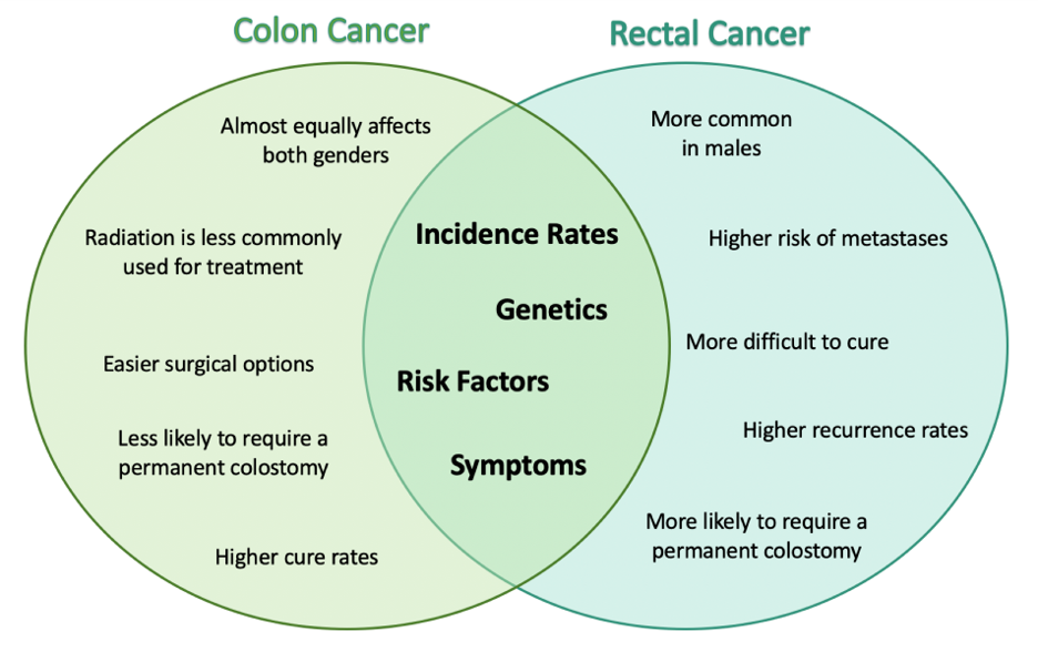
(Selchick, 2023)
Epidemiology
The American Cancer Society (ACS) estimates that there will be 158,850 new cases of CRC (108,860 colon and 49,990 rectal) in the United States in 2026 and 55,230 deaths. Between 2018 and 2022, the CRC incidence rate was 32% higher in males (40.5 per 100,000) than in females (30.7 per 100,000). Most diagnoses occur in adults 65 years and older, as incidence steadily climbs with age. However, over the last two decades, the age at diagnosis has steadily declined (currently 66) as CRC incidence has markedly shifted to a younger patient population. In 2022, 22% of CRCs were diagnosed in people 55 and younger, increasing from 11% in 1995. In 2026, 24,640 cases (16%) will be diagnosed in people younger than 50, and about 30% will occur in people aged 50 to 64 (NCI, 2025a; Siegel et al., 2026).
CRC incidence in the United States from 2018 to 2022 was highest among Alaska Native individuals (AN; 80.9 per 100,000), followed by American Indian individuals (AI; 49.3 per 100,000), non-Hispanic Black individuals (NHB; 40.4 per 100,000), and non-Hispanic White individuals (NHW; 35.2 per 100,000). The incidence rate is lowest among Asian American, Native Hawaiian, and Pacific Islander individuals (AANHPI; 28.5 per 100,000). CRC death rates have decreased by 56% since 1970 (through 2023). As with incidence, CRC mortality also varies significantly by race and ethnicity. The annual age-adjusted CRC mortality rate is 12.7 per 100,000 and is 42% higher in males (15.3 per 100,000) than in females (10.8 per 100,000). Based on data from 2019 to 2023, CRC mortality is highest in AN (31.5 per 100,000), followed by AI (17.7 per 100,000), NHB (16.6 per 100,000), NHW (12.9 per 100,000), and lowest in AANHPI (9.2 per 100,000). Between 2019 and 2023, CRC death rates in AN individuals were more than double those of NHW individuals. Proposed rationales for these racial disparities include several risk factors (e.g., smoking, vitamin D deficiency, diet, and elevated body mass index [BMI]) and potentially Helicobacter pylori infection, a bacterium associated with inflammation and cancer of the stomach, which may also be associated with CRC. AN individuals are less likely to have access to endoscopic services or facilities offering high-quality colonoscopies, contributing to lower screening (Haverkamp et al., 2023; Siegel et al., 2026).
The most important predictor of CRC survival is the cancer stage at diagnosis. CRC is diagnosed at an advanced stage in approximately 22% of patients. Currently, the 5-year relative survival rate is 65%. Survival improves when CRC is diagnosed at earlier stages. The relative 5-year survival rates are as follows:
- 91% for patients with localized-stage disease (i.e., no sign of cancer spread outside the colon or rectum)
- 74% for patients with regional-stage disease (i.e., cancer has spread outside the colon or rectum to nearby structures or lymph nodes)
- 15% for patients diagnosed with distant stage (i.e., cancer has spread to distant organs or lymph nodes; Siegel et al., 2026)
A CRC risk assessment is a vital first step for cancer prevention and early detection. A cancer risk assessment includes a thorough review of the past medical, surgical, social, and family history and an evaluation of any age-appropriate screening tests performed. The family history should include at least a three-generation pedigree, particularly if a hereditary cancer syndrome is suspected. Family history helps identify characteristics of familial cancers and genetic syndromes that require heightened surveillance and screening measures (ACS, 2023a, 2025).
Modifiable Risk and Protective Factors
In the United States, more than half (55%) of CRCs are attributed to modifiable (i.e., lifestyle) risk factors (refer to Table 1); thus, they are potentially preventable. Numerous studies have demonstrated that increased physical activity levels considerably decrease the risk of CRC. According to a 2022 systematic review and meta-analysis, individuals who consistently incorporate a healthier diet, regular physical activity, moderate alcohol consumption, and no tobacco exposure have a 48% lower risk of CRC and a 35% lower risk of CRC-specific mortality than their less-healthy counterparts (Yu et al., 2022). Those who are physically active have a 30% lower risk of developing proximal and distal colon tumors than sedentary people. Male patients with a BMI...
...purchase below to continue the course
Table 1
Modifiable Risk Factors
Modifiable Risk Factors that Increase Risk |
Excess alcohol consumption (average of three or more drinks/day) |
Elevated BMI 30 kg/m2 or more |
Excess consumption of red meat (100 g/day) |
Excess consumption of processed meat (50 g/day) |
Tobacco use (current or former) |
Physical inactivity |
Modifiable Risk Factors that Decrease Risk |
Regular physical activity |
Dairy consumption (400 g/day) |
Whole grain consumption |
Dietary fiber consumption |
(ACS, 2023a, 2025; Siegel et al., 2026)
Several other proposed risks are correlated with CRC, but evidence remains conflicting. While diet is believed to play a dual role in CRC in prevention and promotion through its influence on immune response and inflammation, the precise impact of specific foods on cancer occurrence is not defined beyond those listed in Table 1. Nevertheless, most data describe diets high in refined carbohydrates, processed sugar, and red meat as posing an increased risk for precancerous polyps and CRC. Although diets rich in whole grains and fiber are associated with a reduced risk of CRC due to potentially less carcinogen exposure in higher stool volume and faster transit time, consistent evidence supported by randomized controlled trials is lacking (ACS, 2023a). Prospective studies have proposed that vitamin D deficiency may contribute to CRC incidence, and vitamin D supplementation may protect against CRC, but no evidence confirms that vitamin D supplementation improves outcomes in those with CRC (NCCN, 2026a). Vitamin D might serve as a carcinogenesis inhibitor. Low vitamin D levels are suspected to increase the risk of CRC and may lead to inferior outcomes in patients diagnosed with CRC (i.e., higher mortality). To date, no study has definitively shown that vitamin D supplementation reduces the risk or improves survival in patients with CRC (Macrae, 2026). The NCCN (2026a) does not recommend routine screening for vitamin D deficiency or supplementing vitamin D in patients with CRC.
There is widespread research on the regular use of low-dose acetylsalicylic acid (Aspirin) and other nonsteroidal anti-inflammatory drugs (NSAIDs, such as ibuprofen [Motrin, Advil]) and their role in lowering the risk for CRC and precancerous polyps. However, this data has been challenged, and many questions remain unanswered. It is unclear whether regularly taking NSAIDs and/or acetylsalicylic acid (Aspirin) improves the survival of people with CRC or if it increases harm and serious side effects, such as gastrointestinal (GI) bleeding. While generally antineoplastic in the colon, the effects of acetylsalicylic acid (Aspirin) remain inconsistent, especially in older adults. Specific populations of patients (such as those with adenomas, prior carcinoma who have undergone resection, or high-risk patients with limited screening options) may benefit from regular use. The ACS does not recommend acetylsalicylic acid (Aspirin) for this use. The US Preventive Services Task Force (USPSTF) previously endorsed the regular use of acetylsalicylic acid (Aspirin) as more likely to have a beneficial effect when begun between the ages of 50 and 59, but benefits only become apparent after 10 years of consistent use. However, in 2022, the USPSTF reversed this recommendation, primarily based on data from the Aspirin to Prevent Events in the Elderly (ASPREE) randomized clinical trial. The ASPREE trial unexpectedly demonstrated that acetylsalicylic acid (Aspirin) increased the risk of cancer-related death compared with placebo (Chan, 2025).
Non-Modifiable Risk Factors
Several non-modifiable risk factors are associated with CRC, including advancing age, inflammatory bowel disease (ulcerative colitis and Crohn’s disease), environmental exposures, and inherited or acquired genetic mutations. Most CRCs are sporadic (60% to 70%), meaning healthy cells and genes mutate from genetic and environmental exposures over one’s lifetime. However, a family history of CRC in a first-degree relative (e.g., parent, sibling, child) is the strongest risk factor, increasing the risk by 2 to 4 times compared with those without a family history. About 30% of people with CRC have a family history, with the highest risk occurring when the diagnosis is before age 50 and multiple relatives are affected (ACS, 2023a). About 5% of people diagnosed with CRCs have inherited gene mutations associated with family cancer syndromes (ACS, 2025).
All cancer is fundamentally genetic, as cancer cells contain genetic alterations (mutations) that lead to uncontrolled cell division and growth. Healthy genes comprise DNA sequences with information to guide their proper functioning. Genetic changes in cancer growth typically affect proto-oncogenes, tumor-suppressor genes, and DNA repair genes. Proto-oncogenes create proteins for healthy cellular growth, division, and replication. When mutated, proto-oncogenes become cancer-causing oncogenes, allowing cells to grow and replicate out of control. Likewise, tumor-suppressor genes help regulate healthy cellular growth and division, so mutations in these genes eradicate their ability to control cellular processes, leading to unrestricted cellular division. DNA repair genes are tasked with fixing damaged DNA to prevent cancer growth, as all cells in the human body are prone to DNA damage over time. However, if repair genes are damaged or mutated, the cell loses its ability to repair. Errors cultivate and replicate over time, leading to duplications and deletions of chromosomal components. While cancer can also develop due to spontaneous transformation of the cell’s processes, many are caused by cell damage induced by carcinogens (ACS, 2026b; Yarbro et al., 2018).
The most common inherited causes of CRC include Lynch syndrome (LS), familial adenomatous polyposis (FAP), and inheritance of BRCA1/BRCA2 gene mutations (ACS, 2023a, 2025).
Lynch Syndrome
Lynch Syndrome (LS), or hereditary non-polyposis colorectal cancer (HNPCC), is the most common inherited risk for CRC. LS accounts for up to 4% of all CRCs and also poses an increased risk for several other cancer types, such as ovarian, endometrial (uterine), brain, stomach, and breast (ACS, 2025). Individuals with LS tend to develop colon polyps (benign growths in the colon) at younger ages and typically develop CRC in their 40s or 50s. Affected females have a higher risk of developing CRC than their male counterparts. In the United States, it is estimated that 1 in 279 individuals (1.2 million people) have a gene mutation associated with LS; however, most are undiagnosed since identification primarily depends on a cancer diagnosis (ACS, 2023a). Individuals inherit LS in an autosomal dominant pattern, by which one inherited copy of each cell’s altered gene is sufficient to increase cancer risk (NCI, 2025d; Rogers, 2022).
Changes in the MLH1, MSH2, MSH6, or PMS2 genes are most commonly found in LS. Under physiologic conditions, these genes repair potential errors during DNA replication (i.e., the process during which DNA is copied in preparation for cell division). Collectively, they are referred to as mismatch repair (MMR) genes. Since mutations in any of these genes impede the cell’s ability to fix DNA replication errors, abnormal cells continue to divide. Over time, the accumulated DNA replication errors can lead to uncontrollable cell growth and an increased propensity for developing cancer (NCI, 2025d). Mutations in the MLH1 or MSH2 gene are associated with a higher risk of developing cancer than mutations in the MSH6 or PMS2 genes, which carry a lower risk. Among those with high-risk MLH1 or MSH2 mutations, 18% to 32% will develop CRC by age 50, and 42% to 52% will develop CRC by age 70 (Dominguez-Valentin et al., 2023). Mutations in the epithelial cell adhesion molecule (EPCAM) gene are also associated with impaired DNA repair in LS. Since the EPCAM gene is located next to the MSH2 gene, specific mutations can cause the MSH2 gene to become inactivated, thereby enabling cell growth errors and cancer growth. While gene mutations predispose individuals to cancer, not all people with these mutations will develop cancerous tumors (NCI, 2025d). The NCCN and American Society for Clinical Oncology (ASCO) recommend genetic testing in all patients with CRC. According to the NCCN (2025) guidelines on genetic and familial high-risk assessment for CRC, individuals with a personal history of early-onset colorectal or endometrial cancer, the development of two or more LS-associated tumors, or multiple affected first- or second-degree relatives with LS-associated cancers should consider genetic evaluation and possible testing for LS (NCCN, 2025).
Familial Adenomatous Polyposis
A few types of polyposis syndromes are associated with increased risk for CRC, but familial adenomatous polyposis (FAP) is the most common and accounts for approximately 1% of all CRCs. People with FAP tend to develop multiple benign polyps in their colon in their 20s and 30s. Over time, these patients grow thousands of polyps; unless the colon is removed, these polyps will develop into cancer. The median age of CRC diagnosis in patients with FAP is 39 years. There are two genes associated with FAP that carry different inheritance patterns. Mutations in the adenomatous polyposis coli (APC) gene can cause FAP or attenuated FAP (i.e., polyp growth is delayed). The average age of CRC diagnosis for patients with attenuated FAP is 55 years. Mutations in the APC gene affect the cell’s ability to maintain healthy growth and function, leading to cellular overgrowth. Most people with mutations in the APC gene will develop CRC, but the number of polyps and the time frame in which they become malignant depend on the location of the mutation within the gene. FAP resulting from mutations in the APC gene is inherited in an autosomal dominant pattern (ACS, 2023a; NCI, 2025d; Rogers, 2022).
Mutations in the mutY DNA glycosylase (MUTYH) gene can also cause FAP and may be called MUTYH-associated polyposis (MAP). MUTYH gene mutations prevent cells from repairing errors made during DNA replication. As these errors accumulate, more polyps develop, posing an increased likelihood of cancer. MAP is a more recently recognized syndrome with a large variability in clinical features, but patients usually develop a similar number of polyps as those with attenuated FAP. MAP is inherited in an autosomal recessive pattern, which means that both genes in a pair (one from each patient) must be abnormal to cause the disease. Surgery is the standard of care for cancer prevention in patients with all FAP syndromes. Surgery is typically recommended once polyp growth accelerates beyond control with colonoscopy screenings (ACS, 2023a; NCI, 2025d; Rogers, 2022).
BRCA1/BRCA2 Gene Mutations
All individuals have BRCA1/2 genes, which act as tumor suppressors to prevent cancer by regulating cellular growth and division. Mutations in these genes prevent them from working correctly, increasing the propensity toward cancer development. Most commonly associated with breast, ovarian, and prostate cancers, BRCA1/2 mutations are being explored regarding their role in increasing the risk for several other types of cancers (NCI, 2024). Regarding CRC, the absolute risk associated with BRCA1 and BRCA2 gene mutations is not definitively established (ACS, 2023a). There is insufficient data regarding any increased risk of developing CRC in BRCA mutation carriers, so there are no formal guidelines regarding the necessity or frequency of CRC screening in these patients (NCI, 2025c).
Early Detection and Screening
Although nearly 60% of CRC-related deaths can be mitigated through proper screening and surveillance practices, research demonstrates that at least one in three people is not current with CRC screenings. Since a precancerous polyp can take 10 to 15 years to progress into cancer, screenings at recommended intervals can prevent CRC. Screening offers substantial opportunities to lessen CRC incidence and mortality. CRC screening aims to identify disease in the precancerous or early stage of cancer when it is small, localized, and curable. Various cancer screening guidelines are developed by credible organizations and are grounded in clinical research, evidence, and expert consensus. While there are some variations between the guidelines, they are relatively consistent in their recommendations on CRC screening. The ACS is one of the most widely used, comprehensive, evidence-based resources for cancer care; they publish an annual report summarizing CRC screening recommendations (ACS, 2023a).
Screening Modalities
Screening colonoscopy has led to the widespread detection of asymptomatic CRC carriers, increasing earlier-stage diagnoses. While colonoscopy is the most accurate and commonly used CRC screening test, multiple options exist. CRC screening modalities are divided into two major categories: high-sensitivity stool-based test (collected at home) or visual examination (performed at health care facilities), as described in Table 2 (ACS, 2023a).
Table 2
CRC Screening Tests and Recommended Intervals for Average-Risk Individuals
Stool Tests | Recommended Screening Intervals (Average Risk) | |
Fecal immunochemical test (FIT) |
|
|
High-sensitivity guaiac-based fecal occult blood test (gFOBT) |
|
|
Multitargeted stool DNA test (mt-sDNA) |
| Every 3 years |
Visual Examination Tests | Recommended Screening Intervals (Average Risk) | |
Colonoscopy |
|
|
Computed tomographic colonography (CTC) |
|
|
FSIG |
|
|
(ACS, 2023a)
Figure 3
Colonoscopy
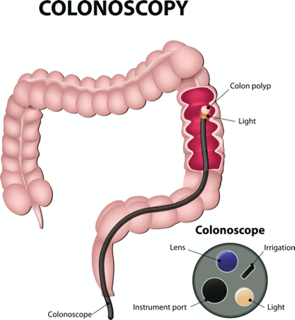
(iStock ID: 530612521)
Test Selection
Test selection depends on patient risk factors, preference, and availability. Research demonstrates that allowing patients options for testing increases adherence to screening. Since at least one-third of all adults and at least 50% of adults aged 50 to 54 are not up-to-date with CRC screening, the ACS (2023a, 2025) and the USPSTF (2021) do not endorse one test over another; they instead stress that all recommended tests can help save lives.
Limitations
All screening tests have limitations; some are potentially harmful, including false-positive and false-negative results. While stool-based tests are noninvasive and less costly, they are more likely to miss polyps or cancers. Furthermore, positive results identified on non-colonoscopy screening tests should be followed by a timely colonoscopy. Delays in the follow-up of abnormal results are associated with an increased risk of advanced disease and mortality. Colonoscopy is also subject to error and may miss adenomas. Although rare, colonoscopy poses a higher risk of complications such as bowel tears, infection, and sepsis. Evidence suggests that potential harms from colonoscopies increase with age but are relatively uncommon. In a review of 67 observational studies including 27,746,669 participants, the rate of major bleeding events and perforation was 14.6 (per 10,000) and 3.1 (per 10,000) colonoscopies, respectively (Lin et al., 2021; USPSTF, 2021).
Summary of CRC Screening Recommendations
The ACS (2026a) screening recommendations are based on risk categories: average risk and increased or high risk. The ACS explicitly defines individuals at high risk to include those with one or more of the following characteristics listed in Table 3. By default, individuals are considered at average risk if they do not have any of the characteristics listed in Table 3.
Table 3
Individuals at Increased or High Risk for CRC
|
(ACS, 2026a)
Average-Risk Screening Recommendations
In response to the increasing incidence of CRC in younger populations, the ACS (2026a) recommends initiating CRC screening at 45 years. The ACS recommends that adults aged 45 years and older undergo regular CRC screening with either a stool test or a visual exam at the testing intervals listed in Table 2. People in good health with at least a 10-year life expectancy should continue regular CRC screening through 75 (ACS, 2026a). The USPSTF (2021) recommends screening for CRC in all adults aged 50 to 75 years (Grade A) and adults aged 45 to 49 years (Grade B). The USPSTF also recommends that clinicians selectively offer screening for CRC in adults aged 76 to 85 years (Grade C), as evidence indicates the net benefit of screening all persons in this age group is small. The decision to perform CRC screening after age 75 should be based on individual cases, considering the patient's overall health, prior screening history, and preferences (USPSTF, 2021). The ACS (2026a) discourages CRC screening in people over 85. All positive results on any non-colonoscopy screening test must be followed up with a timely colonoscopy (ACS, 2026a).
High-Risk Screening Recommendations
People at higher risk for CRC should start screening before age 45. For those with one first-degree relative diagnosed with CRC before age 60, or two first-degree relatives diagnosed at any age, screening should be performed at age 40 or 10 years younger than the earliest age of diagnosis in the family, whichever comes first. Finite screening intervals for those at increased or high risk for CRC vary based on the patient and screening test results. The USPSTF and ACS do not set screening guidelines specifically for those in higher-risk categories due to the high variability between these individuals (ACS, 2026a; USPSTF, 2021).
Pathophysiology
The GI tract extends from the mouth to the anus and consists of several organs. It carries out several digestive processes to perform two primary functions: breaking down food to assimilate nutrients and eliminating waste. Food is processed and mixed with an enzyme called salivary amylase in the mouth before the esophagus propels the food into the stomach. The stomach churns the food and combines it with enzymes, mucus, gastric acids, and other secretions. The food is stored in the stomach until phasic contractions help propel it into the small intestine, where most nutrient absorption occurs. Biochemicals and enzymes secreted by the pancreas and liver break down the food particles into smaller, absorbable nutrients. The nutrients pass through the small intestine walls into blood vessels and lymphatics before reaching the large intestine, where fluid absorption continues. Liquid wastes are transported to the kidneys and excreted from the body in urine. Solid waste travels into the rectum and is eliminated through the anus (Rogers, 2022).
The large intestine is the final passageway of undigested food particles and is subdivided into four regions: the cecum, colon, rectum, and anus. It also includes the appendix. Its primary function is absorbing residual nutrients and water, synthesizing specific vitamins, forming feces (waste), and eliminating it. The cecum is the cul-de-sac, or small pouch, at the beginning of the large intestine. It receives partially digested food (i.e., chyme) from the small intestine and mixes it with bacteria to continue digestion and form feces before transporting it into the colon. A healthy colon is about five feet long and comprises four connecting regions: the ascending, transverse, descending, and sigmoid colon. The ascending colon receives the feces from the cecum, bacteria digest the waste, and the intestinal wall absorbs water, nutrients, and vitamins into the bloodstream. The hepatic flexure is located on the right side of the body near the liver, creating a sharp, right-angle bend in the colon, marking the beginning of the transverse colon. The transverse colon is the longest part of the colon, and most nutrient absorption and feces formation occur in this region. The splenic flexure connects the transverse and descending colon, forming a sharp, right-angle bend on the abdomen's left side. The descending colon walls absorb water and any remaining nutrients from the feces, dehydrating the stool in preparation for elimination. The sigmoid colon is the curved, S-shaped section of the colon that transports and stores the residual fecal matter until it is transported to the rectum. The rectum is the final straight portion of the large intestine and holds fecal matter until the body is ready to eliminate the waste through defecation. The rectum connects the colon to the anus, the GI tract’s final segment. The anus is a short tube at the end of the rectum that terminates at the body’s exterior and through which feces pass during defecation. The six layers of tissue that comprise the colon wall are outlined in Table 4. Figure 4 provides a cross-sectional view of the intestinal wall (Innerbody Research, n.d.; Rogers, 2022).
Figure 4
Cross-Section of Intestinal Wall
(iStock ID: 843960672)
Table 4
Tissue Layers of the Colon
Tissue Layer | Description |
Mucosa |
|
Muscularis mucosa |
|
Submucosa |
|
Muscularis propria |
|
Subserosal adipose tissue |
|
Serosa |
|
(Innerbody Research, n.d.; Rogers, 2022)
While most CRCs arise from polyps, most polyps are not cancerous. Two types of polyps are strongly associated with cancer: hypoplastic and adenomatous (adenoma) polyps. Hypoplastic polyps are typically large (greater than 1 cm), present in multiples (more than 20), and located on the right side of the colon (ascending colon). Adenomas are more closely associated with CRC and comprise two primary growth patterns: tubular and villous. Most adenomas have a combination of both and are therefore called tubulovillous adenomas. Smaller adenomas (less than 1.25 cm) typically follow a tubular growth pattern, whereas larger adenomas have a villous growth pattern. Larger adenomas and those with a villous growth pattern are more likely to have cancerous cells within them. Adenoma subtypes are also characterized by shape (i.e., pedunculated and sessile). As displayed in Figure 5, pedunculated polyps have a mushroom-like appearance as they have a round top attached to the surface by a narrow stalk. Sessile polyps are more common than pedunculated polyps, are flatter, and do not have a stalk. These do not protrude as much from the colon wall, making them harder to identify. The three major types of sessile polyps include papillary, villous, or serrated. Serrated is a term used to describe any polyp with a saw-tooth pattern (ACS, 2023b, 2023c; NCCN, 2026a, 2026b; Rogers, 2022).
Figure 5
Pedunculated and Sessile Polyps
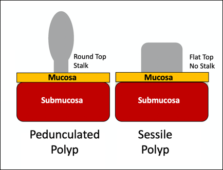
(Selchick, 2020)
CRC Subtypes
CRC is not a solitary tumor type; its pathogenesis ranges from slow-growing to aggressive, rapidly evolving tumors. Histologically, most CRCs are adenocarcinomas. The prefix “adeno” means glands, and “carcinoma” denotes a type of cancer that starts in the cells that form glands producing mucus to line the inner surfaces of the intestines. Adenocarcinomas produce abnormal glands that can infiltrate the submucosa and muscle layer and invade the surrounding tissues. In the submucosa, the cancerous cells can infiltrate lymphatic vessels, spread to nearby lymph nodes, penetrate veins, and metastasize to the liver and other organs. Two less common subtypes of adenocarcinoma include mucinous and signet ring cell. Although rare, other types of CRC include primary colorectal lymphomas, GI stromal tumors, leiomyosarcomas, carcinoid tumors, and melanomas; these account for less than 5% of all cases. CRC subtypes are outlined in Table 5 (ACS, 2023a; Macrae et al., 2026).
Table 5
CRC Subtypes
Subtype | Clinical Features |
Invasive (or infiltrating) adenocarcinoma
|
|
Mucinous adenocarcinoma |
|
Signet Ring Cell adenocarcinoma |
|
Rare types of CRC | |
Carcinoid tumors |
|
Primary colorectal lymphomas |
|
Gastrointestinal stromal tumors (GIST) |
|
Leiomyosarcoma |
|
(Choti, 2022; Lugo-Fagundo & Fishman, 2022; Menon & Cagir, 2025; Tebbi, 2024; Yarbro et al., 2018)
Signs and Symptoms of CRC
Most early-stage CRCs do not have any obvious warning signs and are commonly found during screenings. Symptoms of CRC are typically due to tumor expansion into the lumen or adjacent structures. Some of the most common symptoms of CRC include the following:
- abdominal pain, cramping, gas pains, bloating, or fullness
- rectal bleeding or blood in the stools (bright red, black, or tarry stools)
- new-onset anemia
- unintentional weight loss
- excessive fatigue or unusual tiredness
Rectal tumors can affect the stools’ caliber, causing narrower stools than usual, rectal pain, and bleeding (e.g., dark maroon or bright red blood in the stool). Some patients may experience tenesmus, the persistent inclination to evacuate the bowels, and a sensation that the bowel does not empty completely. Some patients may present with a palpable mass on the digital rectal exam (DRE; Macrae et al., 2026).
Diagnostic Workup
The diagnostic evaluation of patients with CRC depends on the clinical presentation. Patients whose tumors are not identified through screening generally present with any of the suspicious signs and symptoms outlined above or as a medical emergency with intestinal obstruction or acute GI bleeding. Regardless of the presentation, the definitive diagnosis is made by histologic examination of the tissue. In addition to being the most accurate screening test for CRC, colonoscopy is also the most versatile diagnostic test. It allows for the identification and biopsy of lesions throughout the large intestine, the detection of synchronous neoplasms, and the ability to perform polypectomy (polyp removal). Alternatively, a biopsy may be obtained from a surgical specimen. If rectal cancer is suspected, an FSIG may be performed instead. The diagnostic workup also routinely includes a complete blood count to evaluate for anemia, carcinoembryonic antigen (CEA) level, and diagnostic imaging tests (ACS, 2023a; Macrae et al., 2026).
Carcinoembryonic Antigen
Carcinoembryonic antigen (CEA) is the most widely used and diagnostically sensitive tumor marker for patients with CRC. Tumor markers may be produced by the cancer or the body’s response to cancer's presence and measured via a peripheral blood test. However, healthy cells also make CEA in smaller quantities, and thus, it is not sensitive enough to be used as a CRC screening test. Research has demonstrated that the sensitivity of CEA for the diagnosis of CRC is only around 50%. CEA's specificity is low as it is a nonspecific marker and can also be elevated in other cancerous processes, such as pancreatic or breast cancer. CEA can also be elevated in noncancerous conditions such as gastritis, liver disease, peptic ulcer disease, diabetes, and other acute or chronic inflammatory conditions. CEA levels are significantly higher in smokers than in nonsmokers. A preoperative CEA level above 5.0 ng/mL is a poor prognostic indicator, as research has demonstrated that higher preoperative CEA levels adversely impact CRC survival, independent of tumor stage. While most published guidelines, including the NCCN and the ASCO, still recommend CEA testing as part of CRC diagnostic workup and surveillance, serum CEA levels were removed from the American Joint Committee on Cancer (AJCC) Tumor, Node, Metastasis (TNM) staging system, 8th edition, but the group still recommends collecting this information for prognostic reasons (Compton, 2026; Macrae et al., 2026).
Diagnostic Imaging
Imaging tests are part of cancer staging as they evaluate the extent of the tumor, infiltration into surrounding structures, or distant spread to other sites in the body. About 20% of patients in the United States have distant metastatic disease at the time of presentation. The most common CRC metastatic sites are the regional lymph nodes, liver, lungs, and peritoneum. A computed tomography [CT] scan of the chest, abdomen, and pelvis with IV and oral contrast is recommended for most patients undergoing initial CRC workup and staging to assess for distant metastatic disease to the lungs, lymph nodes, liver, peritoneal cavity, and other organs. A positron emission tomography/CT (PET/CT) scan is not routinely indicated but may be considered in select patients with suspected or proven metastatic adenocarcinoma. Magnetic resonance imaging [MRI] of the abdomen and pelvis is considered more clinically valuable for evaluating the extent of primary rectal cancers (NCCN, 2026a, 2026b).
Principles of Pathologic Review
A biopsy sample undergoes a series of tests to determine the cancer’s pathologic features, evaluate its behavior, and select the best treatment options (Yarbro et al., 2018).
Tumor Grade
Tumor grade measures how different the cancer cells look compared with healthy cells under the microscope. It helps predict how likely the cancer is to grow and spread and is defined as follows (ACS, 2023a):
- Grade 1 is well-differentiated (i.e., cancer appears similar to healthy cells) and the least aggressive.
- Grade 2 is moderately differentiated, appears less like healthy cells, and is an intermediate grade (i.e., more aggressive than grade 1).
- Grade 3 is poorly differentiated or undifferentiated (i.e., does not resemble healthy cells), is high-grade, the most aggressive, and tends to grow and spread more quickly.
Gene and Molecular Biomarker Analyses
Kirsten Rat sarcoma and neuroblastoma Rat sarcoma
Rat sarcoma (RAS) is a group of proto-oncogenes often mutated in several cancer types, leading to uninhibited cell division. Kirsten and neuroblastoma RAS (KRAS and NRAS) are commonly mutated in CRCs; KRAS mutations occur in approximately 45%, and NRAS mutations occur in about 55% to 58% of cases. Mutations in KRAS and NRAS have been associated with increased tumor aggressiveness and poorer prognosis. They also have critical clinical implications when devising treatment plans for metastatic CRC. KRAS mutations strongly predict resistance to epidermal growth factor receptor (EGFR) inhibitors. Since EGFR is recognized as an important pathway in CRC growth and progression, several drugs have been developed to block this mechanism, such as cetuximab (Erbitux) and panitumumab (Vectibix; Compton, 2026). According to the NCCN (2026a, 2026b), all patients with metastatic CRC should be tested for KRAS and NRAS mutations.
BRAF V600E
Less than 10% of CRCs have a mutation called B-type rapidly accelerated fibrosarcoma (BRAF) V600E. This mutation is considered a poor prognostic sign, as it causes cancer cells to grow and spread more quickly (Compton, 2026). The NCCN (2026a, 2026b) recommends that all patients with metastatic CRC be tested for BRAF mutations, as they can be targeted with BRAF inhibitors.
MMR/Microsatellite Instability
According to the NCCN (2026a), universal MMR or microsatellite instability (MSI) testing is recommended for all newly diagnosed CRC patients, regardless of stage at diagnosis. Most CRCs do not have high MSI levels or changes in MMR genes; changes in these genes are most common in those with LS. Universal MMR testing helps guide whether patients should be tested for LS and determine if specific treatments (such as immunotherapy) are indicated. While MMR deficiency can be reported as microsatellite instability-high (MSI-H) or mismatch repair deficient (dMMR), they are interpreted to have the same meaning (Compton, 2026; NCCN, 2026a).
Human Epidermal Growth Factor Receptor 2
Human epidermal growth factor receptor 2 (HER2) amplification and overexpression are well-established drivers of malignancy in several tumor types, most commonly breast cancer. In metastatic CRC cancer, only 3% to 5% of patients have HER2 alterations. Still, it has emerged as an actionable target over recent years (Djaballah et al., 2022). The NCCN (2025, 2026a) recommends HER2 testing in all patients with metastatic CRC before starting treatment with EGFR inhibitors, as it is considered another predictive biomarker for poor response to treatment with EGFR inhibitors. However, if there is a known RAS mutation (i.e., KRAS or NRAS), HER2 testing is not indicated. Patients with a HER2 amplification or overexpression may be eligible for treatment with HER2-targeted antibodies such as trastuzumab (Herceptin) and pertuzumab (Perjeta; Djaballah et al., 2022; Dragovich, 2025; NCCN, 2025, 2026a).
Significance of the Anatomical Location of the Tumor
The primary tumor’s anatomical location provides important information about its behavior, unique molecular characteristics, and histological features, which are useful for predicting prognosis, survival, and when devising treatment plans. Specific symptoms may also differ depending on the tumor’s location, although abdominal pain is commonly associated with all sites (Macrae et al., 2026).
Ascending Colon (Right-Sided)
Ascending colon tumors typically present as fungating or polypoid masses that grow out into the open space within the lumen. They are more common in female patients with a genetic predisposition to CRC, such as those with mutations in BRAF V600E or KRAS. Right-sided tumors tend to be diagnosed at more advanced stages, remaining asymptomatic until relatively advanced, then causing discomfort, a palpable mass in the right lower quadrant, and dark red blood mixed with the stool. New-onset anemia, particularly iron-deficiency anemia from unrecognized blood loss, is much more common with right-sided colon tumors (ascending colon), which have a fourfold higher mean daily blood loss than tumors from other sites within the colon (Compton, 2026; Macrae et al., 2026; Rogers, 2022).
Descending Colon (Left-Sided)
Descending colon tumors typically start as button-shaped when small, grow circumferentially, spread along the entire bowel wall, and ulcerate as they penetrate the blood supply. They are more common in male patients and carry a better prognosis overall. Left-sided tumors are associated with an increased risk for intestinal obstruction, abdominal distention, pain, vomiting, and hematochezia (bright red blood on stools’ surface). A change in bowel habits is more commonly a presenting symptom for left-sided CRCs than right-sided. Left-sided tumors are more commonly nonmutated. Therefore, therapy responses are different between these tumor entities, given the distinctions in their molecular profiles (Compton, 2026; Macrae et al., 2026; Rogers, 2022).
Rectal Tumors
Rectal tumors most commonly develop up to 15 cm from the anal orifice and can spread through the rectal wall to surrounding structures, such as the lymph nodes. Rectal tumors most commonly develop in the lower third of the rectum since it lacks serosal covering, and commonly cause hematochezia, tenesmus, rectal pain, and narrower/thinner stool production (Macrae et al., 2026; NCCN, 2026b; Rogers, 2022).
Cancer Staging
CRCs are staged based on the location of origin, extent of disease, depth of invasion into the intestinal wall, and involvement of neighboring and distant structures. CRC staging uses the AJCC TNM (tumor, node, metastases) system to assign cancer a stage. Figure 6 is a graphic depicting the basic stages of colon cancer. Table 6 outlines colon and rectal cancer staging.
Figure 6
The Stages of Colon Cancer
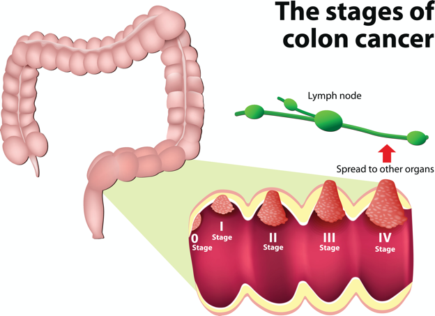
(iStock ID:528814299)
Table 6
Cancer Staging
Colon Cancer Staging | ||
TNM | T: Indicates how far the tumor has grown through the colon wall | |
N: Denotes the presence (or absence) of cancer in the lymph nodes | ||
M: Designates if cancer has spread to distant parts of the body | ||
Stages | Stage 0 | Stage 0 is noninvasive, confined to the colon wall. It has not grown beyond the first layer of the colon wall (carcinoma in situ). |
Stage I | The cancer has grown into the second or third layer of the colon wall but has not spread to nearby lymph nodes or distant sites. | |
Stage II | The cancer has grown into or beyond the fourth layer of the colon wall but has not spread to nearby lymph nodes or distant sites. | |
Stage III | The cancer has spread from the colon to nearby lymph nodes, or there are tumor deposits (small tumors in the fat around the colon). | |
Stage IV | The cancer has metastasized to areas far from the colon. Colon cancer most commonly spreads to the liver and lungs first, but can also spread to other locations, such as the peritoneum (the lining of the abdominal cavity) and the brain. | |
Rectal Cancer Staging | ||
TNM | T: Indicates how far the tumor has grown through the rectum | |
N: Denotes the presence (or absence) of cancer in the lymph nodes | ||
M: Designates if the cancer has spread to distant parts of the body | ||
Stages | Stage 0 | Noninvasive cancer that is contained and has not grown beyond the first layer of the rectal wall. |
Stage I | The cancer has grown into the second or third layer of the rectal wall. There is no cancer in the nearby lymph nodes or distant sites. | |
Stage II | The cancer has grown through the rectal wall and may have attached to or grown into nearby structures or organs. There is no cancer in the nearby lymph nodes or distant sites. | |
Stage III | The cancer has metastasized to nearby lymph nodes, or there are small secondary tumor deposits in the fat around the rectum. | |
Stage IV | The cancer has metastasized to distant sites. Rectal cancer commonly spreads to the liver and lungs first, but may also spread to other areas, such as the peritoneum and prostate. | |
(NCCN, 2026a, 2026b)
Treatment Modalities
Treatment for CRC is often multimodal, as multiple therapies are combined and administered simultaneously (concurrently) or sequentially. CRC treatments are grouped into two major categories of localized and systemic therapies. The optimal treatment strategy depends on several factors, including the pathologic features, stage, gene mutations, underlying health conditions, performance status, and patient preferences (ACS, 2024; NCCN, 2026a, 2026b).
Localized Therapies
Treatments explicitly directed at the tumor without affecting the rest of the body are called localized therapies. Surgery and radiation therapy are the two primary types used for CRC. They are most useful for early-stage cancers that have not metastasized beyond the area of origin. Surgery is the mainstay treatment for early-stage colon and rectal tumors. Since there are fundamental distinctions between surgical interventions for colon and rectal tumors, they will be discussed separately (ACS, 2024; NCCN, 2026a, 2026b).
Surgical Intervention for Colon Cancer
Polypectomy. Most large pedunculated or sessile polyps can be effectively removed by polypectomy, a relatively noninvasive procedure commonly performed during a colonoscopy. The polyp is removed by transecting the stalk with a snare, a wire loop device designed to slip over the polyp. Upon closure of the device, the polyp is removed at the stalk. Snares may or may not use electrocautery to resect polyps en bloc, or a cold snare may be used to remove polyps piecemeal. Polypectomy aims to remove the entire lesion in one piece and avoid leaving residual cancerous cells behind, which could replicate, grow, and spread (ACS, 2024; Seitz et al., 2026).
Colectomy and Hemicolectomy. Surgery to remove all (or most) of the colon is called a total colectomy. A partial colectomy or a hemicolectomy removes a portion of the colon. The type and extent of the surgery depend on the degree of cancer presence and its impact on surrounding structures. A lymphadenectomy commonly occurs during a colectomy and involves removing nearby lymph nodes (ACS, 2024). The NCCN (2026a) advises that a minimum of 12 lymph nodes should be examined to establish an accurate nodal (N) stage. At the end of the procedure, the colon’s remaining ends are adjoined.
Colostomy. More extensive surgery is required if the cancer is advanced or the tumor obstructs the colon. If a large portion of the colon is removed, the surgeon may not be able to reattach the colon. In these situations, a stoma is created in which the end of the intestine is brought to the surface of the abdominal wall to provide an outlet for waste products (stool) to leave the bowel. A colostomy may be temporary or permanent, depending on the part of the colon affected and the stoma’s location. An ileostomy is formed when the end of the small intestine (the ileum) is connected to a stoma. A collection bag is attached to the skin to hold the stool (ACS, 2024).
Surgical Intervention for Rectal Cancer
The type of surgery depends on the anatomical location and extent of the rectal cancer. Early rectal cancers are usually removed by polypectomy, whereas more advanced rectal tumors may require local excision (i.e., healthy surrounding tissue is excised alongside cancerous tissue). It is necessary to evaluate the tumor's proximity to the anus to determine the optimal surgical strategy for rectal cancer. Since rectal tumors are most likely to spread to the anal sphincter (i.e., the ring-like muscles surrounding the anus that prevent stool from leaking out), patients must undergo presurgical MRI imaging of the pelvis. MRI is the imaging modality of choice due to its superior ability to assess the soft tissue structures and depth of invasion into the wall of the rectum (NCCN, 2026b).
Transanal Excision (TAE). Historically, the anus and sphincter were excised along with the rectal tumor. However, removing these structures is associated with significant morbidity, including interference with normal bowel functioning, increased risk for incontinence, and reduced quality of life. Recent advancements in medicine have led to novel surgical techniques that allow experienced surgeons to operate through the anus, removing only the rectal tumors and small amounts of the surrounding tissue, leaving the anus and sphincter intact. Sphincter-sparing TAE surgery is an option for patients with early-stage I rectal tumors near the anal opening that have not spread to the anus or sphincter. During this procedure, the surgeon cuts through the layers of the rectal wall to remove the cancer and some surrounding healthy tissue. The sphincter-sparing TAE technique typically allows patients to retain bowel function and eliminates the need for a permanent colostomy. Lymph nodes are not usually removed during this procedure (ACS, 2024).
Low Anterior Resection (LAR). LAR is a more extensive surgical approach reserved for advanced rectal tumors located in the upper portion of the rectum. The cancerous portion of the rectum is removed, and the lower portion of the colon is then reattached to the remaining part of the rectum. Patients may need a temporary colostomy following a LAR, but most will regain the normal functioning of their bowels (ACS, 2024).
Proctectomy with Coloanal Anastomosis. The surgical removal of the entire rectum is called a proctectomy. Most stage II and III tumors located in the middle or lower third portion of the rectum require a proctectomy, followed by a total mesorectal excision (TME). A TME involves the removal of all surrounding lymph nodes. The colon is then reattached to the anus in a procedure called coloanal anastomosis. Most patients will regain normal functioning of their bowels over time (ACS, 2024).
Abdominoperineal Resection (APR). APR is similar to a LAR, but it is a more extensive surgery. APR is typically indicated for stage II and III rectal tumors that have invaded the sphincter muscles or the levator muscles (i.e., the muscles that control urinary flow). The entire anus and rectum are removed in an APR, necessitating the creation of a permanent colostomy (ACS, 2024).
Pelvic Exenteration. Pelvic exenteration is a radical and complex surgical approach. The rectum and all affected nearby organs are removed, such as the prostate or bladder. Patients require a permanent colostomy, and if the bladder is removed, a urostomy (opening through the skin to allow the elimination of urinary waste) is also required. This surgical technique is not commonly performed as it is associated with significant morbidity, but may be necessary if the cancer has spread into nearby organs (ACS, 2024).
Surgical Risks and Side Effects
The risks and side effects of surgery depend on the size and degree of cancer invasion, the extent of surgery, and the structures removed. All surgeries and invasive procedures are accompanied by risks, such as adverse reactions to anesthesia, bleeding, infection, perforation of the bowel (a hole in the intestines), and life-threatening sepsis. Patients may have complications with newly formed colostomies, such as malfunctioning of the stoma, skin breakdown, and leaking at the site. Adjusting to a colostomy can be challenging, and patients may struggle to properly care for and manage the device. Patients may also be affected by the physical and psychological aspects of body image distortion and lifestyle adjustments. Sexual dysfunction is common among males who undergo LAR or APR, as the nerves that supply blood to the penis may be injured during the surgery. Males may be unable to achieve an erection or orgasm, which can negatively impact interpersonal relationships, quality of life, psychological health, and well-being. APRNs are critical in helping patients acclimate to these life-altering changes by facilitating healthy coping, managing expectations, and referring patients to appropriate support groups or therapists. APRNs should approach these issues with sensitivity, empathy, and compassion, fostering a safe and non-judgmental environment for patients to express their concerns openly (ACS, 2024; Yarbro et al., 2018).
Radiation Therapy
Radiation therapy is a localized treatment that delivers a precisely measured amount of high-energy, highly focused rays of ionizing radiation to the tumor while providing as little injury as possible to surrounding tissue. Radiation causes cellular damage to cancer cells, leading to biological changes in the DNA, rendering cells incapable of reproducing or spreading. All healthy and cancer cells are vulnerable to the effects of radiation and may be injured or destroyed; however, healthy cells can repair themselves and remain functional. The total radiation dose is hyper-fractionated, which means it is delivered to the tumor in small, divided doses, or fractions, rather than all at once. Hyperfractionation allows healthy cells a chance to recover between treatments. The total number of fractions depends on the tumor size, location, patient's overall health, performance status, goals of therapy (e.g., curative intent or palliation of symptoms), and concurrent therapies (Mitin, 2026). Radiation therapy is central in treating CRCs and can be delivered externally or internally; some patients may receive both (ACS, 2024).
External beam radiation therapy (EBRT) delivers radiation from a source outside the body and is the most common type of radiation used for CRC. Historically, radiation beams could only match the tumor’s height and width, exposing more healthy tissue to radiation. Over the last few decades, 3D conformational radiation therapy (3D-CRT) has become the mainstay of EBRT for CRCs. 3D-CRT can reshape the radiation beam to match the tumor’s shape. Further advancements in imaging technology have led to more precise treatment mechanisms. Intensity-modulated radiation therapy (IMRT) is a newer, highly conformal radiation technique that further reduces unintended exposure to healthy tissue. While 3D-CRT and IMRT are very similar in that they both target the tumor while sparing healthy tissue, IMRT allows modulation of the radiation beam’s intensity, delivering a higher radiation dose to an exact location. The enhanced targeting technology of IMRT allows higher doses to reach the tumor, improving clinical outcomes and limiting side effects (Wo et al., 2021). Stereotactic body radiation therapy (SBRT) is a technique in which extremely high biological doses of radiation are administered over a few short treatments. The target area is affected to a higher degree over a shorter period with minimal impact on healthy tissue. In CRC, SBRT is usually reserved for patients with metastatic disease, treating metastatic sites such as pulmonary nodules (Jeong et al., 2024).
Although internal radiation therapy is not widely used for CRC, brachytherapy and intraoperative radiation therapy (IORT) are the most common options. Brachytherapy involves implanting a wire, seed, pellet, or catheter into the body within or near the tumor. In CRC, brachytherapy has been used following EBRT to deliver an added radiation boost to a specified area or to palliate symptoms. IORT is an intensive form of radiation administered directly into the target area during surgery while sparing the surrounding healthy tissues. The role of IORT is limited but may be considered for very close or positive surgical margins following resection as an additional radiation boost for patients with large or recurrent tumors. IORT is also a viable treatment option for patients with CRC that has spread to the liver, causing predominant hepatic disease (NCCN, 2026a, 2026b; Wo et al., 2021).
Radiation Side Effects
Radiation side effects depend on the specific area(s) of the body exposed and the dose received. Superficial skin irritation is common and can include redness, blistering, and inflammation, giving skin the appearance of a sunburn. GI symptoms are common due to the tumor’s anatomical location and the impact of the radiation on surrounding tissues and structures. Common symptoms of GI toxicity include nausea, vomiting, diarrhea, anorexia, bowel incontinence, rectal irritation, rectal bleeding, blood in stools, and pain with defecation. Radiation directed at these regions can lead to fluid volume deficit, electrolyte disturbances, weight loss, and malnutrition. Some patients may require the placement of a feeding tube to ensure adequate nutrition and prevent cachexia. Nutrition is a core component of cancer treatment and largely influences the patient’s tolerance to therapy and toxicities. Radiation can indirectly affect underlying structures located within the radiation field, such as the bladder or prostate, causing cystitis (inflammation of the bladder), dysuria (painful urination), hematuria (blood in the urine), urinary incontinence, and loss of pelvic floor muscular strength. Systemic effects may include fatigue, weakness, dehydration, scarring, fibrosis, and adhesion formation (the radiated tissue sticks together; ACS, 2024; Yarbro et al., 2018).
Systemic Therapies
Chemotherapy
Chemotherapy (cytotoxic or antineoplastic therapy) refers to high-risk, hazardous medications that destroy cancer cells throughout the body. Chemotherapy interferes with the normal cell cycle, impairing DNA synthesis and cell replication, thereby preventing cancer cells from dividing and multiplying (Yarbro et al., 2018). Chemotherapy can be used at various time points during the CRC disease trajectory. Neoadjuvant chemotherapy intends to shrink a tumor so that the surgical intervention is less extensive. Neoadjuvant chemoradiation (i.e., concurrent chemotherapy and radiation therapy) is the gold standard for locally advanced rectal cancer, followed by surgical resection and adjuvant chemotherapy (i.e., additional chemotherapy following surgery). Chemotherapy acts as a radiosensitizer, thereby rendering cancer cells more vulnerable to the toxic effects of radiation. After surgery, adjuvant chemotherapy aims to prevent cancer recurrence, reduce micro-metastases, and eradicate any remaining cancer cells. Palliative chemotherapy aims to relieve or delay cancer symptoms, enhance comfort, reduce symptom burden, and improve quality of life. CRC chemotherapy may include oral and/or IV formulations, with regimens typically consisting of two or more medications (refer to Table 7). Some of the most common chemotherapy agents used for CRC are listed below (ACS, 2024; Dragovich, 2025; Overman & Bednarski, 2026).
- 5-fluorouracil (5-FU)
- capecitabine (Xeloda)
- irinotecan (Camptosar)
- oxaliplatin (Eloxatin)
- trifluridine and tipiracil (Lonsurf; ACS, 2024)
Table 7
Common CRC Chemotherapy Regimens
Regimen Name | Drugs Included |
FOLFOX |
|
FOLFIRI |
|
FOLFOXIRI |
|
CapeOx |
|
Concurrent Chemoradiation |
|
(NCCN, 2026a; 2026b)
Chemotherapy Side Effects
Side effects of chemotherapy are inevitable due to the nonspecific nature of cytotoxic therapy; it simultaneously impacts healthy and cancerous cells. However, side effects vary based on the drug type, dosage, duration of treatment, and underlying health conditions. Not all patients respond similarly, and not all chemotherapy agents pose the same risks. Assessment and education are the most critical components to ensuring timely recognition, intervention, and management of side effects as experienced by each patient. Many side effects, such as nausea, can be primarily thwarted by implementing appropriate prevention strategies and medications. As a group, the most common side effects include reduced blood counts (anemia, thrombocytopenia, neutropenia), fatigue, nausea, anorexia, alopecia (hair loss), mucositis (mouth sores), diarrhea, skin changes, and peripheral neuropathy (damage to the sensory nerves). These agents may also cause palmar-plantar erythrodysesthesia (PPE or hand-foot syndrome, i.e., erythema, tenderness, and skin peeling on the palms/soles) or cardiotoxicity. Table 8 highlights a few of the unique side effects and key considerations associated with each agent (Dragovich, 2025; Overman & Bednarski, 2026).
Table 8
CRC Chemotherapy Agents and Clinical Considerations
Chemotherapy | Clinical Considerations |
5-Fluorouracil (5-FU) |
|
Capecitabine (Xeloda) |
|
Oxaliplatin (Eloxatin) |
|
Irinotecan (Camptosar) |
|
(Olsen et al., 2023; Yarbro et al., 2018)
Targeted Therapy
Targeted agents are devised to attack specific parts of cancer cells to prevent tumor growth or to shrink existing tumors. Proteins (called growth factor receptors) connect the external and internal cellular environments and are essential for healthy cell growth and development. Alterations in genes lead to changes in these proteins, disrupting normal cellular processes and igniting an environment for cancer growth. While each type of targeted therapy has a distinct mechanism of action (refer to Table 9), they all interfere with the cancer cell’s ability to grow, divide, repair, and/or communicate with other cells. By directing their effects on tumor cell growth through specific targets, these therapies are considered less toxic to normal cells than traditional chemotherapy agents. However, cancer cells have the potential to become resistant to them, as they only block specific pathways of cancer growth (Dragovich, 2025; Olsen et al., 2023).
Table 9
Targeted Therapies: Primary Mechanisms of Action
|
|
|
|
|
(Dragovich, 2025; Olsen et al., 2023)
EGFR Inhibitors
EGFR is a protein found on the surfaces of some cells, which causes them to divide when the epidermal growth factor binds to it. EGFR is found at abnormally high levels in certain CRCs, and activation of EGFR accelerates tumor growth. EGFR inhibitors are monoclonal antibodies (i.e., synthetic proteins) that function like human antibodies in the immune system. Scientists analyze specific antigens on cancer cells (target) to determine a protein to match the antigen and then create a specialized antibody to precisely attach to the target antigen, like a key fits a lock. The antibodies bind to the antigen and mark it for destruction by the immune system. Monoclonal antibodies work on cancer cells in the same way natural antibodies work by identifying and binding to the target cells and then alerting other cells in the immune system to the presence of the cancer cells. EGFR inhibitors impede the activation of EGFR, thereby blocking cancer growth. Cetuximab (Erbitux) and panitumumab (Vectibix) are widely used in CRC treatment. They selectively bind to the extracellular component of the EGFR, thereby preventing cellular growth (Clark & Sanoff, 2026; Dragovich, 2025).
Cetuximab (Erbitux) is a chimeric monoclonal antibody, primarily made of a mouse (murine) protein with a (lesser) human protein component. It binds to extracellular EGFR, inhibiting cell growth and inducing apoptosis. Panitumumab (Vectibix) is a fully human monoclonal antibody that inhibits ligand binding to the EGFR, inhibiting cell growth. There is an increased risk of infusion-related reactions with cetuximab (Erbitux) due to its higher murine content, which can induce rigors, chills, and fevers. Rarely, life-threatening hypersensitivity reactions can occur. EGFR inhibitors most commonly affect the skin, causing an acne-like rash on the face and chest. Interval development of this rash typically denotes a therapeutic response to treatment, as patients who develop more severe rashes demonstrate increased survival outcomes. The rash can usually be treated with an antibiotic cream, but requires astute monitoring and intervention to prevent progression. Other side effects include fatigue, diarrhea, and headache (ACS, 2024; Clark & Sanoff, 2026; Dragovich, 2025). As noted earlier, cetuximab (Erbitux) and panitumumab (Vectibix) should not be used in patients with KRAS or NRAS gene mutations, as they are ineffective. Patients with BRAF V600E mutations are also highly unlikely to respond to these agents unless combined with a BRAF V600E inhibitor (NCCN, 2026a, 2026b).
Vascular Endothelial Growth Factor Inhibitors
Vascular endothelial growth factor (VEGF) is a signaling protein that stimulates angiogenesis (i.e., new blood vessel formation) in healthy and cancerous cells. Blood vessels carry oxygen and nutrients to the tissue for growth and survival. Tumors need blood vessels to grow and spread. Angiogenesis inhibitors (VEGF-inhibitors) bind to and inhibit VEGF receptors, blocking the proliferation and formation of new blood vessels. Bevacizumab (Avastin) is a humanized monoclonal antibody and one of the most commonly used VEGF inhibitors. The potential side effects of VEGF inhibitors include bleeding events, headache, hypertension, and proteinuria (protein spilling out in the urine due to increased pressure in the kidneys). Many patients require concurrent treatment with antihypertensives due to the medication’s impact on rising blood pressure levels. VEGF inhibitors are contraindicated within 6 weeks of surgery (preoperatively or postoperatively) due to increased risk for major bleeding events, delayed wound healing, and fistula (an abnormal connection between two hollow spaces within the body). VEGF inhibitors also carry a boxed warning for bowel perforation (i.e., a hole in the intestines). Patients should be counseled to report any sudden onset of severe and diffuse abdominal pain, bloating, nausea, vomiting, or rectal bleeding (Clark & Sanoff, 2026; Dragovich, 2025; Olsen et al., 2023).
BRAF V600E Inhibitors
BRAF inhibitors are only indicated for patients with mutations in the BRAF V600E gene. BRAF-mutated disease is an aggressive subtype of CRC with a poorer prognosis, although the mechanism remains poorly understood. BRAF inhibitors have been extensively studied in malignant melanomas; however, their activity in CRC remains limited. CRC treatment guidelines recommend that BRAF inhibitors are only used as part of a combined therapy modality for CRCs, as they are ineffective when used as monotherapy. The most common BRAF inhibitors used for CRC include encorafenib (Braftovi), vemurafenib (Zelboraf), and dabrafenib (Tafinlar). These oral medications directly attack the abnormal BRAF protein. When given with cetuximab (Erbitux) or panitumumab (Vectibix), the combination has been shown to shrink or reduce the progression of metastatic BRAF V600–mutated CRC and increase survival (Clark & Sanoff, 2026; Dragovich, 2025; NCCN, 2026a, 2026b). Common side effects include skin rash, abdominal pain, diarrhea, anorexia, nausea, joint pain, and fatigue. BRAF inhibitors increase the risk of developing squamous cell skin cancer (SCC). Patients must be educated on the importance of monitoring and reporting new skin lesions, as SCC can be effectively excised (ACS, 2024).
HER2-Based Therapy
HER2-positive CRC is amenable to HER2-directed monoclonal antibody treatments, such as trastuzumab (Herceptin) and pertuzumab (Perjeta). These agents are recombinant DNA-derived humanized monoclonal antibodies that selectively bind with high affinity to the extracellular domain of the HER2 receptor, inducing cell death. They are approved for metastatic CRC whose tumors overexpress HER2. Trastuzumab (Herceptin) and pertuzumab (Perjeta) are IV infusions that may be combined with certain chemotherapy or targeted agents. Both drugs carry boxed warnings for cardiotoxicity, including decreased left ventricular dysfunction (LVEF) or congestive heart failure (CHF). Clinicians are advised to evaluate pre-treatment cardiac studies (i.e., echocardiogram or multiple-gated acquisition [MUGA] scan) to establish a baseline, followed by serial LVEF evaluations at defined intervals during treatment, and then every 6 months up to 2 years after treatment ends. There are specific guidelines for discontinuing or delaying therapy for clinically significant declines in LVEF. In addition, these agents carry boxed warnings for embryo-fetal toxicity, as exposure can result in embryo-fetal death and congenital disabilities. Diarrhea is a common side effect of pertuzumab (Perjeta), with the incidence ranging from 28% to 72%. Clinical studies demonstrate the incidence is highest during the first treatment, decreasing with subsequent cycles. Despite the high incidence of diarrhea in patients on pertuzumab (Perjeta), research demonstrates that it can usually be managed effectively without causing severe toxicity, treatment delays, or drug discontinuation (Djaballah et al., 2022). Tucatinib (Tukysa) is an oral, anti-HER2 targeted agent approved for metastatic CRC when taken along with trastuzumab (Herceptin). It targets HER2 differently than trastuzumab (Herceptin) as it does so intracellularly (i.e., from inside the cancer cell). Combined with trastuzumab (Herceptin), they can more completely block HER2 signals from inside and outside the cell. Tucatinib (Tukysa) is taken by mouth twice daily, and common side effects include diarrhea, rash, fatigue, elevated liver function tests, nausea, abdominal pain, and fever (FDA, 2023).
Immunotherapy
Immunotherapy stimulates the immune system to recognize and destroy cancer cells. Immunotherapy aims to produce antitumor effects by modifying the actions of the body’s natural host defense mechanisms to become more sensitive to cancer cells. Immune-based treatments work differently from chemotherapy as they are highly specialized in their activity. Immune therapy has emerged as an effective treatment strategy for patients with metastatic MSI-H or MMR-deficient (dMMR) CRC who have disease progression (or whose cancer has grown) despite standard chemotherapy. Immune checkpoint inhibitors block the receptors cancer cells use to inactivate immune cells (specifically, T-cells). When this signal is blocked, T-cells can better differentiate between healthy and cancer cells, thereby augmenting the cancer cells' immune response. Checkpoint inhibitors are categorized into two categories: (1) programmed cell death-1 (PD-1)/PD-ligand 1 (PD-L1) inhibitors, and (2) cytotoxic T-lymphocyte–associated antigen 4 (CTLA-4) inhibitors. Pembrolizumab (Keytruda), nivolumab (Opdivo), ipilimumab (Yervoy), and dostarlimab (Jemparli) are four immunotherapy drugs used for metastatic CRC; their mechanism of action is summarized in Table 10. These agents are associated with durable clinical benefits, including improved progression-free and overall survival. Research demonstrates that combining nivolumab (Opdivo) and ipilimumab (Yervoy) leads to increased progression-free survival in these patients. Pembrolizumab (Keytruda) is FDA-approved for patients with unresectable or metastatic, tumor mutational burden-high (TMB-H) solid tumors (including CRC) that have no satisfactory alternative treatment options (Dragovich, 2025; NCCN, 2026a, 2026b; Shoushtari & Johnson, 2026).
Dostarlimab (Jemparli) is the newest anti-PD-1 therapy to join the treatment landscape for rectal cancer. In a phase 2 confirmatory clinical trial, neoadjuvant dostarlimab (Jemparli) demonstrated a 100% clinical complete response rate in 12 patients with dMMR locally advanced rectal cancer who received the drug for 6 months. Although a longer follow-up is necessary to assess the duration of response, this is a very encouraging therapeutic strategy that can potentially improve the clinical outcomes for patients with rectal cancer (Cercek et al., 2023; NCCN, 2026a).
Table 10
Immune Checkpoint Inhibitors for CRC
Drugs | Mechanism of Action |
PD-1/PD-L1 Inhibitors
Pembrolizumab (Keytruda)
Nivolumab (Opdivo) | PD-1 is a transmembrane protein expressed on the surface of circulating T-cells, B-cells, and NK-cells and is used to recognize “self” antigens from “non-self.” PD-1 helps keep the immune response working properly. When PD-1 attaches to PD-L1, an inhibitory ligand expressed on some normal and cancer cells, it acts as an “off switch” to keep them from attacking other cells in the body. The binding of PD-1 to PD-L1 signals T-cells to leave the neighboring cells alone, including cancer cells. Some cancer cells have large amounts of PD-L1, which helps them evade immune attacks. PD-1 inhibitors are monoclonal antibodies developed to prevent the formation of this complex. They disable the immune system’s brakes, and T cells are freed to attack cancer cells. Pembrolizumab (Keytruda) and nivolumab (Opdivo) are humanized monoclonal antibodies that bind with high affinity to PD-1, preventing its interaction with PD-L1 and PD-L2. |
CTLA-4 Inhibitor
Ipilimumab (Yervoy) | The binding of CTLA-4 (a protein receptor that downregulates the immune system) inhibits T-lymphocyte activation, resulting in a negative feedback signal that decreases immune response. Ipilimumab (Yervoy) is a humanized monoclonal antibody that induces T-cell activation by disabling the CTLA-4 feedback inhibition, activating the immune system. |
(Dragovich, 2025; Olsen et al., 2023; Shoushtari & Johnson, 2026)
Immunotherapy Side Effects
Immunotherapy’s most common side effects include fatigue, nausea, anorexia, cough, diarrhea, joint pains, skin rash, and itching. All immunotherapy drugs carry boxed warnings for immune-mediated adverse reactions, which can be fatal if left untreated. An autoimmune response can impact any organ system, inducing nonspecific inflammation throughout the body. The most common immune-mediated complications include enterocolitis, hepatitis, endocrinopathies (inflammation of the thyroid and adrenal glands), nephritis, and uveitis. Pneumonitis, colitis, and Stevens-Johnson syndrome are the most severe and potentially fatal reactions (Postow & Johnson, 2025). Among the immunotherapy agents, PD-L1/PD-1 inhibitors are generally the best tolerated and pose a lower risk for adverse immune-mediated reactions. Compared with drugs that target PD-1 or PD-L1, serious side effects are much more likely with CTLA-4 agents such as ipilimumab (Yervoy). Clinical trials demonstrate that up to 41% of patients experience immune-mediated effects (NCCN, 2023a). When ipilimumab (Yevoy) is combined with nivolumab (Opdivo), there is an even higher risk for severe immune-related toxicities. The most common adverse reactions, occurring in over 20% of patients receiving ipilimumab (Yervoy) plus nivolumab (Opdivo), include fatigue, diarrhea, musculoskeletal pain, abdominal pain, pruritus, and nausea. Most immune-related events are reversible with immunosuppressive steroid treatment (Bristol Myers Squibb, 2026; NCI, 2025b).
Nursing care of the patient receiving immunotherapy requires cautious triage and continuous meticulous assessment to identify signs of potential immune-related adverse events (irAEs), as a timely diagnosis is critical to ensure prompt response and reduce morbidity. Most irAEs are reversible with immunosuppressive steroid treatment but must be graded according to the Common Terminology Criteria for Adverse Events (CTCAE) and managed per specific medication guidelines (NCI, 2025b). Patient education is vital. Patients and caregivers should be taught about the importance of self-assessment and immediately reporting symptoms. With pneumonitis, symptoms can range from mild cough and dyspnea to severe shortness of breath and life-threatening hypoxia. GI toxicity can range from mild diarrhea and abdominal cramping to severe colitis, which can be fatal if not managed. Skin toxicity may present initially as mild pruritus or dermatitis and can progress to Stevens-Johnson Syndrome. This rare but is characterized by a painful systemic red rash that leads to blistering and sloughing of the skin’s top layer. Life-threatening endocrinopathies can cause an abundance of symptoms that may vary widely, such as extreme weakness, excessive fatigue or lethargy, electrolyte disturbances, thyroid inflammation, and pituitary dysfunction (Olsen et al., 2023; Yarbro et al., 2018).
For a more detailed review and understanding of chemotherapy, immunotherapy, and targeted cancer treatments, refer to the following Nursing CE courses:
• Oncology Medication Management Nursing CE Course for APRNs
• Oncology Nursing CE Course Parts 1 and 2
References
American Cancer Society. (2023a). Colorectal cancer: Facts & figures 2023–2025. https://www.cancer.org/content/dam/cancer-org/research/cancer-facts-and-statistics/colorectal-cancer-facts-and-figures/colorectal-cancer-facts-and-figures-2023.pdf
American Cancer Society. (2023b). Your colon or rectal pathology report: Invasive adenocarcinoma. https://www.cancer.org/cancer/diagnosis-staging/tests/pathology-reports/colon-pathology/invasive-adenocarcinoma-of-the-colon.html
American Cancer Society. (2023c). Your colon or rectal pathology report: Polyps (including serrated adenomas). https://www.cancer.org/cancer/diagnosis-staging/tests/pathology-reports/colon-pathology/colon-polyps-sessile-or-traditional-serrated-adenomas.html
American Cancer Society. (2024). Treating colorectal cancer. https://www.cancer.org/content/dam/CRC/PDF/Public/8607.00.pdf
American Cancer Society. (2025). Colorectal cancer risk factors. https://www.cancer.org/cancer/colon-rectal-cancer/causes-risks-prevention/risk-factors.html
American Cancer Society. (2026a). American Cancer Society guideline for colorectal cancer screening. https://www.cancer.org/cancer/types/colon-rectal-cancer/detection-diagnosis-staging/acs-recommendations.html
American Cancer Society. (2026b). Oncogenes, tumor suppressor genes, and DNA repair genes. https://www.cancer.org/cancer/understanding-cancer/genes-and-cancer/oncogenes-tumor-suppressor-genes.html
Bristol Myers Squibb. (2026). Opdivo (nivolumab) and Yervoy (ipilmumab) selected safety profile. https://www.opdivohcp.com/safety/crc/opdivo-yervoy
Cercek, A., Lumish, M., Sinopoli, J., Weiss, J., Shia, J., Lamendola-Essel, M., El Dika, I. H., Segal, N., Shcherba, M., Sugarman, R., Stadler, Z., Yaeger, R., Smith, J., Rousseau, B., Argiles, G., Patel, M., Desai, A., Saltz, L. B., Widmar, M., … Diaz, L. A. (2023). PD-1 blockade in mismatch repair-deficient, locally advanced rectal cancer. New England Journal of Medicine, 386, 2363–2376. https://doi.org/10.1056/NEJMoa2201445
Chan, A. T. (2025). NSAIDs (including aspirin): Role in prevention of colorectal cancer. UpToDate. Retrieved June 2026, from https://www.uptodate.com/contents/nsaids-including-aspirin-role-in-prevention-of-colorectal-cancer
Choti, M. A. (2022). Gastrointestinal stromal tumors (GISTs). https://emedicine.medscape.com/article/278845-overview
Clark, J. W., & Sanoff, H. K. (2026). Initial systemic therapy for metastatic colorectal cancer. UpToDate. Retrieved June 10, 2026, from https://www.uptodate.com/contents/initial-systemic-therapy-for-metastatic-colorectal-cancer
Compton, C. C. (2026). Pathology and prognostic determinants of colorectal cancer. UpToDate. Retrieved June 2026 from https://www.uptodate.com/contents/pathology-and-prognostic-determinants-of-colorectal-cancer
Djaballah, S. A., Daniel, F., Milani, A., Ricagno, G., & Lonardi, S. (2022). HER2 in colorectal cancer: The long and winding road from negative predictive factor to positive actionable target. American Society of Clinical Oncology educational book. American Society of Clinical Oncology: Annual Meeting, 42, 1–14. https://doi.org/10.1200/EDBK_351354
Dominguez-Valentin, M., Haupt, S., Seppälä, T. T., Sampson, J. R., Sunde, L., Bernstein, I., Jenkins, M. A., Engel, C., Aretz, S., Nielsen, M., Capella, G., Balaguer, F., Evans, D. G., Burn, J., Holinski-Feder, E., Bertario, L., Bonanni, B., Lindblom, A., Levi, Z., ... Hovig, E. (2023). Mortality by age, gene and gender in carriers of pathogenic mismatch repair gene variants receiving surveillance for early cancer diagnosis and treatment: A report from the Prospective Lynch Syndrome Database. eClinicalMedicine, 58, 101909. https://doi.org/10.1016/j.eclinm.2023.101909
Dragovich, T. (2025). Colon cancer: Treatment and management. https://emedicine.medscape.com/article/277496-treatment#d11
Haverkamp, D., Redwood, D., Roik, E., Vindigni, S., & Thomas, T. (2023). Elevated colorectal cancer incidence among American Indian/Alaska native persons in Alaska compared to other populations worldwide. International Journal of Circumpolar Health, 82(1), 2184749. https://doi.org/10.1080/22423982.2023.2184749
Innerbody Research. (n.d.). The cecum . Retrieved June 8, 2026, from https://www.innerbody.com/image_dige08/dige44.html
Jeong, J. U., Rim, C. H., Yoo, G. S., Cho, W. K., Chie, E. K., Ahn, Y. C., Lee, J. H., & Korean Oligometastasis Working Group, Korean Cancer Association. (2024). The clinical efficacy of colorectal cancer patients with pulmonary oligometastases by sterotactic body ablative radiotherapy: A meta-analysis. Cancer Research and Treatment, 56(3), 809–824. https://doi.org/10.4143/crt.2023.920
Lin, J. S., Perdue, L. A., Henrikson, N. B., Bean, S. I., & Blasi. P. R. (2021). Screening for colorectal cancer: An evidence update for the U.S. Preventive Services Task Force: Evidence synthesis, no. 202 [Report no. 20-05271-EF-1]. Agency for Healthcare Research and Quality. https://www.ncbi.nlm.nih.gov/books/NBK570913
Lugo-Fagundo, E., & Fishman, E. K. (2022). Colorectal leiomyosarcoma: A case report. Radiology Case Reports, 17(8), 2812–2814. https://doi.org/10.1016/j.radcr.2022.05.023
Macrae, F. A. (2026). Epidemiology and risk factors for colorectal cancer. UpToDate. Retrieved June 8, 2026, from https://www.uptodate.com/contents/epidemiology-and-risk-factors-for-colorectal-cancer
Macrae, F. A., Parikh, A. R., & Ricciardi, R. (2026). Clinical presentation, diagnosis, and staging of colorectal cancer. UpToDate. Retrieved June 8, 2026, from https://www.uptodate.com/contents/clinical-presentation-diagnosis-and-staging-of-colorectal-cancer
Menon, G., & Cagir, B. (2025). Colon cancer. In StatPearls [Internet]. StatPearls Publishing. https://www.ncbi.nlm.nih.gov/books/NBK470380
Mitin, T. (2026). Radiation therapy techniques in cancer treatment. UpToDate. Retrieved June 10, 2026, from https://www.uptodate.com/contents/radiation-therapy-techniques-in-cancer-treatment
Naik, H., Johnson, M. D. D., & Johnson, M. R. (2021). Internet interest in colon cancer following the death of Chadwick Boseman: Infoveillance study. Journal of Medical Internet Research, 23(6), e27052. https://doi.org/10.2196/27052
National Cancer Institute. (2024). BRCA gene changes: Cancer risk and genetic testing. https://www.cancer.gov/about-cancer/causes-prevention/genetics/brca-fact-sheet
National Cancer Institute. (2025a). Age and cancer risk. https://www.cancer.gov/about-cancer/causes-prevention/risk/age
National Cancer Institute. (2025b). CTCAE and adverse event reporting. https://dctd.cancer.gov/research/ctep-trials/for-sites/adverse-events
National Cancer Institute. (2025c). BRCA1 and BRCA2: Cancer risks and management (PDQ®)—Health professional version. Retrieved June 2026, from https://www.cancer.gov/publications/pdq/information-summaries/genetics/brca-genes-hp-pdq
National Cancer Institute. (2025d). Genetics of colorectal cancer (PDQ®)—Health professional version. Retrieved June 2026, from https://www.cancer.gov/types/colorectal/hp/colorectal-genetics-pdq
National Comprehensive Cancer Network. (2025). NCCN clinical practice guidelines in oncology (NCCN guidelines®) genetic/familial high-risk assessment: Colorectal, endometrial, and gastric—Version 1.2025—June 13, 2025. https://www.nccn.org/professionals/physician_gls/pdf/genetics_ceg.pdf
National Comprehensive Cancer Network. (2026a). NCCN clinical practice guidelines in oncology (NCCN guidelines®) colon cancer: Version 2.2026—April 7, 2026. https://www.nccn.org/professionals/physician_gls/pdf/colon.pdf
National Comprehensive Cancer Network. (2026b). NCCN clinical practice guidelines in oncology (NCCN guidelines®) rectal cancer: Version 2.2023—April 7, 2026. https://www.nccn.org/professionals/physician_gls/pdf/rectal.pdf
Olsen, M., LeFebvre, K., & Walker, S. L. (2023). Chemotherapy and immunotherapy guidelines and recommendations for practice (2nd ed.). Oncology Nursing Society.
Overman, M. J., & Bednarski, B. K. (2026). Overview of the management of primary colon cancer. UpToDate. Retrieved June 10, 2026, from https://www.uptodate.com/contents/overview-of-the-management-of-primary-colon-cancer
Postow, M., & Johnson, D. B. (2025). Overview of toxicities associated with immune checkpoint inhibitors. UpToDate. Retrieved June 10, 2026, from https://www.uptodate.com/contents/overview-of-toxicities-associated-with-immune-checkpoint-inhibitors
Rogers, J. L. (2022). McCance & Heuther’s pathophysiology: The biologic basis for disease in adults and children (9th ed.). Elsevier.
Seitz, U., Bohnacker, S., & Binmoeller, K. F. (2026). Endoscopic removal of large colorectal polyps. UpToDate. Retrieved June 2026, from https://www.uptodate.com/contents/endoscopic-removal-of-large-colorectal-polyps
Selchick, F. (2020). Pedunculated and sessile polyp [image].
Selchick, F. (2023). Comparison of colon and rectal cancers [image].
Shoushtari, A. N., & Johnson, D. B. (2026). Principles of cancer immunotherapy. UpToDate. Retrieved June 10, 2026, from https://www.uptodate.com/contents/principles-of-cancer-immunotherapy
Siegel, R. L., Wagle, N. S., Star, J., Kratzer, T. B., Smith, R. A., & Jemal, A. (2026). Colorectal cancer statistics, 2026. CA: A Cancer Journal for Clinicians, 76(2), e70067. https://doi.org/10.3322/caac.70067
Tebbi, C. K. (2024). Carcinoid tumor. https://emedicine.medscape.com/article/986050-overview
U.S. Food & Drug Administration. (2023). Highlights of prescribing information: TUKYSA® (tucatinib) tablets, for oral use. https://docs.seagen.com/TUKYSA_Full_Ltr_Master.pdf
U.S. Preventive Services Task Force. (2021). Colorectal cancer: Screening. https://www.uspreventiveservicestaskforce.org/uspstf/recommendation/colorectal-cancer-screening
Wo, J. Y., Anker, C. J., Ashman, J. B., Bhadkamkar, N. A., Bradfield, L., Chang, D. T., Dorth, J., Garcia-Aguilar, J., Goff, D., Jacqmin, D., Kelly, P., Newman, N. B., Olsen, J., Raldow, A. C., Ruiz-Garcia, E., Stitzenberg, K. B., Thomas, C. R., Jr., Wu, Q. J., & Das, P. (2021). Radiation therapy for rectal cancer: Executive summary of an ASTRO clinical practice guideline. Practical Radiation Oncology, 11(1), 13–25. https://doi.org/10.1016/j.prro.2020.08.004
Yarbro, C. H., Wujcik, D., & Gobel, B. H. (Eds.). (2018). Cancer nursing: Principles and practice (8th ed.). Jones & Bartlett Learning.
Yu, J., Feng, Q., Kim, J. H., & Zhu, Y. (2022). Combined effect of healthy lifestyle factors and risks of colorectal adenoma, colorectal cancer, and colorectal cancer mortality: Systematic review and meta-analysis. Frontiers in Oncology, 12, 827019. https://doi.org/10.3389/fonc.2022.827019
Powered by Froala Editor