About this course:
The purpose of this learning activity is to enhance clinical practice and improve patient outcomes by educating advanced practice registered nurses (APRNs) on the various types of diagnostic radiology imaging tests, ensuring an adequate understanding of the appropriate indications for ordering each exam, as well as the risks, benefits, and critical clinical considerations regarding the use of contrast media.
Course preview
Diagnostic Radiology for APRNs
The purpose of this learning activity is to enhance clinical practice and improve patient outcomes by educating advanced practice registered nurses (APRNs) on the various types of diagnostic radiology imaging tests, ensuring an adequate understanding of the appropriate indications for ordering each exam, as well as the risks, benefits, and critical clinical considerations regarding the use of contrast media.
Upon completion of this module, learners should be able to:
- describe the background of radiology, outline the components of the medical imaging process, and discuss the characteristics that impact image quality
- discuss the various aspects of radiation and the essential components of radiation safety and identify the amount of radiation in some of the most common imaging tests
- differentiate between the general risks of medical imaging tests with radiation exposure and those without radiation exposure
- describe the different types of diagnostic radiology tests and the indications for ordering, key patient teaching points, what to expect during the test, how long the test should take, as well as the associated risks and benefits
- review the different types of contrast media agents regarding their clinical considerations, including risks, contraindications, and monitoring parameters
- outline the signs and symptoms of allergic reactions to iodinated contrast, as well as the management and premedication regimens as guided by the American College of Radiology
- recognize the components of the Appropriate Use Criteria for Advanced Diagnostic Imaging as devised by the Centers for Medicare & Medicaid Services
- discuss the guidelines for diagnostic imaging during pregnancy and lactation
Radiation
Radiation is energy in the form of particles or waves emitted by both natural and synthetic sources. Comprised of two forms, ionizing and non-ionizing, radiation surrounds us in our daily lives. As depicted in Figure 1, radiation spans the electromagnetic (EM) spectrum, from lower-energy microwaves to higher-energy gamma rays. EM energy travels in waves, and the strength of the radiation depends on the frequency (how rapidly the waves move up and down) and the distance the wavelength travels. In general, the smaller the wavelength, the higher the energy of the radiation (Centers for Disease Control and Prevention [CDC], 2024b).
Figure 1
The Electromagnetic Spectrum
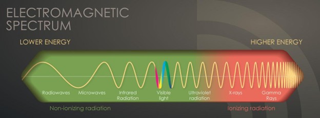
(CDC, 2024g)
Nonionizing radiation is the most prevalent form of radiation present in our environment at low levels. It can heat substances, although it cannot remove electrons from atoms and molecules. The most common types of nonionizing radiation include radiofrequency (RF) waves in many communication and electronic devices, kitchen microwaves, visible light, and lower energy forms of ultraviolet (UV) radiation. Intense, direct exposure to RF and microwave radiation can cause tissue damage from heat. In contrast, overexposure to UV rays can result in skin burns, premature aging, eye damage, and skin cancer. Most skin cancers are directly related to intense, prolonged, and unprotected exposure to UV radiation from the sun and tanning beds. The line dividing ionizing and nonionizing radiation becomes blurred in the UV section of the EM spectrum, as demonstrated in Figure 1. Radiation in the UV section at lower energies is considered non-ionizing, whereas at higher energies, extreme UV radiation becomes more harmful, rendering it ionizing radiation (CDC, 2024c). Magnetic resonance imaging (MRI) and medical ultrasound are the most common sources of non-ionizing radiation exposure in health care (American Association of Physicists in Medicine [AAPM], n.d.-a).
Ionizing radiation removes electrons from atoms and molecules, causing the atom to become ionized (or charged), enabling the wavelengths to pass through air, water, and tissue. Ionizing radiation is considered a carcinogen, or a substance capable of causing cancer, as it can penetrate the human body. When absorbed by living tissue, ionizing radiation can cause harm, especially at high levels. Most humans are exposed to low levels of ionizing radiation daily, whether from natural or artificial sources. Exposure can occur through construction and building materials, terrestrial radiation (radiation from the earth), and cosmic radiation (radiation from space). Radiographs are the most classic example of ionizing radiation (CDC, 2024b). Some people are exposed to higher amounts of natural background radiation, such as those living at higher mountain elevations or engaging in frequent air travel (American College of Radiology [ACR], 2023). Radiographs penetrate the body to visualize underlying structures. Ionizing radiation has sufficient energy to alter molecules within cells of the human body, targeting genetic material or DNA. Radiation interacts directly and indirectly, as it can break bonds within DNA and the water molecules surrounding it, leading to the formation of harmful free radicals (CDC, 2024a; Fazel & Einstein, 2024). When a cell is damaged from radiation exposure, one of the following three events will occur:
- The cell will repair itself and restore its normal function.
- The cell will remain altered, or only partially repair the damage, thereby heightening the risk of future cancer development.
- The cell will die, and the body will recover; however, if there is widespread cell death, this can increase the risk of organ failure (CDC, 2024a).
Health care exposure to ionizing radiation occurs with plain film radiographs, computed tomography (CT) scans, imaging that uses radioactive isotopes, and radiation therapy (AAPM, n.d.-a).
Diagnostic Radiology
Diagnostic radiology is a medical sector that includes various medical imaging technologies widely used throughout the US health care system, with millions of patients undergoing imaging evaluations daily. These are noninvasive and minimally...
...purchase below to continue the course
Radiology is considered one of the most technologically advanced medical fields, dating back to 1895, when Wilhelm Conrad Röntgen first discovered X-rays. Henri Becquerel then discovered radioactivity, in 1896, and Marie and Pierre Curie discovered radium in 1898 by. The field has expanded exponentially over the last few centuries and relies on the collaboration of scientists, medical physicists, radiologists, and imaging technologists. Medical physicists ensure the safe and optimal use of radiologic imaging modalities in patients. Diagnostic radiologists are healthcare providers (HCPs) who undergo specialized training in the analysis and interpretation of medical imaging to draw conclusions that aid in diagnosing, treating, and managing acute and chronic medical conditions and injuries. Interventional radiologists are HCPs who undergo specialized training in medical imaging to perform minimally invasive surgical procedures that diagnose, treat, and cure many conditions. Radiology technologists, also referred to as radiographers, have specialized training in performing digital imaging procedures under the direct supervision of a nurse or HCP. Radiographers are skilled at optimally positioning patients for imaging procedures to ensure accurate, high-quality images. They also serve essential roles in patient education (Elsayes & Oldham, 2014).
Imaging Process
Medical imaging refers to obtaining a permanent image of a particular part of the body to enable measurement and assessment of that body’s function or system. The overall objective of medical imaging is to make a specific area of the patient’s body visible for more detailed evaluation. Radiographs are produced by passing an X-ray beam generated by an X-ray tube through a patient. In its simplest terms, the image represents a shadow of the underlying structures through which the X-ray beam passes. The medical imaging process has five core components: the patient, the imaging system, the system operator (the radiographer), the resultant image, and the observer (the radiologist). Each medical imaging method is devised to reveal distinctive characteristics of the body, and the variability in imaging quality and visibility of structures can differ considerably. Some of the most common factors contributing to the inconsistencies in the resulting images include the quality and characteristics of the imaging equipment, the skill of the operator (including positioning and placement of the patient), specific patient characteristics (such as body habitus, prior surgeries, presence of scar tissue, or prior radiation therapy), as well as the timing of the imaging in relation to the injury or medical condition in question (International Atomic Energy Agency [IAEA], 2014).
Imaging Quality
Obtaining high-quality, high-resolution images is critical for drawing precise conclusions. High-quality images enable radiologists to visualize the body’s structures, evaluate underlying injuries or medical conditions, and make accurate diagnoses. Poor-quality images hinder accurate assessment and evaluation, leading to inconclusive or incorrect diagnoses. This often leads to the need for additional imaging tests, increasing the risks to the patient, contributing to delays in the diagnosis and treatment of the condition, and adding to the high-cost burden to patients and society (Elsayes & Oldham, 2014).
One key aspect of imaging quality is the signal-to-noise ratio, where signal refers to the information obtained from the part of the body being imaged, and noise refers to anything that hinders access to this information. Higher-quality radiographic images have higher signal levels than noise, allowing structures within the body to be observed clearly. Images of low quality have a poor signal-to-noise ratio; in other words, the signal level is similar to or less than the noise level, which causes the structures to become obliterated. Imaging artifacts are a commonly reported contributor to poor imaging quality. An artifact is a visual anomaly or any feature that appears in an image that is not present within the original imaged portion of the body. Artifacts misrepresent structures, can obscure underlying structures, and simulate pathology (Holmes & Griffiths, 2016). Additional characteristics that impact the quality of medical images are highlighted in Table 1.
Table 1
Characteristics that Impact Image Quality
Characteristic | Description |
Magnification and distortion | Areas subject to radiographic imaging are larger than the actual body part being evaluated. For optimal imaging quality, the X-ray source should be as close to the body part in question as possible and positioned parallel to it to provide optimal magnification and minimize image distortion. |
Sharpness and blur | Sharpness is essential for producing a higher-acuity image, whereas image blurring reduces image quality. Factors that affect image sharpness include:
|
Contrast and density | Contrast is the most fundamental characteristic of an image and refers to the difference in brightness (density) between two adjacent structures or between the area of interest and its surroundings. The greater the difference between adjacent tissue types, the easier it is to identify separate structures. Density is defined as the degree of blackening on the film. Four natural tissue densities range from dark to light shades based on their underlying structure. Gas, found in the lungs or throughout the GI tract, appears black on radiographic films. Adipose tissue appears dark gray. Fluid and soft tissues (connective tissue, muscles) appear light gray. Bone is the densest natural tissue, appearing nearly white, while anything metal appears white. |
Equipment | The monitor resolution at which the radiologist views the images and the sophistication of the computer-based picture archiving and communication system (PACS) also affect image quality. Better-quality, more precise images are associated with higher-resolution monitors and more advanced PACS systems. |
(Elsayes & Oldham, 2014; Holmes & Griffiths, 2016)
Risks with Medical Imaging
Many diagnostic radiology imaging tests and procedures involve exposure to ionizing radiation, increasing the risk of harmful effects. Some of the most common imaging tests associated with the highest ionizing radiation exposure include radiographs, CT scans, fluoroscopy, and nuclear medicine scans. Radiation therapy, used to treat malignancies, exposes the patient to even higher doses. The average person’s radiation exposure comes primarily from natural background sources, although up to 48% comes from medical imaging (US Environmental Protection Agency [EPA], 2026). Ionizing radiation can penetrate deep into the body, and repeated exposure from imaging tests increases the risk of cancer later in life. Specific populations such as infants and children, patients with compromised immune systems, and older adults are more vulnerable to the harmful health effects of radiation exposure. Younger age groups have cells dividing rapidly with rapid tissue growth, placing them at higher risk for long-term effects. Also, young people have a longer life span ahead of them, which gives cancer more time to develop. Older adults are at heightened risk from lifelong radiation exposure, impaired organ function, and other factors of aging that already place them at increased risk for cancer development (CDC, 2024a).
Pregnant females are also considered another vulnerable population due to the potential for harming the fetus at various stages of development. Data remains uncertain and inconsistent on the suspected risks in utero and to the newborn due to radiation exposure. Importance is placed on identifying pregnant patients before potential radiation exposure related to medical imaging. Facility policies should address prevention of unnecessary radiation exposure by carefully selecting examinations that limit the potential exposure in pregnant patients. The ACR describes scientific uncertainty, although it is suspected that radiation exposure would have no effect and be too subtle to be clinically detected at levels below 100 milligrays (mGy). At dose levels greater than 100 mGy, there is the potential for harm that could include spontaneous abortion during the first 28 days of the pregnancy, with higher doses potentially causing malformations and risk of intellectual disability during 29–189 days of gestation (ACR, 2023). Providers should use shared decision-making with pregnant patients to determine whether the procedure can be safely postponed until after delivery (CDC, 2024f).
The health effects of ionizing radiation are strongly dose-dependent. The radiation dose or the amount of radiation is the critical factor when evaluating the risk for future unintended health consequences, as the risks are dose-dependent. Harm can increase if radiation exposure is applied to the whole body rather than to a single part. Additionally, a single dose of radiation is more harmful than the same dose spread over a longer period (CDC, 2024a). As described in Table 2, the effects of radiation on the body’s tissues are measured using three methods: absorbed dose, equivalent dose, and effective dose (International Commission on Radiological Protection [ICRP], n.d.).
Table 2
Radiation Doses
Dose | How Radiation Doses Are Measured |
Absorbed dose
|
|
Equivalent dose |
|
Effective dose |
|
(Bell, 2025; ICRP, n.d.; Knipe, 2025, 2026)
The absorbed and equivalent doses are used to evaluate the short-term effect of radiation on tissue, which ranges from weeks to months. When diagnostic imaging is performed correctly, there are generally no short-term effects; therefore, the absorbed and equivalent doses are less meaningful in clinical practice. The effective dose is the most important and valuable dose quantity for most patients, as it reflects long-term effects (Lee & Elmore, 2026). The absorbed radiation dose varies widely depending on the type of examination. Nuclear medicine scans, such as positron emission tomography-computed tomography (PET/CT), have the highest radiation exposure among the most common medical imaging tests. A CT scan exposes the body to up to 1,000 times more radiation than a chest radiograph (EPA, 2026). Table 3 provides a detailed overview of the radiation dose to adult patients associated with common diagnostic radiology imaging examinations, along with a classification of risk levels. Aside from increasing one’s risk for future cancer development, ionizing radiation exposure can also contribute to the development of cataracts. These health effects most commonly result from exposure to very high levels of radiation associated with radiation oncology treatment fields, which require a significantly higher dose of radiation delivered directly to a localized area of the body (Lee & Elmore, 2026).
Table 3
Radiation Dose from Common Diagnostic Imaging Tests
Imaging Modality | Specific Test | Effective Radiation Dose (Approximate) | Comparison to Natural Background Radiation in Years | The Estimated Lifetime Risk of Fatal Cancer* |
Radiograph | Spine radiograph | 1.4 mSv | 6 months | Very low |
Chest radiograph | 0.1 mSv | 10 days | Minimal | |
Extremity radiograph | 0.001 mSv | 3 hours | Negligible | |
Upper gastrointestinal (GI) radiograph | 6 mSv | 2 years | Low | |
Lower GI radiograph | 6 mSv | 2 years | Low | |
Intraoral (dental) radiograph | 0.005 mSv | 1 day | Negligible | |
Intravenous pyelogram (IVP) | 3 mSv | 1 year | Low | |
CT scan | CT head | 1.6 mSv | 7 months | Very low |
CT head (with contrast) | 3.2 mSv | 13 months | Low | |
CT spine | 8.8 mSv | 3 years | Low | |
CT chest | 6.1 mSv | 2 years | Low | |
CT chest (lung cancer screening, low-dose CT scan) | 1.5 mSv | 6 months | Very low | |
CT abdomen and pelvis | 7.7 mSv | 2.6 years | Low | |
CT abdomen and pelvis (with contrast) | 15.4 mSv | 5.1 years | Moderate | |
Coronary CT angiography (CTA) | 8.7 mSv | 3 years | Low | |
Cardiac CT for calcium scoring | 1.7 mSv | 6 months | Low | |
Nuclear medicine | PET/CT scan | 22.7 mSv | 7.6 years | Moderate |
Bone densitometry (DXA) scan | 0.001 mSv | 3 hours | Negligible | |
Breast imaging | Mammography | 0.28 mSv | 34 days | Very low |
*Estimated lifetime risk of fatal cancer from the test’s radiation exposure | ||||
Negligible Less than 1 in 1,000 | Minimal 1 in 1,000,000 to 1 in 100,000 | Very Low 1 in 100,000 to 1 in 10,000 | Low 1 in 10,000 to 1 in 1,000 | Moderate 1 in 1,000 to 1 in 500 |
(EPA, 2025, 2026; Lee & Elmore, 2026; RadiologyInfo.org, 2025)
Radiation Safety
While the potential for increased risk of adverse health effects from diagnostic imaging is clearly described, there is no universally recognized threshold for specific radiation dose and associated effects. Therefore, it has been argued that there is “no safe level” of radiation exposure. The priority is to ensure the potential risks of ionizing radiation are continuously weighed against the benefits derived from the imaging test or procedure. This clinical decision should be made by the ordering provider and the patient, with full disclosure of the potential risks of imaging-related radiation exposure relative to the predicted benefits (ACR, 2023). The Occupational Safety and Health Administration (OSHA) outlines standards for controlling ionizing radiation hazards and preventing radiation exposure to health care workers and patients. Diagnostic radiology departments within hospitals and free-standing diagnostic radiology facilities must implement radiation protection programs managed by a radiation safety officer (RSO), such as a radiologist or a medical physicist who is a qualified expert. Radiology equipment is housed in designated areas within hospitals, usually on the ground floor and is secured behind lead doors to reduce exposure to employees and patients. Radiologists and radiology technicians must undergo specialized training in radiation safety practices, as mandated by state and federal law. OSHA standards require appropriate radiation-caution signage to alert individuals to radiation use or storage in designated areas, such as the bright yellow caution sign used in most medical facilities (OSHA, n.d.).
Keeping each health care worker’s occupational radiation dose as low as reasonably achievable (ALARA) is the key principle for developing workplace radiation protection programs. ALARA is premised on the following safety in three chief components of time, distance, and shielding as follows:
- time: minimize the time spent in areas with elevated radiation levels. This can be accomplished with proper preparation before testing to limit the time the patient and health care worker are in the exposure area
- distance: maximize the distance from sources of radiation, as a worker’s radiation dose of gamma rays and X-rays decreases as the distance from the source increases
- shielding: use shielding for radiation sources between a worker and a radiation source to significantly reduce or eliminate the dose received by the worker. This can be done by inserting the proper lead, concrete, or special plastic shields. Shielding also refers to lead doors and geographic areas within the facility to minimize exposure for individuals who are not directly working in those areas (OSHA, n.d.).
Shielding patients with lead aprons and lead coverings during diagnostic imaging tests has been the standard practice since it was endorsed by the US Food & Drug Administration (FDA) in the US Code of Federal Regulations in 1976. This practice originated due to concerns related to genetic fertility risks, and gonadal shielding was advised and implemented for all radiographs. The AAPM (2019) released a position statement outlining why routine fetal and gonadal shielding is unnecessary, as there is no evidence that the amount of radiation exposure from diagnostic imaging has any adverse effects on reproductive cells. Even after exposure to atomic bombings, no genetic effects have been observed, even three to four generations later. It is now known that the amount of radiation exposure needed to elicit detrimental effects on fertility is 100 times higher than the dose received from diagnostic imaging, further supporting the discontinuation of patient gonadal and fetal shielding in routine practice. On May 30, 2019, the ACR submitted a letter to the AAPM, endorsing their position on patient gonadal and fetal shielding. The ACR incorporated this change into its guidelines, with the objective that this recommendation be universally adopted and become the standard of care across diagnostic radiology (AAPM, n.d.-b). In 2023, the FDA formally rescinded its original ruling on shielding, finding it outdated considering current standards of practice (FDA, 2023b).
Diagnostic Imaging Tests with Radiation Exposure
Imaging studies can be divided into two main categories: planar and cross-sectional. Planar studies produce two-dimensional (2D) images, including basic diagnostic radiology testing such as radiographs and mammography. Cross-sectional imaging techniques capture three-dimensional (3D) aspects of human anatomy by producing more detailed images, often called “slices.” This imaging technology can then create a composite analysis of 2D slices to provide a 3D visualization of the anatomy. Cross-sectional imaging includes CT scans, MRI, and ultrasound (Elsayes & Oldham, 2014).
Radiograph
Radiograph is the most common and readily available type of diagnostic imaging test. Often referred to as a plain film, a radiograph is a quick, noninvasive, and painless imaging modality that produces images of the structures inside the body. In a standard radiograph, a beam of energy is generated by an X-ray generator and aimed at the intended body part. A plate is placed behind the body part to capture the variations of the energy beam. This is the simplest example of how ionizing radiation produces a 2D image. For optimal imaging quality, the X-ray detector (the plate) should be as close to the body part (patient or object being imaged) as possible. The radiation beams should be perpendicular (at a right angle) to the body part, as this helps minimize magnification and enhance the sharpness of the resulting image, thereby producing a more precise result (Elsayes & Oldham, 2014). A single-image radiograph, such as a mammogram, exposes the patient to lower doses of ionizing radiation than continuous fluoroscopy imaging used during cardiac stent placement, because the imaging extends over a longer period (FDA, 2023a).
Radiographs are widely used across various health care domains for several indications. They help diagnose acute bone fractures and cardiopulmonary conditions, such as cardiac enlargement, pneumonia, or pleural effusion. They are also helpful for diagnosing arthritis and identifying foreign bodies or bowel obstructions. In addition, radiographs may assist in fluoroscopy procedures, facilitating the placement of tubes or other devices inside the body. Similarly, radiographs are used to verify the proper placement of a device after surgery or to ensure that no medical devices are left in the body (National Institute of Biomedical Imaging and Bioengineering [NIBIB], 2025b).
Patients are required to remove any clothing or jewelry, especially metal items, which may interfere with the procedure. This standard applies to all forms of diagnostic imaging tests and procedures. The patient should be educated that they are required to either lie, sit, or stand still while the radiograph machine takes images. The patient may be asked to assume several specific positions to obtain the highest-quality imaging results. For instance, when evaluating a patient for pneumonia, the patient is usually asked to take a deep breath and hold it, which helps expand the lung fields and provide a higher-quality image. In addition, several photos may be taken from different viewpoints to facilitate a good view. The entire process should take no more than 15 minutes. As described in Table 2, ionizing radiation exposure from a conventional radiograph is very low to negligible. Adverse effects are rare; however, the risk-to-benefit ratio must be carefully considered before ordering any radiograph (Elsayes & Oldham, 2014).
Fluoroscopy
Fluoroscopy is a medical imaging test used to study the motion of internal body structures. Fluoroscopy uses an X-ray beam that passes continuously through the body to create a real-time video. The video is projected on a monitor, allowing clinicians to evaluate the movement of internal organs or devices in real time. Fluoroscopy-based medical imaging tests are generally noninvasive and are performed to evaluate specific body areas and determine the cause of a particular health problem. Fluoroscopy can help evaluate the bones, muscles, joints, and solid organs, such as the heart, lungs, or kidneys. It can be used alone as a diagnostic procedure or in combination with other procedures. Fluoroscopy plays an essential role in preventing health problems and diagnosing diseases and is used in many diagnostic tests and procedures. Exposure to ionizing radiation during fluoroscopy depends on the test and equipment used (CDC, 2024e). Fluoroscopy is commonly used to:
- evaluate the functioning of the GI tract
- assess swallowing ability
- visualize fractures and determine if the surgical intervention has healed the injury
- perform cardiac catheterization
- locate foreign bodies
- guide medical procedures involving the placement of catheters, stents, or other devices within the body
- guide anesthesia injections into joints or the spine (CDC, 2024e)
Barium Radiographs
Barium radiographs are used to diagnose underlying pathology within the upper and lower GI tract, including ulcers, inflammation, tumors, hernias, or strictures. Barium is a white, chalk-like powder mixed with water to create a liquid that is either ingested by the patient or administered via an enema. The barium coats the lining of the GI tract, providing visualization of the walls of the esophagus, stomach, and intestines. This allows radiologists to evaluate the contours, shapes, sizes, and patency of these structures to identify any underlying pathology. After barium is administered, fluoroscopy allows the radiologist to visualize its movement through the GI tract. There are three barium radiograph procedures: the barium enema or lower GI series, the barium small-bowel meal, and the barium swallow or upper GI series. Barium radiograph tests are typically performed as outpatient procedures, and the defining features and details of each test are described in Table 4 (Gotfried, 2025; Niknejad, 2026).
Table 4
Barium Radiograph Procedures
Procedure | Description |
Barium enema (lower GI series) | Performed as single-contrast or double-contrast: Single-contrast image: the large intestine is filled with barium evaluates for prominent abnormalities or large masses in the large intestine, such as obstruction, diverticulitis, fistulas, or megacolon Double-contrast image: is the preferred mode of examination and involves a small amount of thicker barium being administered into the large intestine, followed by air air prevents barium from filling the intestine and allows it to form a film on the inner surface evaluates for smaller surface abnormalities in the large intestine During the procedure, patients are positioned on an examination table, and a tube is inserted into the rectum to allow for the administration of the barium into the intestines. The fluoroscopy machine is used to obtain images, and the patient may be repositioned as necessary to ensure quality radiograph images are obtained. This exam usually takes 30–60 minutes. After the procedure, some barium will be expelled from the body immediately; the majority will be excreted in the stool over 24–48 hours. |
Barium small-bowel follow- through | Performed by filling the small intestine with barium while radiograph images are taken to evaluate for disorders of the small intestine, such as: ulcers, masses or tumors, and inflammatory bowel disease (IBD, Crohn’s disease, and ulcerative colitis) The patient is given oral barium to drink and then positioned on the exam table. The fluoroscopy machine takes radiographs every 20–30 minutes over 1–2 hours until the entire small bowel is opacified. This exam can take several hours. |
Barium swallow (upper GI series) | performed by swallowing barium and baking soda crystals, which coat the walls of the upper digestive tract It is used primarily to evaluate for disorders of the esophagus and stomach, such as: tumors, ulcers, strictures, pouches, hernias, and swallowing difficulties This test is performed with the patient standing behind the fluoroscopy machine; they may be asked to move in different positions or hold their breath while the images are taken. This exam usually takes 30–60 minutes. |
(Gotfried, 2025; Niknejad, 2026)
Intravenous Pyelogram
IVP, or intravenous urography, is a fluoroscopic procedure that uses iodinated contrast material to assess for abnormalities within the kidneys, ureters, and bladder. The contrast is administered intravenously (IV) and travels through the renal vasculature into the urinary collecting system. The contrast makes these areas appear bright white on the resulting radiograph images, allowing the radiologist to identify any underlying pathology. This test is commonly performed to evaluate the etiology of hematuria or flank pain generated from the kidneys (Mehta & Annamaraju, 2023). IVP is a valuable diagnostic test to assess for the following suspected conditions:
- urinary calculi
- enlarged prostate
- neoplasms of the kidney, ureter, or bladder
- congenital abnormalities of the urinary tract
- complications from surgery on the urinary tract
- scars or urinary strictures (Mehta & Annamaraju, 2023)
Typically performed as an outpatient procedure, patients must empty their bladder immediately before the scan to allow for the best quality images. Following contrast administration, the patient will lie flat on an exam table, and a series of radiograph images will be obtained while the kidneys process the contrast. Depending on the underlying issue, patients may be asked to lie on their side to obtain better images. The exam usually takes up to 1 hour, but in patients with impaired or sluggish renal function, it may take up to 4 hours. Following the procedure, patients are advised to increase oral hydration to flush the contrast out of the renal system (Mehta & Annamaraju, 2023).
Mammography
Mammography is a breast imaging test that uses low-dose X-rays to view the breast tissue to identify abnormalities suspicious of breast cancer. A mammogram is one of the most widely used cancer screening tools that has successfully identified early breast cancer in asymptomatic females and prevented breast cancer deaths. Approximately 33 million screening mammography exams are performed each year. Since the introduction of screening mammography in the late 1980s, the breast cancer mortality rate in the United States has decreased by nearly 40% in individuals ages 40–84 compared to no screening (Grimm et al., 2022).
According to the American Cancer Society (ACS, 2024), the lifetime risk of a female developing breast cancer in their life is 13%, which means that each female in the United States has a 1 in 8 risk level of being diagnosed with breast cancer throughout their lifetime; and this risk increases with age. Randomized clinical trials have demonstrated that routine annual screening mammography can reduce the number of deaths from breast cancer in females aged 40–74 years (Monticciola et al., 2024). No data demonstrate a benefit to routine preventive screening in individuals under 40, although females with known genetic mutations such as BRCA1 or BRCA2 may be advised to screen before age 40 (National Cancer Institute [NCI], 2025).
In modern practice, mammography is performed for one of the following two indications: as a screening modality or as a diagnostic test. Screening mammography is central to detecting precancerous and cancerous breast lesions in females with no symptoms. The recommendations for breast cancer screening are varied, with conflicting guidance from the American College of Obstetricians and Gynecologists (ACOG), ACS, and the US Preventive Services Task Force (USPSTF). The ACS recommends that females at average risk start annual screening at age 45, with the option to start screening at age 40 (ACS, 2023). ACOG revised its recommendations in 2024 to initiate screening mammograms at age 40. ACOG previously recommended starting at age 50, but the rise in invasive breast cancer in females aged 40–49 prompted this revision (ACOG, 2024). The USPSTF (2024) revised recommendations to initiate screening at age 40, with screening every 2 years. This lowered the screening age from age 50 in their prior recommendation.
For additional information regarding the specifics on breast cancer screening for early detection, please refer to the NursingCE course entitled Cancer Prevention and Early Detection.
Diagnostic mammography is ordered for a patient with presenting abnormalities, such as a palpable breast lump, nipple discharge, or skin changes. Diagnostic mammography may also be advised following an abnormal screening mammogram to obtain enhanced and dedicated images of the area of concern. Diagnostic mammograms may use spot compression or focal magnification views to assess potential areas of abnormalities. In many patients, a diagnostic mammogram is performed with an ultrasound of the breast tissue to enhance the interpretation (Slanetz & Lee, 2025).
As depicted in Figure 2, a mammogram is performed by compressing the breast tissue within a machine resembling a rectangular metal box. The patient is instructed to stand facing the mammography machine during the exam, as one breast is placed on the flat surface at a time, and a lever called a compression paddle is lowered to squeeze the breast tissue. Compression of the breast tissue is essential to reduce motion, even out breast thickness, ensure that all breast tissue is visualized, and allow the X-ray beam to penetrate the breast tissue. Further, compressing the breast tissue also allows the use of lower radiation doses when a smaller volume of breast tissue is imaged. The patient will be advised to remain very still during the test and, at times, may be asked to hold their breath to reduce motion and artifact when the X-ray is acquiring the images. Most females describe the exam as uncomfortable due to the pressure on the breast tissue from the compression paddle, but it is generally not considered a painful test. A mammogram usually takes about 30 minutes, and patients can resume normal activities immediately following the test. Once the images are acquired, a radiologist reviews and interprets them (NCI, 2025).
Figure 2
Mammography
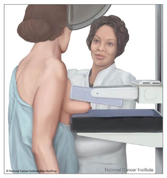
(NCI, 2007)
Standard mammography imaging has evolved significantly over the last few decades, with the development of computer-aided detection (CAD), digital mammography, and breast tomosynthesis. CAD is a form of artificial intelligence designed to search digital mammographic images to help identify potential abnormalities that might otherwise be missed, highlighting them for the radiologist to examine closely. Despite evidence that CAD did not increase diagnostic accuracy, it was being used by 92% of institutions by 2016. CAD has also been associated with increased false positives, resulting in approximately $400 million in unnecessary health care costs (Elmore & Lee, 2022). CAD has been beneficial in diagnostic evaluation by increasing the detection of ductal carcinoma in situ, thanks to its greater sensitivity for detecting calcifications. The clinical benefits and efficacy of CAD continue to be studied; however, its use does not replace the need for a qualified radiologist to interpret mammogram images directly (Slanetz & Lee, 2025).
Some females may opt for digital breast tomosynthesis (DBT), also called 3-D mammography, an advanced imaging modality that captures multiple images of the breast from different angles. This modality is similar to a CT scan and produces higher-quality images by combining multiple thin slices into a 3D image. The radiation dose from some DBT systems is higher than conventional mammography; however, extensive population-based studies have demonstrated improved breast cancer screening detection rates and reduced need for additional views for individuals with dense breasts (Slanetz & Lee, 2025). Although DBT is a newer imaging modality, some of its clinical benefits include the following:
- earlier detection of small breast cancer that may not have been seen on a conventional mammogram
- particularly helpful in patients with dense breasts
- increased likelihood of detecting multiple breast tumors during one imaging test and pinpointing the size, shape, and specific location of the abnormalities
- a reduced number of unnecessary biopsies or additional imaging tests (Slanetz & Lee, 2025)
CT Scans
Computed axial tomography (CAT) scans use a series of radiographs taken from multiple angles along with computer technology to create cross-sectional images of the inside of the body, including bones, blood vessels, organs, and soft tissues. The X-ray detector moves in a circular path around the body to generate multiple views of the body structure being evaluated. For some scans, the table the patient is on moves incrementally with each picture, whereas other tests require continuous table movement. The CT scanner sends X-rays through the body during each scan rotation to form a complete picture in much greater detail than a conventional radiograph (Mafraji, 2025a).
CT scans use ionizing radiation and may be performed with or without intravenous contrast. Some scans use iodine-based contrast, which may be given IV, orally (PO), or both. When contrast is used, patients are typically required to fast or remain nothing by mouth (NPO) for several hours before the scan to improve image quality. Oral contrast is administered before the examination and is most helpful in visualizing the structures of the abdomen and pelvis. When receiving IV contrast, patients will need to have a needle inserted into the arm for the injection (Patel & De Jesus, 2023). Additional details regarding the multifaceted aspects of iodinated contrast administration are described later in this module.
CT scans are better than radiographs at distinguishing soft-tissue densities. As a result, they are the preferred imaging modality for evaluating head and neck, spinal, intra-abdominal, intrathoracic, and intracranial structures. CT scans are used to diagnose injuries from trauma, infections, internal bleeding, tumors, masses, and cancers. They are also used to guide biopsies. The CT scan is considered a first-line screening modality for patients presenting with acute head trauma or stroke since it can quickly and easily evaluate for hemorrhage or an ischemic event. In these cases, the head CT scan is ordered without contrast, as it is obtained emergently. A CT scan without contrast is also the most accurate method for detecting urinary calculi. IV contrast improves imaging and is recommended when malignancy, infection, or soft-tissue trauma is suspected (Mafraji, 2025a).
CT scans are commonly used in the care of oncology patients as part of a cancer staging workup, to evaluate response to cancer treatments, and to monitor for cancer progression or recurrence. CT scans of the face, sinuses, orbits, or neck may also be ordered and are generally performed to evaluate for suspected infection or mass (sinusitis, orbital infection, malignant or benign tumors) in these areas. A chest CT scan is commonly performed to evaluate the lung parenchyma and mediastinum for the presence of pulmonary nodules, masses, pleural effusions, or other signs of lung disease. CT imaging of the abdomen and pelvis has many indications for ordering. Contrast is advised when evaluating suspected appendicitis, diverticulitis, abscess, other infection, and small bowel obstruction. CT angiography (CTA) is a CT scan performed to evaluate blood vessels in a specific area for narrowing, obstruction, or thrombosis. Most commonly, a CTA scan is ordered to evaluate for suspected pulmonary embolism (pulmonary CTA), aortic dissection (thoracic aorta CTA), and brain aneurysm (intracranial CTA). There are numerous additional indications for CT scans. Specific imaging protocols based on the suspected injury or illness to ensure the highest quality of images are obtained, which are beyond the scope of this module (Mafraji, 2025a; Patel & De Jesus, 2023).
Patients should be advised to lie flat and remain still on a table that slides into the scanning machine, which resembles a giant doughnut (refer to Figure 3). The machine rotates around the patient to capture all necessary images, and the scan takes 15–30 minutes. Radiation doses from CT examinations are 100–250 times higher than doses received in plan film radiographs. As explained earlier, patients who undergo CT scans with high doses of radiation or undergo repeated CT scans are at heightened risk of cancer later in life due to the radiation utilized in these scans (CDC, 2024a; EPA, 2026; Lee & Elmore, 2026; Patel & De Jesus, 2023).
Figure 3
CT Scan
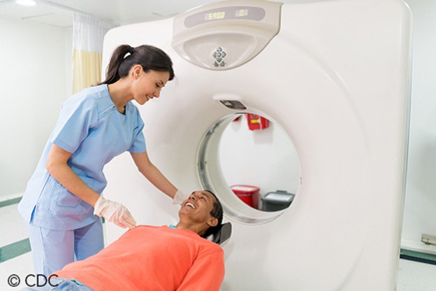
(CDC, 2024d)
Nuclear Medicine Imaging
Nuclear medicine imaging differs from conventional diagnostic imaging in that it can visualize how the body functions at the cellular and molecular levels. Nuclear imaging uses small quantities of radioactive tracers (radiotracers) to diagnose and treat disease. The radiotracers are most commonly injected into a vein but may also be taken orally, inhaled, or directly injected into a specific area. The radiotracer travels through the body, releasing gamma rays that are absorbed by specific tissues and organs. It is then detected by the external scanning device to provide information on organ function and cellular activity (NIBIB, 2025a).
The radiotracers comprise molecules tightly bound to a radioactive atom, and these vary greatly depending on the purpose of the scan. Each radiotracer has a designated half-life, which indicates how long it takes for half of the radioactive substance to decay. Materials used to administer the radiotracer must be disposed of properly. The Nuclear Regulatory Commission (NRC) and the International Commission on Radiological Protection (ICRP) provide regulatory guidance on the use of radioactive materials in nuclear medicine to ensure patient, health care professional, and public safety. Each nuclear medicine imaging test uses a specific radioactive agent (Heston & Tafti, 2024). Radiotracers must meet FDA safety standards for radiation exposure. Various nuclear medicine imaging tests are available and are integral to the care of patients with cancer, heart disease, and bone disorders (NIBIB, 2025a). Some of the most common include:
- PET/CT
- single photon emission computed tomography (SPECT)
- thyroid scintigraphy and radioactive iodine uptake (RAIU) test
- skeletal scintigraphy (Bone Scan)
- bone densitometry scan (DXA)
- multigated acquisition scan (MUGA; NIBIB, 2025a)
Positron Emission Tomography/Computed Tomography (PET/CT)
PET/CT imaging uses the radiotracer fluorine-18 deoxyglucose (FDG) to create 3D images that better localize areas of abnormal cell activity. PET information on cell activity and function is combined with anatomic information from the CT scan (Mafraji, 2025c). There are a few types of PET/CT scans, the most common of which is a full-body PET/CT, which evaluates the internal structures from the midportion of the skull down to the thigh area (“eyes to thighs”), in combination with a low-dose CT scan. This is a hybrid imaging modality; the images acquired from each test are fused using advanced computerized technology to produce higher-quality, enhanced images. The functional imaging from the PET scan illustrates the spatial distribution of metabolic or biochemical activity in the body, which is more closely aligned with the anatomic imaging from the CT scan. PET/CT scans provide superior information for evaluating tissues, staging and restaging cancers, and monitoring the effectiveness of cancer treatments. Since cancer cells take up glucose faster than normal tissue, FDG is a superior radiotracer for evaluating cancerous tissue, as it is chemically similar to glucose. FDG accumulates in the body’s most metabolically active areas, helping differentiate physiologic uptake in healthy tissue from pathologic uptake in diseased tissue (Katal et al., 2022).
The FDG radiotracer is administered IV, and the PET/CT scan produces images that show the radiotracer’s distribution throughout the body to determine whether abnormalities are present. Highly active cancer cells indicate higher FDG uptake, whereas brain cells affected by dementia consume less glucose, as indicated by lower FDG uptake. PET scans that use amyloid imaging agents can detect areas of amyloid plaque in the brain, which may help diagnose Alzheimer’s (NIBIB, 2025a). An example of the images obtained from a PET/CT scan is depicted in Figure 4, depicting a healthy brain on the left and a brain tumor on the right.
Figure 4
PET/CT Scan Images of the Brain
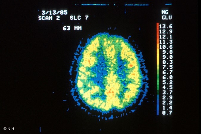
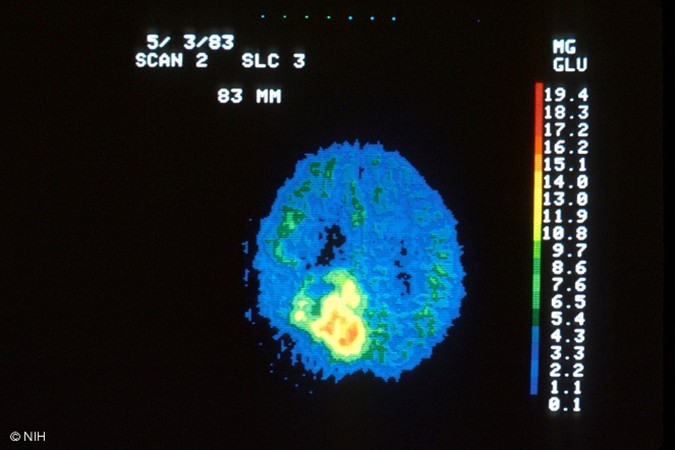
(NCI, 2001a, 2001b)
For 24 hours before the PET/CT scan, patients are advised to avoid strenuous activities such as running or cycling, as these can impair image quality. Since the PET/CT scan measures glucose uptake, strenuous activity can increase radiotracer uptake in strained and recovering muscles, increasing the likelihood of false-positive results. Patients are advised to fast for 6–8 hours before the scan and follow a low-glucose, low-carbohydrate diet for 24 hours before the scan. From midnight until 2 hours before the scan, the patient can drink 12 ounces of water but nothing else. The patient’s fasting blood glucose (FBG) will be obtained on the day of the scan, typically via a fingerstick. The FBG level should be between 70 and 199 mg/dL for the highest-quality imaging results. If the FBG is too high, the scan will be of poor quality, thereby interfering with the clinical benefit and accuracy of the results. Typically, patients with FBG levels below 70 mg/dL or above 200 mg/dL will be referred to the ordering provider for glucose management, and their scan will be rescheduled. Patients with underlying diabetes will receive individualized instructions based on their diabetes management plan. If insulin or diabetic medications are taken too close to the FDG injection time, too much FDG will collect in the muscles rather than flowing throughout the tissues where it should. If a brain PET/CT is being performed, limiting brain activity before testing by avoiding reading or listening to music while waiting for the scan to begin is essential. The PET/CT scanner resembles a standard CT scanner, as depicted in Figure 3. The patient will lie down on the table, usually in the supine position, and should be advised to remain very still. The exam table will move slowly through the scanning ring. The test usually takes 25–40 minutes (Ashraf & Goyal, 2023).
Following the scan, all FDG traces are cleared from the body within 24 hours. There is no concern regarding triggering radiation detection alarms present in some security equipment after that time frame. Patients can request documentation of testing if they will be traveling on the same day as the test (IAEA, n.d.-a). The ACOG (2017) recommendations for breastfeeding following nuclear imaging scans do not definitively state a position for or against the practice, and these guidelines were reaffirmed in 2026. Patients are advised to consult with a lactation specialist. Very low amounts of FDG are known to be excreted into breastmilk during the procedure (Ashraf & Goyal, 2023)
Single Photo Emission Computed Tomography (SPECT)
SPECT is another type of nuclear medicine imaging test, similar to PET/CT scans, which combine CT technology with an intravenously injected radiotracer. The primary distinction between PET/CT and SPECT imaging is the type of radiotracer used. For SPECT imaging, the isotopes commonly used are technetium-99m, iodine-123, and thallium-201. In SPECT imaging, the radiotracer stays within the bloodstream rather than being absorbed by tissues and organs. SPECT imaging focuses primarily on areas of blood flow and is used to evaluate blood flow to surrounding tissues and organs, thereby demonstrating organ function. SPECT studies are most commonly used to diagnose or evaluate heart and brain disorders but may also be used to assess other conditions. Regarding the heart, SPECT imaging can detect blockages in the coronary arteries, damage to the myocardium (heart muscle) from a heart attack, and how well the heart is pumping blood, particularly under stress. In brain function, SPECT studies may be ordered to evaluate for dementia and to assess the location and etiology of a stroke, by visualizing how blood flows through veins and arteries in the brain. It can be used to diagnose areas of ischemia (blood deprivation) within the brain following a stroke or as a result of a tumor. These studies are also used in epilepsy for detecting and identifying seizure activity and localizing epileptic foci. A clinician may also order a SPECT scan to evaluate conditions that are noncardiac or nonneurological, such as parathyroid disease, pulmonary embolism, osteomyelitis, or spondylolysis (Yandrapalli & Puckett, 2022).
During a SPECT scan, the patient is usually placed in the supine position on the examination table and asked to remain very still throughout the examination. After injecting the radiotracer, a gamma camera rotates around the patient. The device accumulates pictures, which are then used to construct 3D images of the radiotracer distribution. The resulting images reveal information about blood flow and target organ function. Patients are advised to fast for at least 3 hours before the scan, which can take up to 2.5 hours to complete. Patients are instructed to avoid caffeine for at least 12 hours before testing, as caffeine can interfere with vasodilatory medications administered during the test. Patients are also advised to discontinue phosphodiesterase-3 inhibitors, such as cilostazol (Pletal), at least 48 hours before testing due to their vasodilatory effects. Following the scan, patients are advised to maintain increased oral hydration for about 2 days to flush the radioactive material from the body. Otherwise, there are no special discharge instructions (Yandrapalli & Puckett, 2022).
Thyroid Scintigraphy and Radioactive Iodine Uptake (RAIU) Test
There are two types of nuclear medicine imaging tests of the thyroid: thyroid scintigraphy, also called the thyroid scan, and the RAIU test. Both scans use a small amount of radioactive iodine, usually I-123, because the thyroid gland is the only tissue in the body that absorbs and retains iodine. The radiation emitted by I-123 is harmless to thyroid cells and can be detected externally through thyroid scanning. Rarely, I-131 may be used with RAIU scans, but I-131 destroys thyroid cells. As a result, it is commonly reserved for treating thyroid disorders such as overactive thyroid, thyrotoxicosis, and thyroid cancer, which are beyond the scope of this module (ACR, 2024).
The thyroid scan is ordered to assess thyroid function and evaluate the gland for abnormalities, such as nodules, masses, or inflammation. Thyroid scans are helpful in, but not limited to, the evaluation of the following:
- location, size, and presence of functioning thyroid tissue
- cause of overt and subclinical thyrotoxicosis
- presence of diffuse thyroid disease or suspected focal masses
- thyroid clinical laboratory tests suggestive of abnormal function
- thyroid nodules detected on clinical examination or other imaging examinations
- congenital thyroid abnormalities
- differentiating types of hyperthyroidism (ACR, 2024)
The RAIU scan is performed to evaluate thyroid function or to determine the etiology of an overactive thyroid gland (hyperthyroidism). It may also be used to plan treatment for thyroid cancer. The RAIU uses a specialized probe to measure the amount of tracer the thyroid gland absorbs from the blood. In most cases, the RAIU scan is performed alongside the thyroid scan to assess whether the radiotracer is evenly distributed within the gland (ACR, 2024). While the RAIU scan does have overlapping indications with the thyroid scan, it is considered most useful in the following situations:
- distinguishing various forms of thyrotoxicosis from hyperthyroidism
- evaluating iodine-131 sodium iodide necessity or dosage to be administered in patients to be treated for hyperthyroidism
- determining the presence of residual functioning thyroid tissue after thyroid resection or radioiodine ablation (ACR, 2024).
Agents containing iodine can decrease iodine uptake in the thyroid gland, leading to inaccurate test results. Iodine is hidden in many commonly used supplements, over-the-counter agents, and certain prescription medications. Therefore, before the test, a comprehensive medication reconciliation should be performed. Patients must be informed to discontinue thyroid hormones, anti-thyroid drugs, and any other medication or dietary supplement containing iodine. Each medicine or supplement has a specified period in which it should be discontinued before the scan. For example, levothyroxine (Synthroid) is a thyroid hormone that must be stopped for 4–6 weeks before the scan. In contrast, iodine-containing cough syrups should be discontinued 2 weeks before the scan (ACR, 2024). Other iodine-based agents that need to be avoided include, but are not limited to, the following:
- iodized salt
- multivitamins
- amiodarone (Pacerone)
- kelp (algae seaweed)
- IV iodinated contrast agents (ICAs)
- sulfonamides
- methimazole (Tapazole)
- high-dose corticosteroids (ACR, 2024 ; Iqbal & Rehman, 2022)
In the 1–2 weeks leading up to the radioactive iodine administration, patients are advised to consume a low-iodine diet, avoiding the highest sources of dietary iodine, including salt, grains, cereals, fish, poultry, and milk products (American Thyroid Association [ATA], n.d.-a). During a thyroid scan, I-123 is either injected into a vein within 30–60 minutes of the scan or administered orally as a pill or liquid. For oral administration, I-123 should be given approximately 24 hours before the scan, allowing the radioactive iodine to reach and saturate the thyroid gland. The oral route is preferred for patients who undergo both thyroid and RAIU scans, as it can be used for both tests and does not require a second radiotracer dose. The thyroid scan is painless, and the patient is usually positioned lying supine on an examination table with their head tilted back to extend the neck. A gamma camera will take thyroid images from at least three different angles, and the patient will be asked to lie very still. A thyroid scan takes about 30 minutes to complete (Iqbal & Rehman, 2022).
The RAIU scan requires the administration of radioactive iodine in liquid or capsule form. The RAIU scan occurs at two distinct time points—usually, 4–6 hours following radiotracer administration and then again at 24 hours post administration. During an RAIU test, the patient is generally seated upright, and a small device called a radioactive detector (uptake probe) is placed against the patient’s neck. The uptake probe measures radioactive iodine uptake, and a gamma camera records images of the thyroid gland. Both instruments detect and record the distribution of radioactive material within the thyroid. The RAIU test usually takes several minutes (RadiologyInfo.org, 2023). Following the test, patients should be advised that most radioactive material is cleared from the body within one to two days. No special precautions are required, as I-123 is harmless to thyroid cells (ACR, 2024). It is safe to use radioactive iodine in patients who report iodinated contrast allergies or seafood allergies, as the reaction is to the compound containing iodine and not the iodine itself (ATA, n.d.-b).
Skeletal Scintigraphy (Bone Scan)
Skeletal scintigraphy, also called a bone scan, is a nuclear medicine imaging test that uses a small amount of radioactive tracer to evaluate and diagnose skeletal disorders associated with abnormal osseous function (Adams et al., 2026). A bone scan is performed to assess for several types of conditions, such as:
- bone fractures, including stress, occult, accidental, and nonaccidental
- primary bone masses or tumors (benign or malignant)
- metastatic bone neoplasms
- underlying bone pain that is otherwise unexplained or unresponsive to conservative treatments, as is the case with chronic low back pain or complex regional pain syndrome
- tumor-like conditions such as Paget’s disease
- infections such as osteonecrosis
- complications from orthopedic hardware or prosthetic joints
- congenital or developmental anomalies
- fibrous dysplasia (ACR, 2021; Adams et al., 2026)
A bone scan is also routinely used to determine if cancer has spread to the bones and to monitor the response to cancer treatment over time. A radiotracer, such as Technetium-99m, is injected into a vein and travels through the bloodstream, emitting gamma radiation. The radiotracers are detected by a specialized gamma camera and fused with computer images to derive an overall picture of the bones at multiple points (Adams et al., 2026). This is demonstrated in Figure 5.
Figure 5
Bone Scan
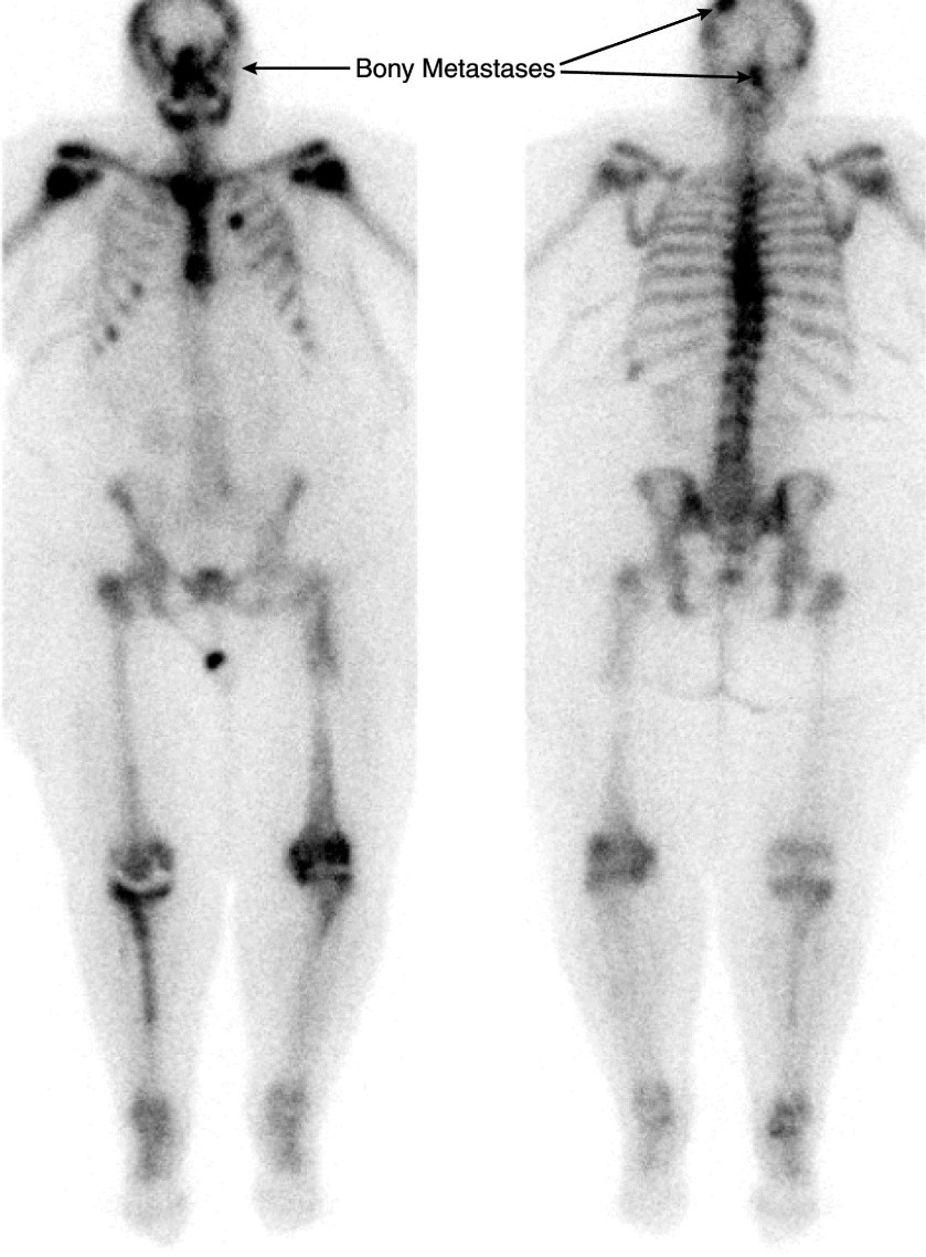
A bone scan can detect molecular changes, enabling early detection of bone disorders. Abnormal areas within the bone will take up more or less of the radiotracer, producing brighter or darker areas in the resulting images (Adams et al., 2026).
In preparation for a bone scan, patients should be counseled to avoid bismuth-containing medications such as bismuth subsalicylate (Pepto-Bismol, Kaopectate) for several days before the scan, as these medications can interfere with the results. In addition, patients should be screened for radiograph tests using barium contrast material, as this can also skew the results (UpToDate Patient Education, n.d.-a).
The radiotracers used in bone scans take a few hours to circulate throughout the body and bind to bones to produce the highest-quality images. Patients should be advised that there will be a 2-to 4-hour period between the injection administration and the scan. During this time, patients will need to consume several glasses of water (usually four to eight) to facilitate the removal of any excess radiotracer from the body. Patients will also be instructed to empty their bladder before the scan, as any residual tracer in the bladder can obscure the view of the underlying pelvic bones. Patients will be asked to lie still on the examination table during the test. A total-body bone scan usually takes about 1 hour to complete. Following the scan, patients may resume normal activities and are advised to increase oral hydration for 1–2 days to help facilitate the removal of any residual radiotracer circulating in their system. It generally takes 48 hours for all the radioactive tracer material to be excreted from the body (Adams et al., 2026).
Dual-Energy X-ray Absorptiometry (DEXA, DXA) Scan
A bone densitometry scan, also called a DXA scan, is used to measure bone density. While it is a nuclear medicine test that uses a small amount of ionizing radiation to generate images of bony structures, it vastly differs from a bone scan. A DXA scan evaluates bone mineral density (BMD), the health and strength of bones, and assesses osteopenia or osteoporosis and fracture risk. According to the World Health Organization (WHO), the gold-standard bone density test is a DXA scan of the central skeleton, including the hip and lumbar spine. BMD is most commonly measured at the spine and hip but can also be measured at the wrist. The degree of bone loss is calculated and classified according to defined diagnostic criteria (Lewiecki, 2025).
Osteoporosis is a chronic, systemic disease characterized by low BMD, bone weakening, and deterioration of bone tissue and architecture. Nationally, approximately 54 million adults have low bone mass (osteopenia) or decreased bone mass at levels of osteoporosis. This accounts for two million bone fractures specifically related to bone mass loss and fragility. Osteoporotic bones are brittle and porous, heightening the risk of fracture (Lewiecki, 2025). It is referred to as a silent disease because the loss of bone mass is not painful and there are generally no warning signs or symptoms preceding a bone fracture. Most people do not know they have osteoporosis until they develop an acute fracture or broken bone, which is the hallmark of the disease. Fractures can occur in any bone within the body, but most commonly occur in the hip bones, vertebrae, and wrist. Osteopenia is a precursor to osteoporosis and is characterized by a lower-than-normal BMD that does not meet the criteria for osteoporosis. People with osteopenia are at higher risk for developing osteoporosis, but when identified early through screening with a DXA scan and appropriate action is taken, progression to osteoporosis can be successfully averted (Rosen & Drake, 2024).
The degree of bone loss is calculated and classified according to defined diagnostic criteria. The DXA scan is a quick, noninvasive, and painless test. The patient is instructed to lie or sit down for less than 10 minutes while the machine scans the body. The test exposes the patient to a very small amount of radiation. DXA test results are reported as a T-score for each measured site, comparing the patient’s BMD to that of healthy young adults with ethnicity- and gender-matched controls. The WHO separates those T-scores into four categories: normal, low bone mass (osteopenia), osteoporosis, and severe or established osteoporosis. A T-score of 0 indicates that the BMD is equal to that of a healthy young adult, a negative T-score indicates that the bones are thinner than average, and a positive T-score denotes that the bones are stronger than average. The difference between a patient’s BMD and the normal range is measured in units called standard deviations. The more standard deviations below 0, denoted by negative numbers, the lower the BMD, the more severe the osteoporosis, and the higher the fracture risk. The WHO classifies osteoporosis as a BMD that is 2.5 standard deviations below normal. Treatment is usually recommended to prevent fractures when the T-score is -2.5 or lower. Table 5 defines T-scores and their corresponding BMD level (Camacho et al., 2020; Lewiecki, 2025).
Table 5
WHO T-Score Interpretation
T-Score | Interpretation |
≥ -1.0 | Normal bone |
-1.0 to -2.5 | Osteopenia |
≤ -2.5 | Osteoporosis |
≤ -2.5, plus 1 or more osteoporotic fractures | Severe or established osteoporosis |
(Camacho et al., 2020; Lewiecki, 2025)
Multigated Acquisition Scan (MUGA)
A MUGA scan may also be called radionuclide ventriculography (RNV), radionuclide angiography (RNA), or gated equilibrium radionucleotide angiography (ERNA). It is a type of nuclear imaging test that evaluates how well the heart is functioning, particularly the left ventricular ejection fraction (LVEF). The LVEF measures the amount of blood pumped out of the heart with each contraction and is expressed as a percentage. The normal LVEF in an adult is 50%–75%. This test may be performed for various reasons but is most commonly ordered as part of a cardiology workup for chest pain, in follow-up to an abnormal electrocardiogram (EKG) or echocardiogram scan, monitoring patients with heart failure, and in evaluating the cardiac function of patients diagnosed with chronic obstructive pulmonary disease (COPD). MUGA scans are also used to monitor patients undergoing potentially cardiotoxic chemotherapy, chest wall radiation, or other cancer treatment regimens that can impair heart function; however, MUGA scans are limited in assessing cardiac function in these individuals (Odak & Kayani, 2023).
During the MUGA scan, small electrodes are placed on the patient’s chest, arms, and legs, similarly to an EKG, to track the patient’s heartbeat and heart rhythm during the test. This is necessary since MUGA scans require tracking R-wave progression to recognize when to begin data collection. The patient’s blood is obtained, mixed with the radioactive tracer, and administered IV. After allowing the radioactive tracer to circulate for 15–20 minutes, a gamma camera captures images of the heart at designated time points during each heartbeat. A MUGA scan may be performed at rest, with the patient lying on the table while the gamma camera images the heart. Alternatively, it may be performed as an exercise scan or stress test, in which the patient walks on a treadmill or rides a stationary bicycle to reach peak activity, then stops and lies on the table. At the same time, the gamma camera takes pictures of the heart. A pharmacologic agent is administered to induce cardiac stress if the individual cannot complete physical activity. The test takes approximately 1–2 hours, and patients can generally resume normal activities following the test. After the scan, patients are advised to drink plenty of water to help flush the radioactive materials through the renal system. MUGA scans have become less common in clinical practice, as echocardiograms are now more readily available (Odak & Kayani, 2023).
Diagnostic Imaging Tests without Radiation Exposure
Magnetic Resonance Imaging (MRI)
MRI is a widely utilized diagnostic imaging modality, with MRI scans performed on an estimated 40 million patients in the United States each year (Shah & Aran, 2023). MRIs are distinct from other forms of diagnostic imaging as they do not use X-rays or ionizing radiation and are considered a very safe imaging test. Instead, MRIs utilize strong EM fields, magnetic field gradients, and radio waves and are essentially giant magnets. They can vary in strength, measured in units called teslas (T). Most modern MRI scanners are 1.5–3T. In context, an MRI of 3T strength is about 60,000 times stronger than the Earth’s magnetic field. Over the last few years, there has been a push to improve and expand MRI imaging capabilities. The current state-of-the-art MRI scanners for neuroimaging have magnetic field strengths of up to 7T. An electric current creates a temporary magnetic field within the patient’s body during an MRI. Radio waves are sent from and received by a transmitter and a receiving device within the machine. These signals are used to generate images of the scanned body area. The signal in an MRI image comes mainly from the protons in fat and water molecules within the body. MRI is at least as good as, and often superior to, CT in distinguishing normal from abnormal soft tissue. MRIs can image nearly any body part; each scan follows a specific protocol depending on the clinical concern and may be performed with or without contrast administration. Gadolinium-based contrast agents (GBCAs) are rare-earth metals administered IV to enhance MRI image contrast. MRI contrast agents differ from CT contrast agents, and patients with CT contrast allergies can typically safely tolerate GBCA injections (FDA, 2021; Ibrahim et al., 2023; Vachha & Huang, 2021).
MRI scans are the preferred diagnostic imaging test for numerous diseases and disorders and are among the most frequently performed intracranial and spinal cord imaging tests. Different MRI types can be used to diagnose aneurysms, multiple sclerosis, strokes, herniated discs, fractures, biliary duct abnormalities, and tumors. While standard MRI scans are superior for evaluating soft tissues and organs, they also offer clinical benefits for imaging the heart and blood vessels, including the detection of structural abnormalities in the aorta, assessment of heart wall thickness, and detection of damage caused by heart disease. MRI is widely used in orthopedics to evaluate bone and joint conditions, including torn ligaments and cartilage. MRI may also be the preferred screening modality in females with dense or fibrotic breasts instead of mammography due to the superior evaluation of dense breast tissue, which can outline the extent of the breast cancer after a positive biopsy (Ibrahim et al., 2023; Mafraji, 2025b).
Patients undergoing MRI scans of the abdomen or soft tissue pelvic structures are usually advised to remain NPO for 6 hours before the scan. Before an MRI of the prostate or to evaluate for a vaginal/rectal fistula, a bowel preparation with an enema may be necessary. However, no special instructions are required for any other MRIs the day before the scan. Patients are advised to lie very still on the table, as motion can create artifacts and reduce image clarity. The table slides into the MRI machine, which resembles a CT scanner, as demonstrated in Figure 3. However, an MRI scanner is deeper and narrower than a CT scanner. Due to this, there is a heightened risk of claustrophobia and anxiety in some patients, who may require a sedative before the scan to ensure relaxation and manage anxiety. Patients should be advised that they will hear loud thumping or banging throughout the examination. Patients should be reassured that these are harmless sounds produced by the MRI magnets. Patients may be given earplugs or headphones to minimize the noise, which can reach up to 120 decibels. An MRI scan can take anywhere from 15 to 45 minutes, depending on the part of the body being imaged; however, some can last longer than 60 minutes (Ibrahim et al., 2023; NIBIB, n.d.).
While there is no risk of radiation exposure with MRIs, there are risks of potential injury and death. Due to strong EM fields, the MRI machine can propel magnetic objects toward its center at dangerous speeds, including medically implanted devices such as cardiac pacemakers and infusion pumps. The RF field can cause tissue heating and burning, particularly in the presence of implanted devices that can heat internally. All patients must undergo screening evaluations before testing to assess for the presence of metal or any implanted hardware. The FDA has received reports of serious adverse events associated with implantable pumps in the MRI setting, such as pump malfunction, including bolus dose, overdose, underdose, or pump failure. According to the FDA, only implantable infusion pumps labeled “MR Conditional” may be safely used in an MRI environment and only under the specified safe-use conditions outlined by the device manufacturer. During the screening, patients must also be asked whether they have ever welded without eye protection or sustained any facial injury from metal. If the patient responds yes, an orbital X-ray must be obtained to ensure there is no hidden metal in the orbits before the MRI (FDA, 2023c; Ghadimi & Thomas, 2025).
Given the strength of the EM fields, MRI use is contraindicated in many cases. These contraindications are categorized as absolute or relative. Devices and objects are designated as MR safe (safe in all environments), MR conditional (safe if specific criteria are met based on the object or device), or MR unsafe (known safety risk). It should be noted that some of these may be compatible at 1.5T or 3T only. Clinicians must refer to a certified MRI safety website or the device manufacturer’s instructions when assessing MRI safety and compatibility (FDA, 2023c; Ghadimi & Thomas, 2025). The following list comprises the most common absolute contraindications of MRI; however, these contraindications change as new advancements are made:
- cardiac implantable electronic devices (CIEDs) such as pacemakers, implantable cardioverter defibrillators (ICDs), cardiac resynchronization therapy (CRT) devices, implantable loop recorders (ILRs), and implantable cardiovascular monitors (ICMs); newer devices may be designated as MR conditional
- metallic intraocular foreign bodies
- implantable neurostimulation systems
- cochlear implants; some may be used with a 1.5T MRI scanner after the battery is removed
- catheters with metallic components (Swan-Ganz catheter)
- metallic fragments such as bullets, shotgun pellets, or shrapnel
- cerebral artery aneurysm clips
- magnetic dental implants
- tissue expanders
- medication patches, hearing aids, body piercing, external drug delivery pumps (insulin pumps), and artificial limbs are all contraindicated and must be removed before the MRI (ACR, 2026; Ghadimi & Thomas, 2025)
Several relative contraindications must be considered before any MRI scan, as the specific patient situation must be evaluated with caution to confirm it is safe to proceed with the exam (Ghadimi & Thomas, 2025). Relative contraindications include patients presenting with any of the following:
- coronary and peripheral artery stents
- nonplastic airway stents or tracheostomy (plastic tracheostomies are safe to proceed with the MRI, but if the composition of the tracheostomy is unknown or unsafe, it must be changed to a plastic source before the MRI)
- intrauterine devices (IUDs) require investigation and confirmation of the make and model; those that are unknown are acceptable only with 1.5T MRI scanners
- ocular prosthesis
- stapes implants
- surgical clips or wire sutures
- certain types of prostheses (joint, penile, etc.)
- if the patient had a colonoscopy procedure within the previous 8 weeks and endoscopy clips were used or placed during the procedure, the scan must be postponed (Ghadimi & Thomas, 2025)
Additional consideration must be given to the following situations before performing the MRI scan:
- patients with programmable shunts must be informed that they must reprogram their shunt with their provider following the MRI
- patients with inferior vena cava (IVC) filters that are of unknown composition must wait 6 weeks following implantation and can be scanned only with a 1.5T MRI scanner
- patients with Harrington rods (a stainless-steel surgical device) can undergo an MRI only on 1.5T scanners
- tattoos should be older than 6 weeks, and ice packs or padding should be used against any tattoo that is in contact with the bore of the scanner or the MRI coil, and patients must be educated to immediately report any warm sensation that develops around the tattoo site (Ghadimi & Thomas, 2025)
Ultrasound
Ultrasound is a safe, noninvasive imaging modality that uses sound waves to generate images of internal body structures. Also known as sonography, ultrasound does not use X-rays or ionizing radiation and can be used for diagnostic and therapeutic purposes. Ultrasound images are obtained by placing a small transducer (i.e., probe) and ultrasound gel on the skin. The transducer produces sound waves at very high frequencies, which exceed the threshold of human hearing. These high-frequency sound waves travel from the probe through the gel and into the body. The reflections of the sound waves off the structure being evaluated are used to generate images on a computer. Images are captured in real time, allowing evaluation of the structures and movement of the body’s internal organs, including blood flow through vessels. Transducers may be placed externally on the skin, as in transabdominal fetal ultrasound, as shown in Figure 6. Some transducers can also be placed directly inside the body via the vagina, GI tract, or blood vessels to optimize image quality. An example is a transducer placed inside the vaginal canal of a nonpregnant female to enhance the visualization of the uterus and ovaries (Baker & dela Cruz, 2023; FDA, 2024).
Figure 6
Fetal Ultrasound
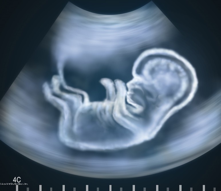
Conventional ultrasound displays images in flat sections of the body. However, technological advancements have led to the development of ultrasound, which allows sound wave data to be formatted into 3-D images. The most widespread use of 3-D ultrasound is to assess a developing fetus, and the distinction between images from a conventional sonogram and a 3-D sonogram is shown in Figure 7. A 4-D ultrasound can also be used to assess fetal characteristics. A 4-D ultrasound is a 3-D ultrasound that is animated (FDA, 2024).
Figure 7
Conventional Sonogram Compared to 3-D Sonogram of Fetus
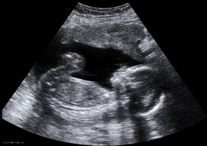

Ultrasound can be performed for diagnostic or therapeutic reasons. Therapeutic ultrasound interacts with tissues in the body, modifying or destroying them. For example, therapeutic ultrasound may be used for ablative functions such as dissolving blood clots or delivering drugs to specific locations within the body. Diagnostic ultrasound is used to evaluate internal organs and structures, tissue and blood movement, tissue or blood velocity, and other abnormalities. Ultrasound may also be used during surgery to help guide the placement of a catheter or other medical device or to guide biopsies. An ultrasound-guided needle biopsy is a standard procedure for evaluating a breast mass for breast cancer (FDA, 2024).
Doppler ultrasound is a specialized technique that evaluates blood flow in the body’s arteries and veins. This type of ultrasound uses the Doppler effect to assess red blood cell movement. Color Doppler is useful for distinguishing arterial from venous flow: color indicates flow toward or away from the transducer, and flow velocity determines the color intensity. Spectral Doppler displays blood flow as a graph rather than a color picture. This provides information on the level of flow restriction, indicating the potential degree of stenosis (Mafraji, 2025d). Doppler ultrasound is used to evaluate the following:
- blockages to blood flow (such as thrombosis, deep vein thrombosis, or other blood clots)
- narrowing of vessels (such as narrowing of the carotid arteries, which increases the risk of stroke)
- vascular tumors or masses
- congenital vascular malformations
- reduced or absent blood flow to organs, such as the testes or ovaries (Mafraji, 2025d)
There is a wide array of indications for ordering a diagnostic ultrasound. Ultrasound can evaluate pain, swelling, or infection symptoms and can be performed safely on nearly every body system. The following list, although not comprehensive, shows some of the most common body parts imaged by ultrasound and indications for ordering a diagnostic ultrasound:
- gallbladder and bile ducts (gallstones and biliary tract obstruction)
- heart and blood vessels (patency of blood flow, evaluate for abdominal aortic aneurysm (AAA), carotid arteries for narrowing)
- liver (cirrhosis, cancer or metastatic tumors, infarction)
- spleen (splenomegaly)
- pancreas (pancreatitis, tumor)
- kidneys, bladder, ureters (masses, cancer, patency, hydronephrosis)
- uterus (fibroids, fetus in pregnant females)
- ovaries (cysts, tumors)
- thyroid and parathyroid glands (masses, enlargement, nodularity)
- scrotum and testes (testicular torsion, swelling, masses, blockage in blood flow; Mafraji, 2025d)
In some cases, ultrasound imaging may also use contrast agents to enhance image quality. Microbubble contrast helps assess blood perfusion in organs, thrombosis, abnormalities in the heart, liver, or kidney masses, and inflammatory activity in IBD (Ahmed, 2026). Microbubble contrast will be discussed later in this module. Most ultrasound exams require little to no special preparation. While ultrasound is considered a safe and painless medical imaging modality suitable for patients of all ages, it has limitations. Air and gas can disrupt ultrasound waves, obscuring images. As a result, ultrasound does not help evaluate air-filled organs and structures, such as the bowels and lungs. Ultrasound cannot penetrate bone but can help identify infections surrounding the bones (NIBIB, 2023). However, ultrasounds ordered to assess the abdominal structures, such as the gallbladder, require patients to fast for 4–8 hours before the scan. For ultrasounds ordered to evaluate the bladder and many prenatal ultrasounds, the patient may be asked to drink four to six glasses of water before their exam and avoid urinating so that the bladder is full and distended for optimal viewing (UpToDate Patient Education, n.d.-b).
Echocardiogram
Transthoracic echocardiography (TTE). A TTE is an ultrasound of the heart that provides a view of the heart obtained from placing the transducer and gel on the chest wall to provide real-time images of the heart in motion. The test is usually performed with the patient lying on their back or left side. Some patients will also be connected to an EKG to monitor their heart rate and rhythm during the exam. It is a commonly used cardiac diagnostic imaging test, considered safe and effective, with no radiation exposure risk and no special preparation required. Utilizing the same sound wave technology outlined in the previous section, an echocardiogram provides critical information about the heart’s structures, including the valves, chambers, walls, and blood vessels, in addition to assessing the function of these individual structures. An echocardiogram may be ordered to evaluate further symptoms of dyspnea, angina (chest pain), as well as to detect any underlying congenital abnormalities. An echocardiogram can provide information about the size, shape, thickness, and movement of the heart, including pericarditis (inflammation of the heart’s lining), stenosis (narrowing of the heart valves), and regurgitation (backward leakage of blood through the heart valves). Like a MUGA scan, an echocardiogram provides information regarding the heart’s systolic function and diastolic filling patterns, as well as LVEF measurement (American Heart Association, 2025; Cascino & Shea, 2023).
Transesophageal echocardiogram (TEE). A TEE is a specialized type of echocardiogram that involves the placement of a small, thin tube into the esophagus to provide a more detailed evaluation of the posterior heart chambers and structures than a standard TTE. It is indicated for use in patients with unexplained hemodynamic disturbance and suspected valvular disease. A tiny transducer is placed at the end of the thin tube and advanced to the midline hypopharynx with the transducer facing anteriorly. The sound waves produce detailed images of the heart, which are translated to a video screen. There are no associated risks of radiation exposure, as ultrasound-based technology is utilized. Patients are typically advised to remain NPO for at least 6 hours before the scan and are given a sedative immediately before the test to promote relaxation. Some patients report a mild sore throat for one to two days following the test. Complications are uncommon, but esophageal perforation is a potential complication of the procedure. A contraindication to performing a TEE would be in patients with known esophageal strictures or varices (Omerovic & Jain, 2023; O'Rourke et al., 2023).
Contrast Media in Medical Imaging
For various diagnostic radiology tests, contrast media are administered to temporarily alter the appearance of body structures on radiographic images, improving the visibility of specific structures. Contrast media alter the absorption of the X-rays to enhance the contrast (or difference) between distinct anatomic structures, thereby generating a higher-quality image of the underlying pathology. Several types of contrast agents are categorized as either positive or negative, as shown in Table 6. Positive contrast media agents include barium compounds, gadolinium, and iodinated agents, which absorb more X-rays than surrounding tissue due to their high density. Positive contrast media increase X-ray attenuation and appear white on the resulting images. Negative contrast media agents, such as air, oxygen, carbon dioxide, and nitrous oxide, are primarily gaseous and absorb fewer X-rays than surrounding tissue due to their low density. Negative agents result in reduced attenuation of X-rays and appear black or gray on the resulting images (ACR, 2025). In 2022, Congress added Section 3621 to the Consolidated Appropriations Act of 2023, requiring the FDA to regulate all contrast agents and radioactive drugs as drugs, not medical devices (117th Congress, 2022).
Table 6
Comparison of Positive and Negative Contrast Media
| Positive contrast media (Radiopaque) | Negative contrast media (Radiolucent) |
Attenuation | Increased | Decreased |
Color on imaging | White | Black or gray |
Common types | Iodinated agents, barium compounds, gadolinium | Gaseous agents (air, oxygen, carbon dioxide, and nitrous oxide) |
(ACR, 2025; Murphy, 2026)
Contrast agents can be administered orally, IV, intra-arterially (IA), or directly into body cavities, such as the urinary or GI tracts, including rectally via enema. Contrast can also be administered intrathecally (into the cerebrospinal fluid in the spinal canal), although this administration route is less common. While contrast agents are used to enhance medical images and their value is widely recognized, they are still considered pharmaceutical agents and are not without risks. Each type of contrast agent has unique properties, side-effect profiles, and risks and is subject to specific monitoring and clinical decision-making based on individual patient factors. Adverse effects from contrast media agents can range from minor side effects to life-threatening clinical emergencies. Therefore, clinicians must be well versed in the potential risks these agents pose before prescribing them and possess a keen awareness of the signs of impending adverse reactions to respond promptly and effectively, thereby reducing morbidity and mortality (ACR, 2025; Murphy, 2026).
Iodinated Contrast Agents
Iodine is a naturally occurring chemical element that is found in many types of contrast agents. Iodine-based contrast agents (ICAs) enhance radiograph and CT images and are most commonly administered by injection through veins, arteries, or other body cavities. ICAs are the most widely used category of contrast agents, with millions of doses administered each year in the United States. Dating back to their initial development in the 1920s, their use has expanded significantly over the last several decades, as has their composition and chemical structure. There are a few different types of ICAs available, grouped by osmolarity (high, low, or iso-), ionicity (ionic or nonionic), and benzene ring number (monomer or dimer). The majority of ICAs are water-soluble with limited use of water-insoluble agents (Murphy, 2026; Sullivan et al., 2023).
Ionic compounds dissociate (dissolve) into positively and negatively charged particles when entering a solution, whereas nonionic compounds do not dissociate. Osmolality is the concentration of particles dissolved in a fluid, where higher osmolality denotes more particles in the serum. Lower osmolality means the serum is more diluted and contains fewer particles. Osmolarity is the concentration of a solution expressed as the total number of solute particles per liter. The main difference between osmolarity and osmolality is that osmolarity is a measure calculated considering the volume of a solution, whereas osmolality is computed considering the mass of a solution (Rogers & Brashers, 2023). These concepts are essential for understanding contrast agents, as the agent’s osmolality affects the incidence of side effects. Initial (first-generation) ICAs were ionic compounds with high osmolality. They had toxic profiles and were accompanied by a high rate of adverse events and allergic reactions in patients. This led to the development of safer, nonionic, lower-osmolality ICAs. Despite this, high-osmolality ICAs are still used for GI and cystourethral administration. Currently, nonionic low or iso-osmolar preparations are considered the gold standard for IV contrast injections and are used nearly exclusively. Nonionic contrast media cause less discomfort (burning) during intravascular administration and are associated with fewer adverse reactions than ionic agents. Further, low-osmolar contrast is associated with significantly lower acute allergic and adverse reaction rates than high-osmolar agents (Bonnemain, 2021; Murphy, 2026). The four classes of iodinated contrast media are grouped by ionization, and an example of each is listed in Figure 8.
Figure 8
Four Groups of Iodinated Contrast Media
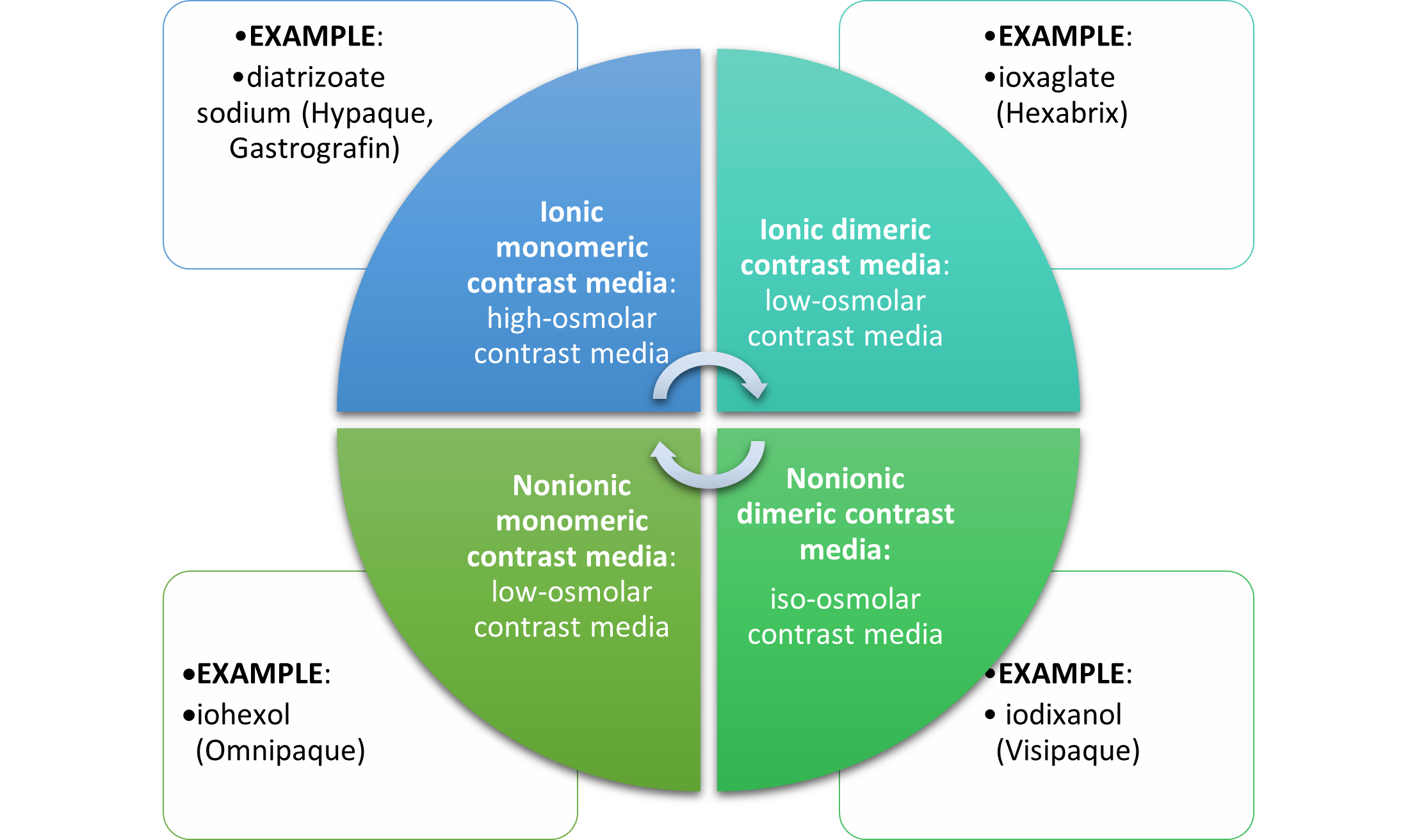
- (Murphy, 2026; Vega et al., 2025)
While the currently used nonionic, low-osmolar, or iso-osmolar ICAs are considered safe and effective when administered correctly, significant risks and clinical considerations need to be considered before they can be prescribed (ACR, 2025; Davenport et al., 2020). Clinicians must balance the potential risks of contrast media with the diagnostic benefits, which are premised on a multitude of factors, including:
- the probability and necessity of an accurate diagnosis
- alternative methods of diagnosis
- risk of misdiagnosis
- expectations about kidney function recovery
- allergic reaction risk and risk of other adverse reactions (Davenport et al., 2020)
One of the most concerning and well-established risks of ICAs is contrast-induced acute kidney injury (CI-AKI) in patients with impaired renal function. This condition is characterized by worsening renal function within 48 hours of IV contrast administration. In 2020, the ACR and National Kidney Foundation (NKF) published a consensus statement on the use of IV contrast media in patients with kidney disease. The statement reveals that the risk of CI-AKI is uncertain primarily due to a lack of control groups sufficient to separate AKI caused by the contrast media (CI-AKI) and contrast-associated AKI (CA-AKI) or postcontrast AKI (AKI coincident to contrast media administration). The consensus statement discloses concerns that the risk of CI-AKI has been overstated and does not apply to all populations; however, it offers specific recommendations based on risk factors and the staging of the patient’s underlying renal disease (Davenport et al., 2020; Rudnick & Davenport, 2026). Table 7 presents the 2012 Kidney Disease: Improving Global Outcomes (KDIGO) staging criteria for AKI and chronic kidney disease (CKD) to inform clinical decision-making regarding IV ICAs. The 2012 AKI guidelines are currently being updated, with the finalized guidelines awaiting release. The KDIGO CKD guidelines were updated in 2024, with changes to the evaluation and management process, but the CKD staging criteria remain the same.
Table 7
KDIGO AKI and CKD Staging
KDIGO AKI Staging | ||
Stage | Serum Creatinine (sCr) Criteria | Urine Output |
1 | 1.5–1.9 times baseline serum creatinine OR Increase in sCr ³ 0.3 mg/dL (³ 26.5 mmol/L) | <0.5 mL/kg/hour for 6-12 hours |
2 | 2.0–2.9 times baseline sCr | <5.0 mL/kg/hour for ≥12 hours |
3 | 3.0 times baseline sCr OR Increase in sCr ³ 4.0 mg/dL (³353.6 mmol/L) OR Initiation of kidney replacement therapy OR Decrease in eGFR to <35 mL/min/1.73 m2 (for patients <18 years old) | <0.3 mL/kg/hour for ≥ 24 hours OR Anuria for ≥12 hours |
KDIGO CKD Staging | ||
Grade | GFR criteria | |
Grade 1 | ³90 mL/min/1.73 m2 with markers of kidney damage | |
Grade 2 | 60–89 mL/min/1.73 m2 with markers of kidney damage | |
Grade 3a | 45–59 mL/min/1.73 m2 | |
Grade 3b | 30–44 mL/min/1.73 m2 | |
Grade 4 | 15–29 mL/min/1.73 m2 | |
Grade 5 | <15 mL/min/1.73 m2 | |
(Davenport et al., 2020; Kidney International, 2012)
Following a thorough review of several extensive, well-controlled research studies, the ACR and NKF report the following findings:
- CA-AKI
- the risk of CA-AKI increases with each stepwise increase in the CKD stage, with risk percentages as follows:
- 5% risk with eGFR ³ 60
- 10% risk with eGFR 45–59
- 15% risk with eGFR 30–44
- 30% risk with eGFR less than 30 mL/min/1.73 m2
- risk factors for CA-AKI include: eGFR, diabetes mellitus, exposure to other nephrotoxic agents, hypotension, hypovolemia, congestive heart failure, and impaired kidney perfusion
- there was no clinically significant difference in the risk of CA-AKI between types of contrast administered
- the risk of CA-AKI increases with each stepwise increase in the CKD stage, with risk percentages as follows:
- CI-AKI
- the risk of CI-AKI is cited as substantially less than CA-AKI; the authors caution that the actual risk remains undetermined in those patients with severe kidney disease; several large controlled observational studies have demonstrated no evidence of CI-AKI regardless of the CKD stage
- the risk percentages of CI-AKI are as follows:
- 0% risk with eGFR ³ 45
- 0%–2% risk with eGFR 30–44
- 0%–17% risk with eGFR less than 30 mL/min/1.73 m2
- there are limited studies linking patient-related factors to CI-AKI, and the primary risk factor was found to be eGFR, without any other risk factors confirmed
- no studies have directly compared the risk of CI-AKI between the various types of contrast, and there is no clinically significant difference in risk based on the type of contrast use (Davenport et al., 2020)
Table 8 summarizes key recommendations for clinicians, as outlined in the consensus statement on IV ICA in patients with CKD and those at risk for AKI.
Table 8
Guidelines for IV ICA in Patients with AKI/CKD
Recommendations |
|
|
|
|
|
|
Prophylaxis |
|
(Davenport et al., 2020; Rudnick & Davenport, 2026)
Patients with renal disease are not the only ones at risk for complications from ICAs. Diabetic patients taking metformin (Glucophage) and other biguanides are at risk for lactic acidosis. Lactic acidosis is characterized by a buildup of lactic acid in the bloodstream (greater than 2 mmol/L), which can lead to severe symptoms, including extreme exhaustion, muscle cramps, body weakness, abdominal pain, diarrhea, headache, jaundice, and breathing changes. Since the kidneys are primarily responsible for excreting metformin (Glucophage), patients with underlying renal insufficiency who take this medication are at heightened risk for this severe complication (ACR, 2025; Chu & Stolbach, 2025). The FDA has archived its 2016 recommendation regarding IV contrast administration in a patient taking metformin (Glucophage) and has not provided more recent recommendations. Current practice refers to the ACR contrast media guidelines:
- in high-risk patients, metformin (Glucophage) should be temporarily discontinued at the time of testing and withheld for 48 additional hours
- resumption of metformin (Glucophage) dosing can occur when the eGFR is >30 mL/min/1.73m2 (ACR, 2025; Rudnick & Davenport, 2026)
According to the ACR (2025), patients taking metformin (Glucophage) who are not high risk, have no evidence of AKI, and have an eGFR >30 mL/min/1.73 m2 do not need to discontinue metformin (Glucophage) before or after ICA administration. There is no obligatory need to re-evaluate the patient’s renal function following the test or procedure. However, for patients with AKI or severe CKD (stage 4 or 5), metformin (Glucophage) should be temporarily discontinued at the time of or before the procedure. The ACR (2025) notes that for patients who meet the criteria for withholding metformin (Glucophage), renal function (eGFR) must be reassessed 48 hours after the imaging procedure before resuming metformin (Glucophage). Further, patients with severe cardiac disease, such as symptomatic congestive heart failure, angina, pulmonary hypertension, cardiac arrhythmias, or severe but compensated cardiomyopathy, may also be at risk of cardiac events. While the ACR (2025) determines that these patients are at a modestly increased risk, it does not advise restricting ICAs solely based on the patient’s cardiac status. However, the risks and benefits should be considered individually, and the ACR clarifies several common misconceptions regarding the use of ICAs in the following patient groups:
- beta-blockers: patients on beta-blockers do not need to discontinue their medication(s) before contrast administration
- sickle-cell trait/disease: contrast should not be restricted in patients with sickle-cell trait/disease, as there is no evidence that iodinated contrast increases the risk for acute sickle crisis
- pheochromocytoma: there is no evidence that contrast use increases the risk of hypertensive crisis in patients with pheochromocytoma
- myasthenia gravis: there is a questionable relationship between the use of iodinated contrast and exacerbations of myasthenia gravis due to limited studies in a small number of patients; it remains controversial whether iodinated contrast should be considered a relative contraindication in these patients
- hyperthyroidism: patients with hyperthyroidism can develop thyrotoxicosis following exposure to iodinated contrast, although it is a rare complication. The ACR (2025) does not recommend the restriction of contrast medium solely based on a history of hyperthyroidism; however, it makes the following recommendations for two specific circumstances:
- “In patients with acute thyroid storm, iodinated contrast medium exposure can potentiate thyrotoxicosis; in such patients, iodinated contrast medium should be avoided. Corticosteroid premedication in this setting is unlikely to be helpful;
- In patients considering radioactive iodine therapy or patients undergoing radioactive iodine imaging of the thyroid gland, administration of an iodinated contrast medium can interfere with the treatment and diagnostic dose uptake. If iodinated contrast medium is administered, a washout period is suggested to minimize this interaction. The washout period is ideally 3–4 weeks for patients with hyperthyroidism, and 6 weeks for patients with hypothyroidism” (ACR, 2025, p. 6).
Allergic Reactions
ICAs pose a risk of allergic reactions, which can have variable presentations ranging from acute to delayed, as outlined in Table 9. Acute reactions are further categorized by severity from mild to severe. The frequency of allergic reactions following the administration of iodinated contrast media has decreased with the shift from high-osmolality to nonionic low-osmolality contrast media. Patients can have an allergic reaction to initial exposure to the ICA, mediated by anaphylactic mechanisms, with symptoms developing within minutes. Anaphylactic reactions are usually unpredictable but are the most clinically significant reactions to ICAs, as they involve the release of histamine and other biologic mediators. Most reactions occur within 5–60 minutes after ICA administration. However, delayed reactions can occur up to 1 week following administration. Delayed reactions are more common in young adults, females, and patients with a history of contrast allergy, and they tend to present as cutaneous urticaria or a rash (ACR, 2025; Rogers & Tadi, 2023).
Table 9
Allergic Reactions to Iodinated Contrast Media
Severity | Description |
Mild
| Symptoms are self-limited without evidence of progression. |
| |
Moderate
| Symptoms are more pronounced and usually require medical management; they may become severe if left untreated. |
| |
Severe
| Symptoms are often life-threatening and can result in permanent morbidity or death if not managed appropriately. |
| |
Delayed | Urticaria or rash that develops hours to 7 days following ICA administration Delayed reactions are usually limited to dermatologic manifestations. |
(ACR, 2025; Rogers & Tadi, 2023; Vega et al., 2025).
While ICA reactions are sporadic and often unpredictable, certain factors are associated with an increased risk. The ACR (2025) advises that prior allergic-like or unknown-type reactions to the same class of contrast medium are considered the most significant risk factor for predicting future adverse events. Additional risk factors for ICA allergic-like reactions include the following:
- Patients with a prior allergic reaction (or an unknown type of reaction) to an ICA have a 5-fold risk of developing a future allergic-like reaction with subsequent exposure to contrast.
- Patients with a history of asthma are at increased risk for an allergic reaction and may be more likely to develop bronchospasm, but those with well-controlled asthma may not be at increased risk.
- Patients with multiple allergies (such as dermatitis and urticaria) have an increased risk of a severe reaction to contrast media.
- Patients with mild allergies (such as seasonal allergies) are at unclear risk (ACR, 2025; Rogers & Tadi, 2023; Vega et al., 2025).
Despite popular misconception, it is now well established that there is no link between shellfish allergy and allergy to ICAs. Patients with shellfish or povidone-iodine (Betadine) allergies are at no higher risk from iodinated contrast than those with other allergies. Furthermore, there is no cross-reactivity between the different contrast classes; therefore, a patient with an ICA allergy is not at increased risk for a gadolinium-based allergy (ACR, 2025; Vega et al., 2025).
Treatment of Allergic Reactions
According to ACR, all facilities in which ICAs are injected must be equipped with emergency supplies, and staff must be trained in basic life support in the event of a life-threatening allergic reaction. While most allergic reactions are mild, self-limiting, and do not worsen, they can sometimes become more severe. Therefore, it is advised that any patient who displays signs of a reaction be observed for at least 20–30 minutes, or longer if needed, to ensure clinical stability and recovery. Mild reactions may require symptomatic treatment but often do not require any treatment at all. Moderate reactions are usually not life-threatening; severe reactions are rare but potentially fatal. Severe reactions require prompt recognition and action by qualified healthcare providers (HCPs) to prevent morbidity and death (ACR, 2025).
Clinicians should maintain IV access, monitor vital signs, and assess the patient’s appearance for any signs of reaction. The ACR (2025) lists the following five immediate assessments that should be performed upon recognition of a potential allergic reaction:
- What is the patient’s general appearance?
- Can the patient speak? How does their voice sound?
- What is the quality of the patient’s breathing?
- What is the patient’s pulse?
- What is the patient’s blood pressure?
Clinicians caring for these patients are advised to prioritize a focused assessment and to evaluate the patient’s level of consciousness, skin appearance, phonation quality, lung auscultation, blood pressure, and heart rate. These key assessment points help quickly determine the severity of the reaction, enabling effective treatment to be promptly and successfully administered. In free-standing outpatient facilities, staff should be trained to activate emergency response systems and to know when to elevate care, such as by calling 911. These facilities should also be equipped with the necessary equipment, including, but not limited to, oxygen, an automated external defibrillator (AED), suction equipment, and medications. In addition to preserving IV access, monitoring vital signs, and delivering supplemental oxygen by mask as needed (at a rate of 6–10 L/min) is essential. Table 10 concisely outlines guidelines for additional interventions to manage acute reactions to ICAs in adult patients (ACR, 2025).
Table 10
Management of Acute Reactions to ICAs in Adults
Clinical Manifestation | Treatment |
HIVES (URTICARIA) | |
Mild Scattered or transient | No treatment is needed, but if symptomatic, may use:
|
Moderate Numerous or symptomatic |
|
Severe Widespread or progressive |
|
* Note: Second-generation antihistamines may be beneficial for patients who need to drive themselves home | |
DIFFUSE ERYTHEMA | |
With hypotension | Administer 1,000 mL of IV fluids rapidly:
|
With profound hypotension (unresponsive to fluids) |
|
*Note: For the remainder of this treatment algorithm, follow the epinephrine administration dosing guidelines and instructions as outlined in this section, based on the administration route (IV, IM, or auto-injector); IV administration is preferred in hypotensive patients due to poor perfusion to the extremities | |
BRONCHOSPASM | |
Mild |
|
Moderate |
|
Severe |
|
LARYNGEAL EDEMA | |
|
|
HYPOTENSION Systolic blood pressure (SBP) <90 mm Hg | |
|
|
Severe hypotension with bradycardia (pulse < 60 bpm) Vasovagal Reaction |
|
Severe hypotension with tachycardia (pulse > 100 bpm) Anaphylactoid Reaction |
|
HYPERTENSIVE CRISIS Diastolic blood pressure (DBP) > 120 mmHg; SBP > 200 mm Hg Symptoms of end-organ compromise | |
|
|
PULMONARY EDEMA | |
|
|
SEIZURES/CONVULSIONS | |
|
|
HYPOGLYCEMIA | |
If the patient can swallow safely: |
|
If the patient is unable to swallow safely and IV access IS available: |
|
If the patient is unable to swallow safely and IV access is NOT available: |
|
(ACR, 2025)
Premedication Regimen for Patients with an ICA Allergy
While well-controlled studies and definitive data are not available on the efficacy of premedication in preventing allergic reactions in high-risk patients, the ACR (2025) believes that premedication does reduce the likelihood of a reaction in high-risk patients receiving low-osmolality iodinated contrast medium. While there is no formal consensus on the exact premedication regimen, oral premedication is preferable to IV premedication due to lower cost, greater convenience, and stronger evidence in the literature. The majority of data exist on the use of oral methylprednisolone (Medrol) or oral prednisone (Deltasone), with or without oral diphenhydramine (Benadryl). The ACR (2025) supports the use of the following premedication regimens in patients at high risk, at the discretion of the ordering clinicians.
Standard 12- or 13-hour premedication regimen:
- For use in outpatient, emergency department, and inpatient settings for patients at high risk in whom medication use is not anticipated to delay care decisions or treatment adversely.
- Regimen #1: prednisone (Deltasone) 50 mg PO at 13 hours, 7 hours, and 1 hour before contrast administration, plus diphenhydramine (Benadryl) 50 mg IV, IM, or PO 1 hour before contrast administration, OR
- Regimen #2: methylprednisolone (Medrol) 32 mg PO 12 hours and 2 hours before contrast administration, and diphenhydramine (Benadryl) 50 mg may be added as in regimen #1, OR
- Regimen #3: (for patients who are unable to take oral medication) is the same as regimen #1, but hydrocortisone (Cortef) 200 mg is given as a replacement for each dose of prednisone (Deltasone), plus diphenhydramine (Benadryl) 50 mg IV 1 hour before contrast administration (ACR, 2025)
Accelerated IV premedication regimen:
- For use in outpatient, emergency department, and inpatient settings for patients at high risk in whom the use of 12- or 13-hour premedication is anticipated to delay care or treatment adversely.
- Regimen #1: (usually 4–5 hours in duration) methylprednisolone sodium succinate (Solu-Medrol) 40 mg IV or hydrocortisone sodium succinate (Solu-Cortef) 200 mg IV immediately, and then every 4 hours until contrast administration, plus diphenhydramine (Benadryl) 50 mg IV 1 hour before contrast administration, OR
- Regimen #2: (usually 4–5 hours in duration) dexamethasone sodium sulfate (Decadron) 7.5 mg IV immediately, and then every 4 hours until contrast administration, plus diphenhydramine (Benadryl) 50 mg IV 1 hour before contrast administration, OR
- This is the preferred regimen for patients with an allergy to methylprednisolone (Solu-Medrol)
- Regimen #3: methylprednisolone sodium succinate (Solu-Medrol) 40 mg IV or hydrocortisone sodium succinate (Solu-Cortef) 200 mg IV, plus diphenhydramine (Benadryl) 50 mg IV, each 1 hour before contrast administration
- Any regimen less than 4–5 hours in duration (including this regimen) has no evidence of efficacy but may be considered in emergent situations where there are no alternatives (ACR, 2025)
Special Considerations for Patients who are Pregnant or Breastfeeding
The effects of ICAs on the human embryo and fetus remain incompletely understood. Studies have demonstrated that ICAs can cross the placenta and enter the fetus in measurable amounts, but the effects are unknown, and long-term data are still being collected. The ACR (2025) does not recommend routine screening for pregnancy before ICA administration. The FDA supports this recommendation, and most ICAs are category B medications, except for diatrizoate meglumine and diatrizoate meglumine sodium, which are classified as category C. The literature base regarding the safe use of ICA in breastfeeding patients is also limited; however, many studies have demonstrated that the routine dose of ICA absorbed by the infant’s gut from ingested breast milk is extremely low. While the ACR states that available data suggest it is safe for the lactating individual and infant to continue breastfeeding after receiving ICA, the individual should be allowed to make an informed decision after fully disclosing the available information. For individuals who abstain from breastfeeding temporarily, the recommendation is to express and discard breastmilk for 12–24 hours following ICA administration. There is no benefit to refraining from breastfeeding beyond 24 hours (ACR, 2025; Perelli et al., 2022). The ACOG Committee on Obstetric Practice recommends that iodinated contrast be used only when required to obtain essential additional diagnostic information that will affect the care of the fetus or pregnant individual during the pregnancy. The ACOG also supports the continuation of breastfeeding without interruption following the use of iodinated contrast materials, with these guidelines reaffirmed in 2026 (ACOG, 2017).
Gadolinium
GBCAs are the primary contrast agents used to enhance MRI image quality. Administered IV, GBCAs are paramagnetic and work by altering the magnetic properties of neighboring water molecules, thereby enhancing the quality of the MRI images. The FDA approved the first GBCA for use in 1988. Currently, seven types of MRI contrast agents are available. Similar to ICAs reviewed in the prior section, the osmolarity and viscosity of the GBCAs can also vary widely and are generally higher for the ionic than the nonionic agents. Gadolinium is a metal in the Lanthanide series, and when used alone, it is toxic to humans. Gadolinium in GBCAs has undergone chelation, in which other chemical ions are mixed with gadolinium to mitigate its harmful effects on the body. The majority of GBCAs used in the United States are gadolinium chelates, which have a positive magnetic susceptibility and function to produce increased signal on the resulting images. The GBCA is injected into the patient before the MRI and is eliminated from the body primarily via the kidneys, with a small amount of liver excretion (Ibrahim et al., 2023; Rogers & Tadi, 2023).
GBCAs are typically well tolerated and are much less likely to produce an allergic reaction than ICAs. The incidence of adverse events when administered at clinical doses of 0.1–0.2 mmol/kg is low, ranging from 0.07% to 2.4%. Most reactions are mild and physiologic, including coldness, warmth, pain at the injection site, nausea, vomiting, headache, paresthesia, and dizziness. Allergic-like reactions are uncommon, ranging from 0.004% to 0.7%, and symptoms are generally very mild and transient, usually limited to hives, other skin reactions, or itchy eyes. While manifestations of allergic-like reactions to GBCA can be similar to those of ICAs, they are rare, with life-threatening anaphylactic reactions occurring at a low rate of 0.001% or 0.01%. For example, in a survey of 20 million GBCA doses, only 55 severe reactions were reported. While fatal reactions to GBCAs are possible, they are rare and are often due to cardiovascular or respiratory effects. When managing GBCA-related allergic reactions, clinicians should follow the same ICA guidelines outlined in Table 10 (ACR, 2025).
In 2015, the FDA published a safety alert after residual gadolinium was found in the brains of patients who had received multiple doses of GBCAs over their lifetimes. Research has demonstrated that gadolinium deposits appear to occur preferentially in specific areas of the brain, even in the absence of clinically evident disease and with intact blood-brain barriers. The etiology of this remains unclear and is a relatively undefined clinical phenomenon. Legal suits have been brought in recent years due to this phenomenon, despite a lack of clinical correlation (ACR, 2025). Clinicians must balance the potential risks of GBCAs with the diagnostic benefits, which are also premised on a multitude of patient-specific factors, such as:
- the probability and necessity of an accurate diagnosis
- alternative methods of diagnosis
- risk of misdiagnosis
- the clinical benefit of the diagnostic information against the unknown, yet the potential risk of gadolinium deposition in the brain
- expectations regarding the patient’s risk for the development of nephrogenic systemic fibrosis (Harvey et al., 2020)
Nephrogenic systemic fibrosis (NSF) is a rare systemic but potentially life-threatening complication with an unknown etiology unique to GBCAs. It is characterized by thickening of skin, organs, and other tissues in patients with severe preexisting renal disease. The initial symptoms typically include skin thickening and pruritus, which can then rapidly progress to contractures and joint immobility. In extreme cases, the condition can be fatal. The time between GBCA administration and the onset of NSF is usually 2–10 weeks. However, rare case reports of symptoms have appeared years after the last GBCA exposure. NSF seems to have first appeared around 2000, but radiology guideline changes did not occur until 2006, at which time clinicians began withholding GBCAs in patients with AKI or severe CKD when the GFR was <30 mL/min/1.73 m2. While this led to a decline in NSF cases, further investigation showed that the risk of NSF was higher with specific GBCAs. As a result, modern-day radiology algorithms group GBCAs based on their reported associations with NSF in vulnerable patient populations, as outlined in Table 11 (ACR, 2025; Ibrahim et al., 2023).
Table 11
Classification of GBCAs in Relation to Reported Cases of NSF with ACR Recommendations for Use
Group | Risk Interpretation | Agents |
Group I
Associated with the highest number of NSF cases | The ACR considers patients exposed to Group 1 GBCAs to be at risk for developing NSF if any of the following conditions apply: patients on dialysis (any form), those with CKD grade 4 or 5 without dialysis, or those with AKI. |
|
Group II
Associated with few (if any) unconfounded cases of NSF | The ACR considers the risk of NSF among patients exposed to standard or lower than-standard doses of Group II GBCAs to be sufficiently low or possibly nonexistent. |
|
Group III Data are insufficient regarding the risk for NSF, but few (if any) unconfounded cases of NSF have been reported | There are insufficient real-life data to determine the likelihood of NSF from administering group III GBCAs. Therefore, the ACR considers Group III agents to have the same risk level as Group I GBCAs. |
|
Summary ACR Recommendations for GBCA Use in Patients at Risk for NSF | ||
| ||
(ACR, 2025)
The ACR (2025) advises clinicians to follow the algorithm shown in Figure 9 for evaluating renal function before GBCA administration. Regarding this clinical decision-making algorithm, a history of renal disease, including AKI, is considered a risk factor, as well as any of the following:
- prior or current dialysis
- renal transplantation
- single kidney
- kidney surgery
- renal cancer
- hypertension requiring medical therapy
- diabetes mellitus (ACR, 2025)
Figure 9
Clinical Decision Algorithm for Evaluating Renal Function Before Group I or Group III GBCA Administration
(ACR, 2025)
The ACR (2025) makes several recommendations regarding the use of GBCAs in the following patient groups:
- patients with CKD 4 of 5 not on dialysis: group I GBCAs are contraindicated, and group II GBCAs should be used instead
- patients with AKI: group I GBCAs should be avoided if possible, and group II agents are preferred
- patients on metformin (Glucophage): it is not necessary to discontinue metformin (Glucophage) before GBCA administration when the standard dose of gadolinium (0.1–0.3 mmol/kg) is being used
- sickle cell disease/trait: the risk to patients with sickle cell disease at approved dosages is very low or nonexistent, and withholding contrast or premedicating patients with no other risk factors is not recommended (ACR, 2025)
Special Considerations for Patients who are Pregnant or Breastfeeding
No known adverse effects have been reported in fetuses when standard doses of GBCAs were administered to pregnant females; however, similar to ICAs, the data is limited, and long-term studies are lacking. There are no reported cases of NSF related to GBCAs in pregnant females. However, gadolinium chelates can accumulate in the amniotic fluid, which suggests a potential risk for NSF in the pregnant individual or fetus. Since it remains unclear how GBCAs can affect the fetus, it is recommended that these agents be administered with caution in pregnant patients. GBCAs should be used only if considered critical and the potential benefits justify the potentially unknown risks to the fetus. The lowest possible dosage for diagnostic results should be used in these cases. The ACOG Committee on Obstetric Practice acknowledges that the use of gadolinium contrast is limited and should not be withheld if it significantly improves the predicted diagnostic performance and outcomes for the fetus or pregnant individual. The ACOG and ACR also recommend that breastfeeding not be interrupted following gadolinium administration, as less than 0.04% of the administered dose is excreted into breast milk. However, the individual should be allowed to make an informed decision after fully disclosing the available information. For individuals who temporarily abstain from breastfeeding, the recommendation is to express and discard breast milk for 12–24 hours after GBCA administration. There is no benefit to refraining from breastfeeding beyond 24 hours. These guidelines were reaffirmed in 2026 (ACOG, 2017; ACR, 2025).
A 2019 study found an increased prevalence of GBCA administration during the first trimester of pregnancy. This was attributed to a lack of pregnancy screening before diagnostic testing. To avoid GBCA administration during pregnancy, the ACR recommends that facilities that conduct imaging with GBCA administration have a pregnancy screening protocol. Unfortunately, no pregnancy screening technique is 100% effective, and false negatives may result if testing occurs early in the pregnancy; therefore, all individuals able to become pregnant should be counseled on the potential risks and lack of complete understanding of the ramifications of fetal exposure to GBCA (ACR, 2025).
Barium Sulfate
Barium sulfate contrast agents (BSCAs) are preferred for evaluating and visualizing the GI tract. They can only be administered orally or rectally. They are composed of a suspension of insoluble barium sulfate particles not absorbed by the gut. They are ideal for illuminating the GI tract because they have excellent coating properties for the GI mucosa, as demonstrated in Figure 10. They are the most common type of orally administered contrast material and enhance visualization of the GI tract on CT scans, abdominal and pelvic radiographs, and specific MRI scans. They are also commonly used for fluoroscopic studies, such as upper gastrointestinal (UGI) examinations or small bowel evaluations (ACR, 2025; Gotfried, 2025).
Figure 10
Visualization of the Intestinal Tract Following Administration of Barium Contrast
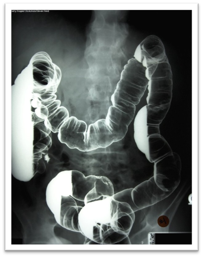
Patients scheduled to undergo a scan using an oral BSCA should be advised to remain NPO for 4–8 hours before the exam, depending on the type of scan and the institution’s policy. If the contrast material is to be administered rectally, the patient will usually be instructed to follow a clear liquid diet the day before and require a colon preparation with bisacodyl (Dulcolax), polyethylene glycol (Miralax), or magnesium citrate to clean the colon before the exam (ACR, 2025; Gotfried, 2025).
While the ACR (2025) explicitly states that there are no absolute contraindications to the use of BSCAs, it recommends avoiding their use in patients suspected or known to have bowel perforations or prior barium allergy. Older adults or those unable to change positions may have difficulty turning themselves to distribute the barium properly. Patients who have a suspected fistula, paralytic ileus, severe dehydration, or a history of cystic fibrosis (which is associated with an increased risk for blockage in the small bowel) are at greater risk for having an adverse reaction to or complication from BSCA administration. Barium sulfate is generally well tolerated orally, although some patients describe a mildly unpleasant taste. When barium sulfate is administered via enema, patients may experience temporary abdominal fullness and mild discomfort. It is recommended to warm the BSCA to room temperature, which helps enhance patient tolerability and reduce colon spasms (ACR, 2025; Campos, 2024; Gotfried, 2025).
Adverse reactions to BSCAs are rare and almost always mild. The most common symptoms include nausea, vomiting, abdominal cramping, or discomfort, which can occur during or after the examination. Patients can have vasovagal reactions, which are more common with rectal contrast administration after the colon is distended. Following completion of a BSCA procedure, patients should be instructed to increase oral fluid intake to facilitate and expedite the excretion of contrast material in the feces. Patients should be informed that their stool may be white for a few days as the contrast material is eliminated. It is common for some patients to experience changes in their routine bowel habits for the first 12–48 hours, such as diarrhea or constipation. Barium enemas are also used therapeutically in some instances to remove feces from the bowel. This procedure may be recommended for patients with severe constipation or blockages to prevent surgical intervention and reduce the risk of bowel perforation. There have been rare reports of appendicitis following the use of a barium enema. This phenomenon, known as barium-induced appendicitis, is thought to result from barium retention and subsequent obstruction of the appendiceal lumen. Perforation may also occur during barium administration; this adverse effect is most commonly observed in patients with toxic megacolon after a barium enema (Campos, 2024; Zaccarini et al., 2022).
Allergic-like reactions to BSCA are uncommon, with an incidence rate of 1 in 750,000 examinations. If an allergic-like reaction does occur, the manifestations are usually very mild and limited to transient rashes, urticaria, pruritus, or mild bronchospasm. Moderate to severe allergic reactions are rare, occurring in about 1 out of 2.5 million patient exposures. Clinical manifestations of moderate to severe allergic reactions may include more extensive dermatologic changes, respiratory symptoms, and vascular events such as hypotension. Rarely, angioedema of the stomach and small bowel and toxic epidermal necrolysis (TEN) have been described. Although some believe there is a correlation between asthma and allergic-type reactions to barium, no conclusive evidence supports this claim (ACR, 2025).
Although this is infrequent, direct barium toxicity has been reported in patients receiving oral and rectal BSCAs. The etiology is based on the concept that any barium dissociating from the stable barium compound may be absorbed through the bloodstream, leading to toxicity. Symptoms of barium toxicity usually present rapidly and include watery diarrhea, nausea, and vomiting. Absorption of barium can cause electrolyte imbalances, including rapid hypokalemia, which, if left untreated, can cause muscle weakness, respiratory or cardiac arrest, and death. The treatment is limited to supportive therapy, rehydration, and managing electrolyte disturbances (ACR, 2025).
Microbubble Contrast Agents
Microbubble contrast agents (MCAs) are most commonly used to enhance ultrasonography images, particularly heart exams. Currently, microbubbles are the only approved contrast agent for use with ultrasounds. MCAs are microscopic, typically 1–8 µm in diameter, highly compressible, gas-filled, and coated with a phospholipid or protein outer layer. MCAs have a high degree of “echogenicity,” or the ability to reflect ultrasound waves; structures with higher echogenicity appear brighter than surrounding tissues on ultrasound images. These tiny gas bubbles can be safely injected IV through a peripheral or central access line or instilled into hollow structures, such as the urinary bladder. Once injected, they provide a transient improvement in ultrasound visualization, allowing detection of differences between the gas in the microbubbles and the surrounding tissues. They enhance the ultrasound signal from blood and improve visualization of blood flow without subjecting the patient to the risks associated with other contrast media (ACR, 2025; Yusefi & Helfield, 2022).
These agents are approved for administration via slow IV infusion or bolus injection. Since the microbubbles dissolve rapidly, usually within 15 minutes, image acquisition should be completed within 10 minutes post-injection before the microbubbles spontaneously rupture and dissolve. As the microbubbles dissolve, they release gas, which is eliminated mainly by the lungs through exhalation. This is a valuable and practical alternative to ICAs or GBCAs for patients with AKI, CKD, or allergies (Yusefi & Helfield, 2022).
According to the ACR (2025), there are currently three FDA-approved MCAs for IV administration in adults undergoing echocardiography to improve visualization of the left ventricular cavity and endocardial borders. These three agents include perflutren lipid microspheres (Definity), sulfur hexafluoride lipid-type A microspheres (Lumason; SonoVue), and perflutren protein-type A (Optison). Sulfur hexafluoride lipid-type A microspheres (Lumason) is additionally approved for liver imaging and imaging of the pediatric urinary tract during voiding ultrasonography (ACR, 2025; Yusefi & Helfield, 2022). While their current FDA-approved indications are limited to the above, these agents have also been successfully used off-label for the following assessments and procedures:
- blood flow in tumors
- benign cysts and solid tumors in the kidney
- blood perfusion in organs
- abnormalities in the heart
- thrombosis, such as in myocardial infarction
- bowel wall inflammation or inflammatory activity in inflammatory bowel disease, as well as liver and kidney masses
- endoleaks following AAA repair
- abscesses and phlegmons
- ultrasound-guided interventions and ablative therapies (ACR, 2025; Yusefi & Helfield, 2022)
MCAs have a reasonably favorable safety profile, with an adverse event rate lower than that of ICAs or GBCAs. MCAs have been administered millions of times worldwide and have demonstrated an excellent safety profile. A meta-analysis of microbubble tolerance found that the most severe adverse effect is pseudoanaphylaxis, with a rate of 0.004%–0.009%. Adverse events are usually mild and physiologic. The most common symptoms include headaches, flushing, warmth, nausea, and altered taste, with the most severe reactions occurring within 30 minutes of MCA administration. The risk of severe cardiopulmonary reactions increases in patients with a preexisting and unstable cardiac condition, such as an acute myocardial infarction or congestive heart failure. There is no reported renal toxicity with MCAs. MCAs should not be used intra-arterially and are contraindicated in patients with prior allergic reactions to these agents (ACR, 2025; Yusefi & Helfield, 2022).
Special Considerations for Patients Who Are Pregnant or Breastfeeding
No well-controlled studies evaluating the risks of MCAs when administered to pregnant females are available. Therefore, the ACR (2025) recommends using these agents only in pregnant females when necessary and when the prospective benefits outweigh the unknown but potential risks to the fetus. Breastfeeding individuals should be counseled to temporarily pump and discard breastmilk for up to 24 hours following administration.
Saline Contrast Media
Agitated saline contrast (ASC) is considered a distinct type of contrast within the MCA family. It is administered intravenously and provides an air microbubble contrast to the right side of the heart. Like the aforementioned MCAs, ASC is transient and diffuses into the lungs. This technique helps examine the right side of the heart and identify intracardiac or extracardiac (pulmonary arteriovenous) shunts, particularly right-to-left shunts. The primary safety concern following ASC administration is cerebral air embolism. There have been reports of ischemic complications, including transient ischemic attack and stroke. This extremely low risk has been attributed to a technical error where the ASC was not mixed or injected correctly. When ischemic events occur, the effects are often mild and short-lived (Ahmed, 2026; Millington et al., 2023).
Gaseous Contrast Agents
Gaseous agents (such as oxygen [O2] and carbon dioxide [CO2]) can also serve as contrast agents in specific imaging tests. Similar to MCAs used in ultrasonography, these agents are absorbed rapidly by the body and eliminated through exhalation. Research is limited on these contrast agents, and the ACR manual of contrast media omits any reference to or discussion of these agents entirely. While air and oxygen can be dangerous due to the risk of gas emboli during specific procedures, CO2 does not pose a risk of gas emboli and can be used with relative safety. CO2 has the most research data supporting its clinical benefits and use as an intravascular contrast agent. It is used as an alternative to ICA or GBCAs in patients with renal dysfunction, as it has no adverse effects on the kidneys and is not associated with known ICA allergies. It is the least expensive contrast medium, readily available, has a lower molecular weight than ICAs, and is less viscous than blood. These characteristics make it an ideal agent for imaging small collateral vessels and various vascular beds and chambers. It displaces blood from vessels and acts as a negative contrast agent. However, administering CO2 as a contrast agent requires extreme care as it is a colorless, odorless, and significantly compressible gas. It is also less sensitive and specific than ICAs or GBCAs (Gupta et al., 2020; Young & Mohan, 2023). CO2 has been successfully used as a contrast agent in the following clinical situations:
- angiography in patients with peripheral arterial disease who are at risk for contrast-induced nephropathy
- aortic aneurysm repairs
- aortography
- renal artery angiography
- inferior vena cava imaging and filter placement
- portal vein imaging
- splenoportography
- oncologic tumor embolization procedures
- upper extremity venography
- dialysis fistula or graft repair
- assessment of gastrointestinal bleeding (Gupta et al., 2020; Young & Mohan, 2023)
Contraindications to the use of CO2 include the concurrent use of nitrous oxide as a sedating agent. This is due to the decreased solubility of CO2 when mixed with nitrous oxide. CO2 arteriography should not be performed above the diaphragm due to an increased risk of cerebral air embolism leading to stroke or death. When possible, the patient should be placed in the Trendelenburg position to decrease the risk of embolism. CO2 can also become trapped in the pulmonary artery or right side of the heart, leading to obstruction of venous return and subsequent hypotension and bradycardia. This phenomenon can be alleviated by placing the patient in a left lateral decubitus position to float the gas in a layer above the blood until the gas molecules dissolve in the bloodstream entirely (Young & Mohan, 2023).
Diagnostic Imaging During Pregnancy and Lactation Overview
According to the ACOG Committee on Obstetric Practice, diagnostic imaging during pregnancy and lactation is controversial, with various opinions. The confusion regarding the safety of diagnostic imaging often results in unnecessary avoidance of needed testing and interruption of breastfeeding. The most common documented adverse effects from high-dose radiation exposure on the developing fetus include growth restriction, microcephaly, and intellectual disability. The ACOG Committee on Obstetric Practice (2017) makes the following recommendations regarding diagnostic imaging procedures during pregnancy and lactation, with reaffirmation of these recommendations in 2026:
- Ultrasound and MRI are the preferred imaging techniques for patients. Still, their use should be confined only to those occasions when it is strongly predicted to provide medical benefit to the female parent or fetus.
- Most radiation exposure through CT scans or nuclear medicine imaging is provided at a dose much lower than the exposure associated with fetal harm. If these techniques are deemed medically necessary, they should not be withheld solely based on pregnancy.
- It is recommended that iodinated contrast be used only if required to obtain essential additional diagnostic information that will affect the care of the fetus or the female parent during the pregnancy. Breastfeeding can be continued without interruption following the use of iodinated contrast materials. For individuals who abstain from breastfeeding temporarily, the recommendation is to express and discard breastmilk for 12–24 hours following administration.
- The use of gadolinium contrast with MRI should be limited and used only if it significantly improves the predicted diagnostic outcomes for the fetus or female parent. Breastfeeding should not be interrupted following gadolinium administration.
- Radioactive iodine-123 (I-123) and iodine-131 (I-131) readily cross the placenta, and use should be avoided in pregnancy. If a thyroid scan is essential, it is recommended that the radioactive isotope technetium 99m be used.
- Since specific nuclear materials excreted into breastmilk can have deleterious effects on the infant, consultation with experts in breastfeeding and nuclear medicine is recommended when these compounds are used in lactating females (ACOG, 2017; ACR, 2025)
The Burden of Overuse and Misuse of Medical Imaging
The rate of diagnostic imaging and associated radiation exposure has expanded tremendously since the 1990s. Health care spending increased to $4.3 trillion in 2021, representing approximately a 60% increase over the prior decade. This increased spending affects both Medicare and employer-sponsored insurance plans. Medical imaging accounted for 8.85% of health care costs in 2021, a 35.9% overall increase from the prior decade. The adoption of preventive scans in line with published guidelines may have led to increased imaging, although imaging examinations in emergency department settings also showed an increased trend. Imaging examinations, including ultrasound, CT, and MRI, increased in 2021 compared to the prior decade (Horný et al., 2024). Projections estimate that imaging utilization will continue to rise exponentially with the aging of the US population, to a level of up to 26.9% higher in 2055 than current usage. Medicare fee-for-service recipients have historically had higher utilization rates than Medicare Advantage recipients. Projections have factored in the continued rise in Medicare Advantage consumers (Christensen et al., 2025).
There has been a rise in radiation exposure secondary to increased CT scans and higher radiation doses per scan. Due to technological advancements, modern CT scans are multiphase, meaning they are performed with repeated scans before and after injection of a contrast agent. The multiphase scanning process produces more precise, higher-resolution images, enabling greater diagnostic accuracy. However, they expose patients to 30%–50% more radiation than older CT scanners (Agency for Healthcare Research and Quality [AHRQ], 2025b).
The increased use of diagnostic imaging has led to increased adverse effects, including incidental findings, overdiagnosis, overtreatment, and higher health care costs. It is estimated that unnecessary diagnostic imaging accounts for between $75.7 and $101.2 billion annually in wasted health care spending in the United States (Chalmers et al., 2021). While radiation exposure is a widely recognized and well-described health risk of medical imaging, there is the risk of false-positive and false-negative test results. False-positive results occur when an abnormality is identified on a scan but no pathology is present in the patient. False positives often lead to additional diagnostic workup, including further radiology imaging tests that carry additional radiation exposure or invasive tests, such as a biopsy. False-negative results indicate that no abnormality was identified on the imaging test, despite the presence of an underlying pathological process in the patient. False-negative results lead to delays in treatment and a false sense of security for the affected patient (Elmore & Lee, 2026).
Incidental findings are conditions identified unintentionally during evaluation for another disease or ailment. It is estimated that up to 30% of all diagnostic imaging reports include at least one incidental finding, with CT scans showing the highest rate. Incidental findings pose several medical and ethical dilemmas for clinicians and patients, often leading to additional diagnostic workup and monitoring and, depending on the identified condition, contributing to further unnecessary radiation exposure. There are times when incidental findings can be lifesaving, such as when a pancreatic tumor is identified during imaging for another reason. However, most incidental findings contribute to the overuse of medical imaging services and increase the cost burden on patients, the health care system, and the economy (Davenport, 2023).
Similarly, overdiagnosis and overtreatment are well described in medical imaging literature. Emergency room providers and radiologists agree that diagnostic imaging is overused in the evaluation of emergency room patients. Time constraints and limited patient information during emergency room evaluations are felt to contribute to the overuse of imaging, as well as fear of malpractice claims (Kwee et al., 2024). Overdiagnosis also has concerns. Overdiagnosis leads to additional imaging studies and potentially unnecessary biopsies or surgeries. Ductal carcinoma in situ (DCIS) is frequently overdiagnosed on screening mammograms, requiring further imaging or intervention. The overdiagnosis of DCIS is statistically the largest contributor to breast cancer overdiagnosis (Poelhekken et al., 2025).
To reduce the burden of unnecessary medical imaging, the American Board of Internal Medicine (ABIM) Foundation launched the Choosing Wisely campaign in 2012. The campaign has compiled evidence-based recommendations from various medical specialty societies about potentially unnecessary medical testing and procedures. It is intended to guide clinicians on the proper ordering of diagnostic imaging and to promote conversations between clinicians and patients. Choosing Wisely provides information to help patients make healthcare choices that are supported by evidence and truly necessary, while avoiding unnecessary testing (ABIM Foundation, n.d.). A survey commissioned by the ABIM Foundation, in preparation for Choosing Wisely, confirmed HCPs’ view that unnecessary testing is a significant problem. HCPs reported that patients commonly request unnecessary testing and that patient insistence, along with the fear of malpractice, is the main reason for ordering unnecessary testing. More current research continues to support these original provider views, with reports of difficulty accessing prior records as an additional reason for overuse. Patient surveys indicated that most testing lacks sufficient risk to outweigh the perceived additional diagnostic value of the test results (ABIM Foundation, 2014; Lyu et al., 2017; Rudin et al., 2022). Approximately one-fourth of the Choosing Wisely recommendations address unnecessary imaging testing (Horný et al., 2024).
Expanding on the Choosing Wisely campaign, the Centers for Medicare & Medicaid Services (CMS) established the Protecting Access to Medicare Act of 2014, Section 218(b), to increase the rate of appropriate advanced diagnostic imaging services provided to Medicare beneficiaries. This legislation led to the development of the Appropriate Use Criteria (AUC) for Advanced Diagnostic Imaging, which serves as a resource modality to increase the rate of appropriate advanced diagnostic imaging services while decreasing the risk of medically unnecessary imaging. The AUC information is available to clinicians through an electronic, interactive tool called a Clinical Decision Support Mechanism (CDSM). The CDSM tool supports clinical decision-making and can be accessed through electronic health record (EHR) systems, private-sector mechanisms, or CMS-established mechanisms. The objective is to ensure that clinicians order the most patient-appropriate test for the specific clinical condition (AHRQ, 2025a). Initially, participation was voluntary, but on January 1, 2023, the payment penalty phase was implemented. CMS released a statement removing the voluntary participation clause, informing providers ordering Medicare Part B advanced diagnostic imaging services to access CDSM and review the AUC before ordering the test. This requirement applies to CT, MRI, nuclear medicine, and PET scans for all Medicare patients to be rendered in outpatient settings and emergency departments. CMS uses data collected from the AUC program to identify outlier ordering clinicians, who are then subject to a prior authorization requirement. On January 1, 2024, the AUC system was rescinded and is under further evaluation (CMS, 2026). Despite this, the recommendations when ordering medical imaging tests are summarized as follows (IAEA, n.d.-b):
- Justification: Each diagnostic radiology 24imaging test and procedure must be justified and performed only when necessary. The benefits and risks of the intended test must be considered, and alternatives that pose less risk should be contemplated.
- Optimization and ALARA: Tests should be performed using the minimal effective radiation dose required to achieve quality images, thereby reducing the radiation exposure as much as possible.
- Avoid unnecessary repeat testing: Repeat testing is often necessary to monitor disease status and evaluate for treatment response, such as with cancer; however, all unnecessary repeat testing should be avoided.
References
Adams, C., Jensen, J., & Leslie, S. W. (2026). Bone scan. In StatPearls [Internet]. StatPearls Publishing. Retrieved March 25, 2026, from https://www.ncbi.nlm.nih.gov/books/NBK531486
Agency for Healthcare Research and Quality. (2025a). Clinical decision support systems. https://psnet.ahrq.gov/primer/clinical-decision-support-systems
Agency for Healthcare Research and Quality. (2025b). Radiation safety. https://psnet.ahrq.gov/primer/radiation-safety#Radiation-Risks-Associated-With-Diagnostic-Imaging
Ahmed, H. (2026). Contrast echocardiography: Contrast agents, safety, and imaging technique. UpToDate. Retrieved March 12, 2026, from https://www.uptodate.com/contents/contrast-echocardiography-contrast-agents-safety-and-imaging-technique
American Association of Physicists in Medicine. (n.d.-a). How do we protect ourselves from unwanted radiation exposure and accidents? Retrieved March 13, 2026, from https://www.medicalradiationinfo.org/radiationandmedicine/radiation-safety/protect-unwanted-radiation-exposure-accidents/
American Association of Physicists in Medicine. (n.d.-b). Patient gonadal and fetal shielding in diagnostic imaging: Frequently asked questions. Retrieved March 13, 2026, from https://www.aapm.org/org/policies/documents/CARES_FAQs_Patient_Shielding.pdf
American Association of Physicists in Medicine. (2019). AAPM position statement on the use of patient gonadal and fetal shielding: PS 8-A. https://www.aapm.org/org/policies/details.asp?id=2552
American Board of Internal Medicine Foundation. (n.d.). Choosing Wisely®: Promoting conversations between patients and clinicians. Retrieved March 23, 2026, from https://abimfoundation.org/what-we-do/choosing-wisely
American Board of Internal Medicine Foundation. (2014). Unnecessary tests and procedures in the health care system: What physicians say about the problem, the causes, and the solutions. https://www.choosingwisely.org/files/Final-Choosing-Wisely-Survey-Report.pdf
American Cancer Society. (2023). American Cancer Society recommendations for the early detection of breast cancer. https://www.cancer.org/cancer/types/breast-cancer/screening-tests-and-early-detection/american-cancer-society-recommendations-for-the-early-detection-of-breast-cancer.html
American Cancer Society. (2024). Breast cancer facts & figures 2024–2025. https://www.cancer.org/content/dam/cancer-org/research/cancer-facts-and-statistics/breast-cancer-facts-and-figures/2024/breast-cancer-facts-and-figures-2024.pdf
American College of Obstetricians and Gynecologists. (2017). ACOG committee opinion: Guidelines for diagnostic imaging during pregnancy and lactation. Obstetrics & Gynecology, 130(4), e210–e216. https://www.acog.org/-/media/project/acog/acogorg/clinical/files/committee-opinion/articles/2017/10/guidelines-for-diagnostic-imaging-during-pregnancy-and-lactation.pdf
American College of Obstetricians and Gynecologists. (2024). ACOG updates recommendation on when to begin breast cancer screening mammography. https://www.acog.org/news/news-releases/2024/10/acog-updates-recommendation-when-to-begin-breast-cancer-screening-mammography
American College of Radiology. (2021). ACR–ACNM-SNMMI-SPR practice parameter for the performance of skeletal scintigraphy (bone scan). https://gravitas.acr.org/PPTS/GetDocumentView?docId=127
American College of Radiology. (2023). ACR-SPR practice parameter for imaging pregnant or potentially pregnant patients with ionizing radiation. https://gravitas.acr.org/PPTS/GetDocumentView?docId=23
American College of Radiology. (2024). ACR-ACNM-SNMMI-SPR practice parameter for the performance of scintigraphy and uptake measurements for benign and malignant thyroid disease. https://gravitas.acr.org/PPTS/GetDocumentView?docId=60
American College of Radiology. (2025). ACR manual on contrast media. https://edge.sitecorecloud.io/americancoldf5f-acrorgf92a-productioncb02-3650/media/ACR/Files/Clinical/Contrast-Manual/ACR-Manual-on-Contrast-Media.pdf
American College of Radiology. (2026). ACR manual on MR safety. https://edge.sitecorecloud.io/americancoldf5f-acrorgf92a-productioncb02-3650/media/ACR/Files/Clinical/Radiology-Safety/Manual-on-MR-Safety.pdf
American Heart Association. (2025). What is an Echocardiogram? https://www.heart.org/-/media/Files/Health-Topics/Answers-by-Heart/What-Is-Echocardiography.pdf
American Thyroid Association. (n.d.-a). Low iodine diet. Retrieved March 17, 2026, from https://www.thyroid.org/low-iodine-diet
American Thyroid Association. (n.d.-b). Radioactive iodine. Retrieved March 17, 2026, from https://www.thyroid.org/radioactive-iodine
Ashraf, M. A., & Goyal, A. (2023). Fludeoxyglucose (18F). In StatPearls [Internet]. StatPearls Publishing. Retrieved March 27, 2026, from https://www.ncbi.nlm.nih.gov/books/NBK557653/
Baker, M., & dela Cruz, J. (2023). Ectopic pregnancy, ultrasound. In StatPearls [Internet]. StatPearls Publishing. Retrieved March 27, 2026, from https://www.ncbi.nlm.nih.gov/books/NBK482192/
Bell, D. J. (2025). Absorbed dose. Radiopaedia. https://radiopaedia.org/articles/absorbed-dose?lang=us
Bonnemain, B. (2021). History of contrast media: Celebrating the centenary of the use of Lipiodol® in radiology. Journal of Clinical Practice Research, 43(6), 626–630. https://doi.org/10.14744/etd.2021.76429
Camacho, P. M., Petak, S. M., Binkley, N., Diab, D. L., Eldeiry, L. S., Farooki, A., Harris, S. T., Hurley, D. L., Kelly, J., Lewiecki, E. M., Pessah-Pollack, R., McClung, M., Wimalawansa, S. J., & Watts, N. B. (2020). American Association of Clinical Endocrinologists/American College of Endocrinology clinical practice guidelines for the diagnosis and treatment of postmenopausal osteoporosis—2020 update. Endocrine Practice, 26, 1–46. https://www.endocrinepractice.org/article/S1530-891X(20)42827-7/fulltext
Campos, A. (2024). Barium sulfate contrast medium. Radiopaedia. https://radiopaedia.org/articles/barium-sulfate-contrast-medium?lang=us
Cascino, T., & Shea, M. J. (2023). Echocardiography. Merck Manual Professional Version. https://www.msdmanuals.com/professional/cardiovascular-disorders/cardiovascular-tests-and-procedures/echocardiography
Centers for Disease Control and Prevention. (2024a). About health effects of radiation. https://www.cdc.gov/radiation-health/about/health-effects-of-radiation.html
Centers for Disease Control and Prevention. (2024b). About ionizing radiation. https://www.cdc.gov/radiation-health/about/ionizing-radiation.html
Centers for Disease Control and Prevention. (2024c). About non-ionizing radiation. https://www.cdc.gov/radiation-health/about/non-ionizing-radiation.html
Centers for Disease Control and Prevention. (2024d). Facts about computed tomography (CT) scans [image]. https://www.cdc.gov/radiation-health/data-research/facts-stats/ct-scans.html
Centers for Disease Control and Prevention. (2024e). Facts about fluoroscopy. https://www.cdc.gov/radiation-health/data-research/facts-stats/fluoroscopy.html
Centers for Disease Control and Prevention. (2024f). Radiation in healthcare: Imaging procedures. https://www.cdc.gov/radiation-health/features/imaging-procedures.html
Centers for Disease Control and Prevention. (2024g). The electromagnetic spectrum: An overview [image]. https://www.cdc.gov/radiation-health/about/electromagnetic-spectrum.html
Centers for Medicare & Medicaid Services. (2026) Appropriate use criteria program. https://www.cms.gov/Medicare/Quality-Initiatives-Patient-Assessment-Instruments/Appropriate-Use-Criteria-Program
Chalmers, K., Smith, P., Garber, J., Gopinath, V., Brownlee, S., Schwartz, A. L., Elshaug, A. G., & Saini, V. (2021). Assessment of overuse of medical tests and treatments at US hospitals using Medicare claims. JAMA Network Open, 4(4), e218075. https://doi.org/10.1001/jamanetworkopen.2021.8075
Christensen, E. W., Drake, A. R., Parikh, J. R., Rubin, E. M., & Rula, E. Y. (2025). Projected US imaging utilization, 2025 to 2055. Journal of the American College of Radiology, 22(20), 151–158. https://doi.org/10.1016/j.jacr.2024.10.017
Chu, J., & Stolbach, A. (2025). Metformin poisoning and toxicity. UpToDate. Retrieved March 18, 2026, from https://www.uptodate.com/contents/metformin-poisoning-and-toxicity
Davenport, M. S. (2023). Incidental findings and low-value care. American Journal of Roentgenology, 221(1), 117–123. https://doi.org/10.2214/AJR.22.28926
Davenport, M. S., Perazella, M. A., Yee, J., Dillman, J. R., Fine, D., McDonald, R. J., Rodby, R. A., Wang, C. L., & Weinreb, J. C. (2020). Use of intravenous iodinated contrast media in patients with kidney disease: Consensus statements from the American College of Radiology and the National Kidney Foundation. Radiology, 294(3), 660–668. https://doi.org/10.1148/radiol.2019192094
Elmore, J. G., & Lee, C. I. (2022). Artificial intelligence in medical imaging—Learning from past mistakes in mammography. JAMA Health Forum, 3(2), e215207. https://doi.org/10.1001/jamahealthforum.2021.5207
Elmore, J. G., & Lee, C. I. (2026). Screening for breast cancer: Evidence for effectiveness and harms. UpToDate. Retrieved March 19, 2026, from https://www.uptodate.com/contents/screening-for-breast-cancer-evidence-for-effectiveness-and-harms
Elsayes, K. M., & Oldham, S. A. A. (2014). Introduction to diagnostic radiology. McGraw-Hill Education. https://mhmedical.com/book.aspx?bookID=1562
Fazel, R., & Einstein, A. J. (2024). Radiation risk to healthcare workers from diagnostic and interventional imaging procedures. UpToDate. Retrieved March 12, 2026, from https://www.uptodate.com/contents/radiation-risk-to-healthcare-workers-from-diagnostic-and-interventional-imaging-procedures
Ghadimi, M., & Thomas, A. (2025). Magnetic resonance imaging contraindications. In StatPearls [Internet]. StatPearls Publishing. Retrieved March 25, 2026, from https://www.ncbi.nlm.nih.gov/books/NBK551669
Gotfried, J. (2025). Radiographs and other imaging contrast studies of the gastrointestinal tract. Merck Manual Professional Version. https://www.merckmanuals.com/professional/gastrointestinal-disorders/diagnostic-and-therapeutic-gastrointestinal-procedures/radiographs-and-other-imaging-contrast-studies-of-the-gastrointestinal-tract
Grimm, L. J., Avery, C. S., Hendrick, E., & Baker, J. A. (2022). Benefits and risks of mammography screening in women ages 40 to 49 years. Journal of Primary Care & Community Health, 13. https://doi.org/10.1177/21501327211058322
Gupta, A., Dosekun, A. K., & Kumar, V. (2020). Carbon dioxide-angiography for patients with peripheral arterial disease at risk of contrast-induced nephropathy. World Journal of Cardiology, 12(2), 76–90. https://doi.org/10.4330/wjc.v12.i2.76
Harvey, H. B., Gowda, V., & Cheng, G. (2020). Gadolinium deposition disease: A new risk management threat. Journal of the American College of Radiology, 17(4), 546–550. https://doi.org/10.1016/j.jacr.2019.11.009
Heston, T. F., & Tafti, D. (2024). Nuclear medicine safety. In StatPearls [Internet]. StatPearls Publishing. Retrieved March 25, 2026, from https://www.ncbi.nlm.nih.gov/books/NBK603730/
Holmes, K., & Griffiths, M. (2016). Chapter 14: Image quality. Radiology Key. https://radiologykey.com/image-quality
Horný, M., Chang, D., Christensen, E. W., Rula, E. Y., & Duszak, R. (2024). Decomposition of medical imaging spending growth between 2010 and 2021 in the US employer-insured population. Health Affairs Scholar, 2(3), qxae030. https://academic.oup.com/healthaffairsscholar/article/2/3/qxae030/7636360
Ibrahim, M. A., Hazhirkarzar, B., & Dublin, A. B. (2023). Gadolinium magnetic resonance imaging. In StatPearls [Internet]. StatPearls Publishing. Retrieved March 25, 2026, from https://www.ncbi.nlm.nih.gov/books/NBK482487
International Atomic Energy Agency. (n.d.-a). Radiation protection of patients during PET/CT scanning. Retrieved March 27, 2026, from https://www.iaea.org/resources/rpop/health-professionals/nuclear-medicine/pet-ct/patients
International Atomic Energy Agency. (n.d.-b). X-rays: What patients need to know. Retrieved March 12, 2026, from https://www.iaea.org/resources/rpop/patients-and-public/x-rays
International Atomic Energy Agency. (2014). Diagnostic radiology physics: A handbook for teachers and students. IAEA Publications. https://www-pub.iaea.org/MTCD/Publications/PDF/Pub1564webNew-74666420.pdf
International Commission on Radiological Protection. (n.d.). Absorbed, equivalent, and effective dose. Retrieved March 27, 2026, from https://icrpaedia.org/Absorbed,_Equivalent,_and_Effective_Dose
Iqbal, A., & Rehman, A. (2022). Thyroid uptake and scan. In StatPearls [Internet]. StatPearls Publishing. Retrieved March 25, 2026, from https://www.ncbi.nlm.nih.gov/books/NBK555978
Katal, S., Eibschutz, L. S., Saboury, B., Gholamrezanezhad, A., & Alavi, A. (2022). Advantages and applications of total-body PET scanning. Diagnostics, 12(2), 426. https://doi.org/10.3390/diagnostics12020426
Knipe, H. (2025). Equivalent dose. Radiopaedia. https://radiopaedia.org/articles/equivalent-dose?lang=us
Knipe, H. (2026). Effective dose. Radiopaedia. https://radiopaedia.org/articles/effective-dose
Kidney International. (2012). KDIGO clinical practice guideline for acute kidney injury. Kidney International Supplements, 2(1), 1–141. https://kdigo.org/wp-content/uploads/2016/10/KDIGO-2012-AKI-Guideline-English.pdf
Kwee, R. M., Toxopeus, R., & Kwee, T. C. (2024). Imaging overuse in the emergency department: The view of radiologists and emergency physicians. European Journal of Radiology, 176, 111536. https://doi.org/10.1016/j.ejrad.2024.111536
Lee, C. I., & Elmore, J. G. (2026). Radiation-related risks of imaging. UpToDate. Retrieved March 12, 2026, from https://www.uptodate.com/contents/radiation-related-risks-of-imaging
Lewiecki, E. M. (2025). Overview of dual-energy x-ray absorptiometry. UpToDate. Retrieved March 13, 2026, from https://www.uptodate.com/contents/overview-of-dual-energy-x-ray-absorptiometry
Lyu, H., Xu, T., Brotman, D., Mayer-Blackwell, B., Cooper, M., Daniel, M., Wick, E. C., Saini, V., Brownlee, S., & Makary, M. A. (2017). Overtreatment in the United States. PLoS One, 12(9), e0181970. https://doi.org/10.1371/journal.pone.0181970
Mafraji, M. A. (2025a). Computed tomography (CT). Merck Manual Professional Version. https://www.merckmanuals.com/professional/special-subjects/principles-of-radiologic-imaging/computed-tomography
Mafraji, M. A. (2025b). Magnetic resonance imaging (MRI). Merck Manual Professional Version. https://www.merckmanuals.com/professional/special-subjects/principles-of-radiologic-imaging/magnetic-resonance-imaging
Mafraji, M. A. (2025c). Positron emission tomography (PET). Merck Manual Professional Version. https://www.merckmanuals.com/professional/special-subjects/principles-of-radiologic-imaging/positron-emission-tomography-pet
Mafraji, M. A. (2025d). Ultrasound. Merck Manual Professional Version. https://www.merckmanuals.com/professional/special-subjects/principles-of-radiologic-imaging/ultrasonography
Mehta, S. R., & Annamaraju, P. (2023). Intravenous pyelogram. In StatPearls [Internet]. StatPearls Publishing. Retrieved March 25, 2026, from https://www.ncbi.nlm.nih.gov/books/NBK559034
Millington, S. J., Mayo-Malasky, H., & Koenig, S. (2023). Agitated saline contrast injection in patients with severe hypoxemia. Journal of Intensive Care Medicine, 38(5), 479–486. https://doi.org/10.1177/08850666231159019
Monticciola, D. L., Hendrick, R. E., & Helvie, M. A. (2024). Outcomes of breast cancer screening strategies based on cancer intervention and surveillance modeling network estimates. Radiology, 310(2), e232658. https://doi.org/10.1148/radiol.232658
Murphy, A. (2026). Iodinated contrast media. Radiopaedia. https://radiopaedia.org/articles/iodinated-contrast-media-1
National Cancer Institute. (2001a). PET scan of a brain tumor [image]. https://visualsonline.cancer.gov/details.cfm?imageid=1755
National Cancer Institute. (2001b). PET scan of a normal brain [image]. https://visualsonline.cancer.gov/details.cfm?imageid=1753
National Cancer Institute. (2007). Mammography in progress [Image]. https://visualsonline.cancer.gov/details.cfm?imageid=4361
National Cancer Institute. (2025). Mammograms. https://www.cancer.gov/types/breast/mammograms-fact-sheet#what-are-the-benefits-and-potential-harms-of-screening-mammograms
National Institute of Biomedical Imaging and Bioengineering. (n.d.). Magnetic resonance imaging (MRI). Retrieved March 27, 2026, from https://www.nibib.nih.gov/science-education/science-topics/magnetic-resonance-imaging-mri
National Institute of Biomedical Imaging and Bioengineering. (2023). Ultrasound. https://www.nibib.nih.gov/science-education/science-topics/ultrasound
National Institute of Biomedical Imaging and Bioengineering. (2025a). Nuclear medicine. https://www.nibib.nih.gov/science-education/science-topics/nuclear-medicine
National Institute of Biomedical Imaging and Bioengineering. (2025b). Medical x-rays. https://www.nibib.nih.gov/science-education/science-topics/medical-x-rays
Niknejad, M. T. (2026). Barium swallow. Radiopaedia. https://radiopaedia.org/articles/barium-swallow
Occupational Safety and Health Administration. (n.d.). Ionizing radiation. Retrieved March 12, 2026, from https://www.osha.gov/ionizing-radiation/control-prevention
Odak, M., & Kayani, W. T. (2023). MUGA scan. In StatPearls [Internet]. StatPearls Publishing. Retrieved March 25, 2026, from https://www.ncbi.nlm.nih.gov/books/NBK564365
Omerovic, S., & Jain, A. (2023). Echocardiogram. In StatPearls [Internet]. StatPearls Publishing. Retrieved March 25, 2026, from https://www.ncbi.nlm.nih.gov/books/NBK558940
117th Congress. (2022). H.R.2617—Consolidated Appropriations Act, 2023. https://www.congress.gov/bill/117th-congress/house-bill/2617
O'Rourke, M. C., Goldstein, S., & Mendenhall, B. R. (2023). Transesophageal echocardiogram. In StatPearls [Internet]. StatPearls Publishing. Retrieved March 25, 2026, from https://www.ncbi.nlm.nih.gov/books/NBK442026
Patel, P. R., & De Jesus, O. (2023). CT scan. In StatPearls [Internet]. StatPearls Publishing. Retrieved March 25, 2026, from https://www.ncbi.nlm.nih.gov/books/NBK567796
Perelli, F., Turrini, I., Giorgi, M. G., Renda, I., Vidiri, A., Straface, G., Scatena, E., D'Indinosante, M., Marchi, L., Giusti, M., Oliva, A., Grassi, S., De Luca, C., Catania, F., Vizzielli, G., Restaino, S., Gullo, G., Eleftheriou, G., Mattei, A., … Cavaliere, A. F. (2022). Contrast agents during pregnancy: Pros and cons when really needed. International Journal of Environmental Research and Public Health, 19(24), 16699. https://doi.org/10.3390/ijerph192416699
Poelhekken, K., Greuter, M. J. W., van der Vegt, B., Dorrius, M. D., & de Bock, G. H. (2025). Overdiagnosis of ductal carcinoma in situ by grade and definition in population-based screening: A modeling study. The Breast, 84, 104594. https://doi.org/10.1016/j.breast.2025.104594
RadiologyInfo.org. (2023). Thyroid scan and uptake. https://www.radiologyinfo.org/en/info/thyroiduptake
RadiologyInfo.org. (2025). Radiation dose. https://www.radiologyinfo.org/en/info/safety-xray
Rogers, D. C., & Tadi, P. (2023). Intravenous contrast. In StatPearls [Internet]. StatPearls Publishing. Retrieved March 25, 2026, from https://www.ncbi.nlm.nih.gov/books/NBK557794
Rogers, J. L., & Brashers, V. L. (Eds.). (2023). McCance and Huether’s Pathophysiology: The biologic basis for disease in adults and children (9th ed.). Elsevier.
Rosen, H. N., & Drake, M. T. (2024). Clinical manifestations, diagnosis, and evaluation of osteoporosis in postmenopausal women. UpToDate. Retrieved March 20, 2026, from https://www.uptodate.com/contents/clinical-manifestations-diagnosis-and-evaluation-of-osteoporosis-in-postmenopausal-women
Rudin, R. S., Thakore, N., Mulligan, K. L., & Ganguli, I. (2022). Addressing the drivers of medical test overuse and cascades: User-centered design to improve patient-doctor communication. Joint Commission Journal on Quality and Patient Safety, 48(4), 233–240. https://doi.org/10.1016/j.jcjq.2022.01.005
Rudnick, M. R., & Davenport, M. S. (2026). Prevention of contrast-induced acute kidney injury associated with computed tomography. UpToDate. Retrieved March 12, 2026, from https://www.uptodate.com/contents/prevention-of-contrast-induced-acute-kidney-injury-associated-with-computed-tomography#H1762264800
Shah, A., & Aran, S. (2023). A review of magnetic resonance (MR) safety: The essentials to patient safety. Cureus, 15(10), e47345. https://doi.org/10.7759/cureus.47345
Slanetz, P. J., & Lee, C. I. (2025). Breast imaging for cancer screening: Mammography and ultrasonography. UpToDate. Retrieved March 13, 2026, from https://www.uptodate.com/contents/breast-imaging-for-cancer-screening-mammography-and-ultrasonography
Sullivan, C. M., Paul, N. S., & Rieder, M. J. (2023). Bridging the gap between bench and clinic: the importance of understanding the mechanism of iodinated contrast media hypersensitivity. British Journal of Radiology, 96(1141), 20220494. https://doi.org/10.1259/bjr.20220494
UpToDate Patient Education. (n.d.-a). Patient education: Bone scan (The basics). UpToDate. Retrieved March 27, 2026, from https://www.uptodate.com/contents/bone-scan-the-basics
UpToDate Patient Education. (n.d.-b). Patient education: Ultrasound (The basics). UpToDate. Retrieved March 27, 2026, from https://www.uptodate.com/contents/ultrasound-the-basics
U.S. Environmental Protection Agency. (2025). Frequent Questions: Radiation in medicine. https://www.epa.gov/radiation/frequent-questions-radiation-medicine
U.S. Environmental Protection Agency. (2026). Radiation sources and doses. https://www.epa.gov/radiation/radiation-sources-and-doses
U.S. Food & Drug Administration. (2021). MHRA Safety guidelines for magnetic resonance imaging equipment in clinical use. https://assets.publishing.service.gov.uk/government/uploads/system/uploads/attachment_data/file/958486/MRI_guidance_2021-4-03c.pdf
U.S. Food & Drug Administration. (2023a). Medical x-ray imaging. https://www.fda.gov/radiation-emitting-products/medical-imaging/medical-x-ray-imaging
U.S. Food & Drug Administration. (2023b). Radiological health regulations; amendments to records and reports for radiation emitting electronic products; amendments to performance standards for diagnostic x-ray, laser, and ultrasonic products. https://www.federalregister.gov/documents/2023/01/20/2023-00922/radiological-health-regulations-amendments-to-records-and-reports-for-radiation-emitting-electronic
U.S. Food & Drug Administration. (2023c). Testing and labeling medical devices for safety in the magnetic resonance (MR) environment: Guidance for industry and food and drug administration staff. https://www.fda.gov/media/74201/download
U.S. Food & Drug Administration. (2024). Ultrasound imaging. https://www.fda.gov/radiation-emitting-products/medical-imaging/ultrasound-imaging
U.S. Preventive Services Task Force. (2024). Breast cancer: Screening. https://www.uspreventiveservicestaskforce.org/uspstf/recommendation/breast-cancer-screening
Vachha, B., & Huang, S. Y. (2021). MRI with ultrahigh field strength and high-performance gradients: Challenges and opportunities for clinical neuroimaging at 7T and beyond. European Radiology Experimental, 5, 35. https://doi.org/10.1186/s41747-021-00216-2
Vega, F., van der Molen, A. J., Ruiz-Calatayud, M., Alberdi, A., Catalá, M., Blanco-Mota, C., van de Ven, A. A. J. M., Laguna, J. J., & Sebastia, C. (2025). Allergological evaluation of hypersensitivity reactions after administration of contrast agents: What the radiologist needs to know. European Journal of Radiology, 190, 112194. https://doi.org/10.1016/j.ejrad.2025.112194
Yandrapalli, S., & Puckett, Y. (2022). SPECT imaging. In StatPearls [Internet]. StatPearls Publishing. Retrieved March 25, 2026, from https://www.ncbi.nlm.nih.gov/books/NBK564426
Young, M., & Mohan, J. (2023). Carbon dioxide angiography. In StatPearls [Internet]. StatPearls Publishing. Retrieved March 25, 2026, from https://www.ncbi.nlm.nih.gov/books/NBK534244
Yusefi, H., & Helfield, B. (2022). Ultrasound contrast imaging: Fundamentals and emerging technology. Frontiers in Physics, 10, 791145. https://doi.org/10.3389/fphy.2022.791145
Zaccarini, D. J., Lubin, D., Sanyal, S., & Abraham, J. L. (2022). Barium sulfate deposition in the gastrointestinal tract: Review of the literature. Diagnostic Pathology, 17, 99. https://doi.org/10.1186/s13000-022-01283-8
Powered by Froala Editor