About this course:
This module explores the epidemiology, pathophysiology, risk factors, clinical manifestations, diagnosis, and treatment modalities of endometrial (uterine) cancer to promote early diagnosis, improve clinical outcomes, and provide optimal care, patient education, and support throughout the disease trajectory.
Course preview
Endometrial (Uterine) Cancer
This module explores the epidemiology, pathophysiology, risk factors, clinical manifestations, diagnosis, and treatment modalities of endometrial (uterine) cancer to promote early diagnosis, improve clinical outcomes, and provide optimal care, patient education, and support throughout the disease trajectory.
By the completion of this module, learners will be able to:
- explore the epidemiology of endometrial (uterine) cancer in the United States and risk factors for the development of the disease
- discuss the anatomy of the uterus and the pathophysiology leading to the development of endometrial (uterine) cancer
- differentiate the endometrial (uterine) cancer subtypes
- describe the clinical manifestations, diagnostic workup, and core components of cancer staging
- describe the management of endometrial (uterine) cancer, including an overview of treatment risks, side effects, and the elements of patient education
Endometrial (uterine) cancer is the most common gynecologic malignancy in the United States. According to the American Cancer Society (ACS), approximately 68,270 individuals will be diagnosed with this malignancy, and about 14,450 will die annually. The condition is commonly referred to as endometrial cancer (EC), as approximately 95% of these cancers begin in the endometrium (the lining of the uterus), compared to only 5% that begin in the myometrium or stroma, known as uterine sarcomas. Historically considered a disease solely affecting postmenopausal individuals, this cancer has become increasingly prevalent in younger, premenopausal individuals. This increased incidence is linked to the rising number of individuals with a body mass index (BMI) of 30 kg/m2 or higher. EC is a highly treatable condition when diagnosed early and managed effectively, and more than 600,000 survivors are currently living in the United States. Healthcare providers (HCPs) must remain informed about the disease’s clinical features and warning signs to expedite diagnosis and reduce morbidity and mortality (ACS, 2025g, 2026a; Centers for Disease Control and Prevention [CDC], 2024c).
*Module disclaimer: Since most uterine cancers are endometrial in origin, the information in this course refers to EC unless otherwise specifically stated.
Epidemiology
EC represents 3.2% of all new cancer diagnoses and 2.3% of all cancer deaths in the United States. According to the latest data from the Surveillance, Epidemiology, and End Results Program (SEER), the annual age-adjusted incidence of EC based on 2019 to 2023 data is 28.7 per 100,000 people, and the death rate was 5.4 per 100,000 based on the data from 2020 to 2024. Approximately 3.1% of females are affected during their lifetime; in 2023, it was estimated that 890,295 females were living with EC. Although EC can be diagnosed among females over the age of 20, it is most frequently diagnosed in postmenopausal females aged 55 to 64 (31.3%) and 65 to 74 (30.9%). The median age at diagnosis is 64. Similarly, EC rates can vary by race/ethnicity. EC is most common among Indigenous females (32.0 per 100,000), followed by non-Hispanic Black females (31.1 per 100,000), Hispanic females (29.0 per 100,000), non-Hispanic White females (27.7 per 100,000), and non-Hispanic Asian/Pacific Islander females (24.3 per 100,000; SEER, 2026).
EC is the eighth most common cause of death from cancer among females in the United States, with the highest deaths among females aged 65 to 74 (35.3%), followed by females aged 75 to 84 (25.4%), females aged 55 to 64 (19.8%), and females over the age of 84 (11.6%). The median age at death is 71. The 5-year relative survival rate of EC is 84%, which can vary based on the cancer stage. More specifically, the 5-year survival rate for localized EC (i.e., cancer that has not spread outside the uterus) is 94.9%, declining to 70.1% for regional (spread to regional lymph nodes) and 19.9% for those with distant metastases (i.e., cancer spreading to distant organs or sites outside the uterus). Similar to diagnosis rates, death rates vary by race/ethnicity. Non-Hispanic Black females have the highest death rate (9.9 per 100,000), followed by non-Hispanic White females (4.9 per 100,000), Hispanic females (4.6 per 100,000), Indigenous females (4.5 per 100,000), and non-Hispanic Asian/Pacific Islander females (3.8 per 100,000). Research has shown improved cancer survival rates across almost all cancers over the last five decades, except for endometrial and cervical cancers. From 2010 to 2024, the age-adjusted rates of new EC cases increased by 2.2%, and the death rates increased by 1.0% annually. A lack of significant treatment advances for recurrent and metastatic disease accounts for these survival rates (SEER, 2026; Somasegar et al., 2023).
Anatomy and Physiology
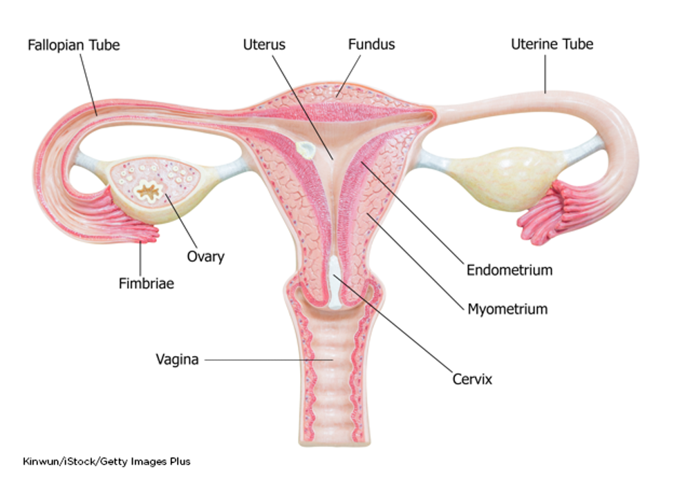
The uterus is the hollow, pear-shaped organ positioned between the urinary bladder and the rectum that protects and supports the developing fetus. It is a thick-walled and muscular structure composed of three major parts: the fundus, the uterine corpus (body), and the cervix. The basic anatomy of the female reproductive system is displayed in Figure 1. The fundus is the curved uppermost region that serves as a connection point for the fallopian tubes. The corpus is the largest part of the uterus, responsible for the bulk of its size, and is the usual site of implantation. The cervix is the fibromuscular lower portion that connects the uterine cavity to the vagina (Ameer & Peterson, 2025; Hinkle et al., 2025; Rogers, 2022).
Figure 1
Female Reproductive System
The cervix has two narrow openings at each end, as demonstrated in Figure 2. It enables the passage of sperm into the uterine cavity through dilation of the external os (external orifice) and the internal os (internal orifice). During labor, the cervix opens (dilates) to allow for the passage of the neonate through the birth canal. The uterus often varies in size and shape depending on the female’s reproductive phase and response to sex hormones. The average nonpregnant adult uterus measures 6 to 8 cm in length, 5 cm in width, and 2.5 cm in thickness, but it can enlarge up to five times its size during pregnancy. The uterus is substantially larger in parous individuals (i.e., those who have given birth) than in nulliparous individuals (i.e., those who have never carried a pregnancy to term). After menopause (the permanent cessation of menstruation), the uterus shrinks to its nulliparous size (Ameer & Peterson, 2025; Hinkle et al., 2025; Rogers, 2022). According to the American College of Obstetricians & Gynecologists (ACOG, 2025b), the average age of menopause is 51 years.
Figure 2
...purchase below to continue the course The uterus comprises three major inner layers: the endometrium, myometrium, and perimetrium, as outlined in Table 1 and depicted in Figure 1. Table 1 Inner Layers of the Uterus Layer Composition and Function Endometrium Myometrium Perimetrium (Ameer & Peterson, 2025; Gasner & Aatsha, 2023; LibreTexts, n.d.) The uterine tubes (also called fallopian tubes) are ducts that transport the oocyte (immature egg cell) from the ovary to the uterus. Several ligaments uphold the uterus’s position within the pelvic cavity. The broad ligament is the primary support, extending laterally on both sides of the uterus and attaching to the pelvic wall. The round ligament is embedded within the broad ligament, extends downward to the vagina, and pulls the uterus forward to maintain its anteverted positioning. Most females have an anteverted uterus tilted forward at the cervix toward the abdomen. The uterosacral ligaments extend from the posterior aspect of the cervix and vagina and support the uterus and pelvic organs posteriorly. The uterine artery is the main blood supply to the uterus, with a smaller supply from the ovarian artery. The uterine artery arises from the internal iliac artery and divides into ascending and descending branches. The ascending branch supplies the corpus and tubes. The descending (vaginal) branch feeds the cervix and upper vagina in conjunction with the vaginal artery, which arises on the lateral wall of the vagina (Ameer & Peterson, 2025; Gasner & Aatsha, 2023; LibreTexts, n.d.; Rogers, 2022). Aside from implantation, pregnancy, and labor, the uterus is also responsible for menstruation. A balance between estrogen and progesterone primarily controls these functions as part of the hypothalamic-pituitary-gonadal (HPG) axis. The HPG axis is a tightly regulated feedback system between the hypothalamus, pituitary gland, and ovaries (Figure 3). Hormonal changes during puberty signal the hypothalamus to release increased levels of gonadotropin-releasing hormone. In response, the anterior pituitary produces follicle-stimulating hormone (FSH) and luteinizing hormone (LH). FSH and LH signal the ovaries to release increased estrogen, progesterone, and a smaller testosterone supply. In individuals of childbearing age, ovulation occurs when a surge of LH triggers the ovary to release a mature egg into the fallopian tube. Ovulation occurs once per month, signaling the endometrium to thicken in preparation for fertilization. Estrogen's role is to promote the growth and health of the uterine lining to prepare for implantation. Progesterone maintains that lining so the fertilized egg can implant (or attach) itself to the endometrium’s thickened walls. If a fertilized egg does not adhere to the uterine lining, progesterone levels decline, and the unfertilized egg is expelled through the vagina during menstruation. After menopause, the ovaries stop making estrogen and progesterone; however, a small amount of estrogen is still generated naturally in adipose (fat) tissue (ACOG, 2025b; Hinkle et al., 2025; LibreTexts, n.d.; Rogers, 2022). Figure 3 Female Hypothalamic-Pituitary-Gonadal (HPG) Axis Pathophysiology Endometrial hyperplasia (EH) is a pathological condition in which the endometrial cells crowd together, become unusually thick, and may appear abnormal. Hyperplasia is the increase in the number of cells in an organ or tissue. These cells are not cancerous but can develop into dysplasia (precancerous cells). Dysplasia can be mild, moderate, or severe, depending on how the cells appear under a microscope and how much tissue or organ is affected (Singh et al., 2024; Urban, 2025). This process is demonstrated in Figure 4. Figure 4 Progression of Epithelial Cells to Cancer Cells EH is a well-established predisposing factor for EC. It is most commonly caused by excess estrogen with inadequate progesterone levels to oppose it. This imbalance leads to the proliferation (overgrowth) of the endometrial glands, which become variably shaped and irregularly distributed. EH most commonly occurs in postmenopausal individuals due to ovulation cessation (since progesterone is not generated). However, it may also occur during the perimenopausal period (transition period to menopause) due to ovulation irregularity. If ovulation does not occur, progesterone is not secreted, and the endometrium is not shed, leading to an overgrowth of the endometrial lining. Other causes of EH include polycystic ovarian syndrome (PCOS), a BMI of 30 or above, estrogen-secreting ovarian tumors, and menopausal hormone therapy (MHT, previously referred to as hormone replacement therapy [HRT]). The World Health Organization classifies EH as having two categories: benign hyperplasia without atypia and atypical hyperplasia/endometrial intraepithelial neoplasia (EIN). The absence or presence of atypia (abnormal-appearing cells) is the most crucial feature of EH. Benign hyperplasia does not demonstrate any abnormal cells (atypia) and carries a minimal risk (about 1%) of progression to invasive cancer. In contrast, atypical EIN is marked by an overgrowth of atypical cells and is a precancerous condition. Without treatment, EIN will develop into EC (ACOG, 2025b, 2026; Singh et al., 2024). Risk Factors While some individuals with EC do not have any identifiable risk factors, the two most substantial risk factors include estrogen exposure and a BMI of 30 or above. As described earlier, the balance of estrogen and progesterone plays a significant part in developing these conditions. Therefore, anything that increases endogenous (internal) or exogenous (external) estrogen exposure heightens risk (ACS, 2025b; Chen & Berek, 2026b; Ramirez & Salvo, 2026; Singh et al., 2024). The most common risk factors are outlined in Table 2. Table 2 Risk Factors for Endometrial Cancer (EC) (ACOG, 2025b, 2026; ACS, 2025b; Creasman, 2026a; Mahdy et al., 2024) Tamoxifen Tamoxifen (Soltamox), a selective estrogen receptor modulator (SERM), is the oldest hormonal treatment for breast cancer, initially approved by the US Food & Drug Administration (FDA) in 1977. It is a nonsteroidal antiestrogen agent that functions as a partial agonist by blocking estrogen receptors on cancer cells. It is also the most common agent used for chemoprevention in patients at high risk for breast cancer and is highly effective. However, it carries an increased risk for EC as it has an estrogen-like effect on the uterus. Tamoxifen (Soltamox) only increases the risk for EH, EC, and uterine sarcoma in postmenopausal individuals. Premenopausal individuals are not at increased risk for these conditions. Several studies have found that the increased risk for developing EC is two to three times higher in postmenopausal individuals taking tamoxifen (Soltamox) than in age-matched controls not taking the medication. The risk is time-dependent, meaning the risk increases with a longer duration of therapy. HCPs must counsel patients on these risks and the need to follow up with a gynecologist regularly. Individuals must report abnormal vaginal bleeding, bloody vaginal discharge, staining, or spotting, the most common early warning signs of EH and EC. All patients taking tamoxifen (Soltamox) are at increased risk for thromboembolic events, such as stroke, deep vein thrombosis, and pulmonary embolism. Since thromboembolic events can lead to significant morbidity and mortality, resulting in life-threatening consequences, early identification and intervention are essential (ACOG, 2024; FDA, 2018). HCPs are responsible for counseling patients regarding the risks associated with tamoxifen (Soltamox) and providing adequate education on monitoring precautions and immediate reporting of suspicious symptoms (ACOG, 2024; Chen & Berek, 2026b; Creasman, 2026a; FDA, 2018). For information regarding the diagnosis and management of thromboembolic events, refer to the following: Lynch Syndrome Lynch syndrome (LS), also called hereditary nonpolyposis colorectal cancer (CRC), is a common genetic risk factor for CRC that also increases the risk for EC. In the United States, it is estimated that 1 in 279 individuals (1.2 million people) have a gene mutation associated with LS; however, most are undiagnosed since identification depends on a cancer diagnosis. In addition to CRC and EC, additional LS-related cancers include gastric, ovarian, pancreatic, urothelial, glioblastoma, biliary tract, and small intestine cancers. Changes in the protein expression of MutL homolog 1 (MLH1), MutS homolog 2 (MSH2), MSH6, or postmeiotic segregation 2 (PMS2) genes are most commonly found in LS. Under physiologic conditions, these genes repair potential errors during DNA replication (the process during which DNA is copied in preparation for cell division); collectively, they are known as mismatch repair (MMR) genes. Since mutations in any of these genes impede the cell’s ability to repair the DNA replication errors, abnormal cells continue to divide. Over time, the accumulated DNA replication errors can lead to uncontrolled cell growth and an increased propensity for cancer development. Mutations in the MLH1 or MSH2 gene are associated with a higher risk of developing cancer than mutations in the MSH6 or PMS2 genes, which carry a lower risk. While gene mutations predispose individuals to cancer, not all people with these mutations will develop cancerous tumors (ACS, 2024; CDC, 2026a; Hall & Neumann, 2025; Villano, 2025). Individuals inherit LS in an autosomal dominant pattern, which means one inherited copy of each cell’s altered gene is sufficient to increase cancer risk. Females with LS are at a higher lifetime risk (up to 60%) for EC. LS causes approximately 1,600 new diagnoses of EC annually, or about 3% of EC cases. The National Comprehensive Cancer Network (NCCN) guidelines advise that patients who meet any of the following criteria should be evaluated for LS: Immunohistochemistry (IHC) and microsatellite instability (MSI) are screening tests performed to help identify patients at higher risk for LS. The NCCN guidelines on LS added a new recommendation statement encouraging the universal screening of all CRCs and ECs for mismatch repair deficiency (dMMR) to “maximize sensitivity for identifying individuals with LS and to simplify care processes” (LS-A, p. 6). Greater than 90% of LS-related cancers are microsatellite instability-high (MSI-H) or lack expression of at least one of the MMR proteins; therefore, MMR testing helps guide whether the patient should be tested for LS. While MMR deficiency can be reported as mismatch repair deficient (dMMR) or MSI-H, they have the same meaning. All patients with MSI-H or dMMR should be referred to a genetic counselor to undergo formalized genetic testing. LS can only be confirmed by a specialized gene panel blood test. Diagnosing LS in EC patients is critical. It allows for the testing and early diagnosis of relatives who may also be affected by the condition, thereby reducing morbidity and mortality associated with LS-related cancers (CDC, 2026a, 2026b; Hall & Neumann, 2025; NCCN, 2026a, 2026b). BMI of 30 or Greater Roughly 60% of EC cases in the United States are attributed to elevated BMI and, therefore, are potentially preventable. Elevated BMI is linked to up to 80% of all EC cases worldwide and remains a well-known global health challenge. In the United States, elevated BMI has become an epidemic. Based on data from the CDC’s National Center for Health Statistics, the age-adjusted prevalence of elevated BMI in adults across the United States from 1999 through 2020 increased from 30.5% to 41.9%. It is estimated that there are currently 100 million adults with an elevated BMI and 22 million adults with a severely elevated BMI (40 or greater) in the United States. Despite its adverse effects on health, elevated BMI is expected to rise substantially over the next several decades (CDC, 2024a; Crosbie et al., 2022; Ordeanu et al., 2025). EC was one of the first malignancies linked to elevated BMI, dating back to 1966 when an epidemiological study proposed that weight reduction was the most practical preventative measure for the disease (Wynder et al., 1966). According to the World Cancer Research Fund/American Institute for Cancer Research (WCRF/AICR, 2018), there is compelling evidence that elevated BMI throughout adulthood increases the risk of at least 13 different tumor types. Of these 13 tumor types, EC has the strongest link to elevated BMI. In a meta-analysis of 26 studies by the WCRF/AICR (2018), the researchers found that for every 5 kg/m2 increase in BMI, there was a 50% increase in the risk of developing uterine cancer. The mechanisms underlying BMI as a core driver of EC are premised partly on the increased amount of circulating estrogen carried in adipose tissue, thereby igniting a series of molecular mechanisms contributing to the pathogenesis of EC. As noted earlier, the ovaries stop making estrogen after menopause, but a small amount of estrogen is generated in adipose tissue. Therefore, estrogen supplied in adipose tissue significantly impacts postmenopausal individuals. Excess adipose tissue leads to an overproduction of estrogen (hyperestrogenism), unopposed by progesterone. Even small amounts of circulating estrogens are not adequately counterbalanced in postmenopausal individuals. An elevated BMI also induces insulin resistance, increased bioavailability of steroid hormones, and inflammation, collectively generating a metabolic state that drives tumorigenesis. Once diagnosed with EC, those with a BMI of 30 or above have poorer long-term health outcomes and less successful treatment courses. Research has found that individuals with an elevated BMI have more perioperative complications, such as an increased risk for infection, more antibiotic use, and higher rates of wound complications (Chen & Berek, 2026b; Constantine et al., 2019; Mahdy et al., 2024; Ordeanu et al., 2025). Several studies have demonstrated a significant association between a BMI of 30 or above and an increase in all-cause mortality compared to those with a BMI below 25. In a systematic review, Kokts-Porietis and colleagues (2021) evaluated 46 studies and found that a higher BMI was associated with higher all-cause mortality and cancer recurrence rates. Despite the clear evidence linking EC and an elevated BMI, there is limited public awareness of this relationship. Studies have demonstrated that at least half of individuals diagnosed with EC were unaware of the impact that an elevated BMI has on cancer risk (Derbyshire et al., 2022; Ordeanu et al., 2025). Risk Reduction and Prevention Unfortunately, there are currently no routine screening tests to identify EC. Aside from lifestyle choices geared toward maintaining a BMI below 25, additional factors that protect against or lower the risk of EC include pregnancy, oral contraceptive pills (OCPs), and specific intrauterine devices (IUDs). Since hormonal balance shifts toward progesterone during pregnancy, multiparous individuals (those having several pregnancies) are at lower risk for EC. Researchers have found an inverse relationship between the number of pregnancies and the risk of EC. In addition, each subsequent pregnancy was associated with increased risk reduction. Older age during pregnancy and the last birth have also been found to be a protective factor for EC, although the exact mechanism is unknown; it may be related to the increased progesterone throughout pregnancy. It is estimated that individuals whose last birth was between 35 and 39 years of age had a 32% risk reduction for EC. Breastfeeding has also been identified as a protective factor, with an 11% risk reduction when breastfeeding occurred for at least 3 months compared to no breastfeeding (ACS, 2025b; Chen & Berek, 2026b; National Cancer Institute [NCI], 2025). OCP use is associated with a 30% to 40% risk reduction of EC compared to no OCP use. Researchers have proposed that using OCPs effectively lowers the risk of EC by suppressing endometrial cell proliferation and regulating the balance of the female sex hormones. The risk reduction is strengthened with OCP use over an extended period, with the lowest risk among those who have taken OCPs for over 10 years. The protective effects of long-term OCP use persist for at least 10 years after OCPs are discontinued. The risk reduction for EC has been associated with long-term OCP users who also smoked, had an elevated BMI, or rarely exercised. According to the ACOG, all females who take estrogen after menopause should take progesterone to counteract the excess estrogen. If menses are irregular, OCPs (or alternate forms of progesterone) may be recommended to help balance hormones. Although many HCPs focus on the impact of estrogen increasing the risk of EC, the lack of progesterone is equally important (ACOG, 2025b; Chen & Berek, 2026b; Jahanfar et al., 2024). Females who have used an IUD seem to have a lower chance of getting EC; however, information regarding the protective effects of IUDs is primarily limited to those that do not contain hormones. The benefit of IUD use as a risk reduction strategy for EC is less clear than OCP use. However, progesterone-only releasing IUDs (i.e., levonorgestrel [Mirena]) have effectively managed complex atypical hyperplasia or early-grade ECs. Studies have shown that levonorgestrel (Mirena) IUD therapy for the conservative treatment of these conditions has resulted in a return to normal histology in most patients. Therefore, IUD use may be beneficial as they are increasingly utilized to treat EH. Diet and exercise also serve as protective factors against EC. Increased physical activity is associated with decreased BMI, central adiposity, and changes in metabolic hormone levels and growth factors that reduce EC risk (Bolling et al., 2026; Chen & Berek, 2026b; Mariani et al., 2026; NCI, 2018). Signs and Symptoms Since there are no screening tests for EC, patients need to recognize the warning signs. The most common sign of EH and EC is abnormal uterine bleeding (AUB), presenting in 75% to 90% of cases. AUB may include bleeding during the menstrual cycle that is heavier or lasts longer than usual, spotting between periods, and postmenopausal bleeding (PMB). AUB also includes menstrual cycles that are shorter than 21 days (counting from the first day of the menstrual period to the first day of the next menstrual period) or unusually heavy and prolonged menstrual bleeding characterized by blood clots larger than a quarter and lasting more than 7 days. The risk of EC is significantly less for premenopausal individuals with AUB than for postmenopausal individuals. PMB is the most ominous sign of EC and should always raise clinical suspicion and prompt further workup. As many as 6% to 19% of individuals with PMB have EC. Other symptoms concerning for EC may include watery or blood-tinged vaginal discharge, pelvic pain, dyspareunia (painful intercourse), dysuria (painful urination), and urinary frequency. Patients can also experience abdominal bloating and distention, early satiety, and associated weight loss. Laboratory findings are usually normal unless significant bleeding results in anemia (CDC, 2024b; Chen & Berek, 2026a; Chi et al., 2024; Ramirez & Salvo, 2026). A significant warning sign of EH and EC is a thickened endometrial lining, particularly in postmenopausal individuals. The endometrium changes in appearance and thickness throughout the menstrual cycle. In premenopausal individuals, there is significant variation in the endometrium thickness based on different stages of the menstrual cycle; thickness can range from 2 to 16 mm. The endometrium in postmenopausal individuals should be smooth, homogeneous, and less than 4 mm. Endometrial abnormalities are detected using transvaginal ultrasound (TVUS) or magnetic resonance imaging (MRI). Abnormal signs include endometrial thickness greater than 4 mm, edema (fluid), and irregularity of the lining with heterogeneous enhancement. Although expert opinions may vary, HCPs should consider sampling the endometrium for postmenopausal patients without uterine bleeding with an endometrial thickness greater than 4 mm (Chen & Berek, 2026a; Feldman & St. Laurent, 2026). HCPs should obtain a detailed history and physical exam for anyone with any concerns related to menstruation, specifically AUB (Kaunitz, 2026; Mikes et al., 2025). Refer to Table 3 for specific aspects of the history and physical exam that should be completed. Table 3 History and Physical Examination for Abnormal Uterine Bleeding Component Specific Aspects of History or Physical Examination Menstrual history Sexual and reproductive history Associated symptoms Other pertinent components Physical exam (Kaunitz, 2026; Mikes et al., 2025) Differential Diagnosis Aside from EH and EC, the HCP should consider several other uterine conditions in diagnosing patients presenting with AUB (Kaunitz, 2026; Mikes et al., 2025). The International Federation of Gynecology and Obstetrics (FIGO) generated a universally utilized and widely adopted acronym to classify the underlying etiologies of AUB called PALM-COEIN (Table 4). Table 4 PALM-COEIN Acronym PALM (Visually Objective Structural Criteria) COEIN (Nonstructural Issues) P: Polyp C: Coagulopathy A: Adenomyosis O: Ovulatory dysfunction L: Leiomyoma E: Endometrial disorders M: Malignancy and hyperplasia I: Iatrogenic (Jain et al., 2023; Kaunitz, 2026; Mikes et al., 2025; Pinkerton, 2026) While one or more of the above conditions can contribute to AUB, the five most common noncancerous uterine pathologies that may induce AUB are described in Table 5 (Kaunitz, 2026; Mikes et al., 2025). Table 5 Common Uterine Pathologies Condition Description Leiomyoma Polyp Adenomyosis Endometriosis Endometritis (Jain et al., 2023; Kaunitz, 2026; Mikes et al., 2025; Pinkerton, 2026; Rogers, 2022) Diagnostic Workup According to the ACOG Committee on Gynecologic Practice (2023), PMB requires prompt and efficient evaluation. The committee supports TVUS as the initial evaluation of PMB and as part of the diagnostic workup of AUB in menstruating individuals. TVUS is a safe, noninvasive imaging modality that uses sound waves to generate images of the internal structures within the vagina and pelvis. It is capable of evaluating and measuring the thickness of the endometrium. A small probe (transducer) is placed directly inside the vagina. The transducer produces sound waves at very high frequencies, which exceed the human hearing threshold, and are used to generate images on a computer. Images are captured in real-time, which allows for evaluating the structures and movement of the body’s internal organs (Nahlawi et al., 2022; National Institute of Biomedical Imaging and Bioengineering, 2023). Abnormalities identified on TVUS and/or persistent bleeding should prompt histologic evaluation of the endometrium as the essential next step. The only way to definitively diagnose EC is by evaluating the endometrial cells under a microscope. An endometrial biopsy (EMB) is the most common procedure to obtain a sample of the endometrial tissue. An EMB is usually performed in the outpatient setting by a gynecologist or gynecologic oncologist. It is a very accurate (sensitivity is greater than 90%) and straightforward procedure in which a thin, rod-like tool (uterine sound) is inserted through the cervix to induce dilatation of the external os. The uterine sound is removed, and a thin, flexible catheter is inserted through the cervical opening into the uterus. The catheter is rotated, and a small amount of the endometrial tissue is removed using suction. The procedure typically takes less than 15 min; resulting discomfort and pelvic cramping are similar to menstrual cramps. Although an EMB is a very safe and effective procedure, rare but possible complications may include bleeding, infection, and perforation of the uterine wall (the tip of an instrument passes through the uterine wall, creating a hole; Chen & Berek, 2026a; Chi et al., 2024; Creasman, 2026c; Ramirez & Salvo, 2026). Alternatively, endometrial tissue can be obtained by dilation and curettage (D&C) with or without a hysteroscopy. If the EMB did not provide enough tissue or the results were unclear, D&C is the next step in the diagnostic workup. A D&C is a surgical procedure typically performed in an outpatient surgery center or hospital under mild sedation. Like an EMB, the cervix is dilated, and a thin instrument (curettage) is inserted into the uterus to remove tissue. D&C is a reasonable initial procedure for patients who cannot tolerate an EMB, those with heavy bleeding, and those at high risk for EC (i.e., patients with LS; ACOG, 2025a; Chen & Berek, 2026a; Chi et al., 2024; Creasman, 2026c; Ramirez & Salvo, 2026). A hysteroscopy examines the uterine cavity using a thin tube with a light at the end called a hysteroscope (Figure 5). A biopsy may be performed for any abnormalities identified. Although rare, complications following a D&C or hysteroscopy are the same as those of an EMB and can include bleeding, infection, or uterine perforation (ACOG, 2025a; Chi et al., 2024; Creasman, 2026c; Ramirez & Salvo, 2026). Figure 5 Hysteroscopy HCPs must ensure that patients are educated on postprocedure care. It is normal to have some mild cramping and vaginal spotting for a few days following an EMB or D&C. Symptoms are typically well controlled with over-the-counter analgesics such as anti-inflammatories (e.g., ibuprofen [Motrin, Advil]) or acetaminophen (Tylenol). Many patients find adequate relief from nonpharmacological analgesics, such as applying heat or ice. Patients should not place anything inside the vagina for at least 3 days after the procedure; this includes avoiding tampons, douching, and abstaining from sexual intercourse (ACOG, 2025a; Chi et al., 2024; Creasman, 2026c; Ramirez & Salvo, 2026). Once EC is diagnosed, the NCCN (2026b) guidelines endorse the diagnostic workup components outlined in Table 6. Table 6 Components of Diagnostic Workup complete history and physical laboratory tests (complete blood count, comprehensive metabolic panel, including liver function tests, and renal function studies) imaging studies to evaluate for other sites of disease as follows: referral to a reproductive endocrinologist for fertility concerns if indicated (Mahdy et al., 2024; NCCN, 2026b; Ramirez & Salvo, 2026) Cancer Subtypes Approximately 95% of all ECs are adenocarcinomas, a cancer of the glandular cells (epithelial cells that produce mucus or fluid). EC is classified into two major groups: Type I and Type II. Type I tumors are most common and account for 80% of all ECs. These tend to be early-stage, low-grade tumors that typically develop from precursor lesions (e.g., EH). These are linked to excess estrogen (estrogen-dependent) and elevated BMI and are more likely to develop in younger individuals. They are typically less aggressive, do not commonly spread to other tissues, and usually carry a favorable prognosis. Endometrioid adenocarcinoma is the most common Type I tumor. Type II ECs are more likely to develop in older individuals. They are primarily diagnosed at advanced stages and are usually high-grade tumors (grade 3). They are not linked to excess estrogen, are more aggressive, faster-growing, and more likely to spread. Type II tumors carry a poorer prognosis, are more likely to recur, and are associated with higher mortality, accounting for at least 40% of EC-related deaths. Type II subtypes include papillary serous carcinoma, clear cell carcinoma, and carcinosarcoma. They are more likely to grow and spread outside the uterus and require more aggressive treatment (Chi et al., 2024; Huvila & McAlpine, 2025; Mahdy et al., 2024; NCCN, 2026b). Uterine Sarcoma Uterine sarcoma is a rare type of uterine cancer that develops in the myometrium or connective tissues rather than the endometrium. It accounts for 5% of all uterine cancer diagnoses and is more aggressive. It spreads to other body parts, most commonly the lungs, more rapidly than EC subtypes. Uterine sarcoma is more aggressive and harder to treat. Regardless of the subtype, uterine sarcomas are more common in Black females. Sarcomas include three major subtypes as follows: Cancer Staging The cancer stage at diagnosis guides treatment options and strongly influences overall survival. EC staging is done surgically with a total hysterectomy (TH), bilateral salpingo-oophorectomy (BSO), and lymph node evaluation. Lymph node evaluation is an important part of the staging procedure because it provides prognostic information and helps determine the need for adjuvant therapy. The choice of approach for lymph node evaluation can range from sentinel lymph node dissection to total lymphadenectomy. The chosen approach varies widely, but guidance from the NCCN recommends sentinel lymph node dissection to reduce the risk of lymphedema associated with a total lymphadenectomy. The decision should be based on the probability of lymph node metastasis, the feasibility of assessing the lymph nodes, and the risk for lymphedema. Two detailed cancer-staging systems are used: the FIGO system and the American Joint Committee on Cancer’s (AJCC) Tumor, Node, Metastasis (TNM) staging system, 8th edition. Both systems describe specific characteristics used to assign stages I through IV, as outlined in Table 7 and demonstrated in Figure 6. The FIGO system was most recently updated in 2023 to account for some ECs that are more aggressive than others. Cancer staging reflects the cell type, tumor grade, anatomical location of the tumor, and extent of malignancy. Within the TNM staging system, T denotes the size of the tumor and whether it has grown into nearby tissue, N refers to the presence of cancer in the lymph nodes, and M indicates whether cancer has metastasized to other parts of the body beyond the site of origin. Tumor grade measures how different the cancer cells look compared to healthy cells under the microscope. It is based on cell differentiation and varies from low grade (grade 1) to high grade (grade 3). Grade 1 is well differentiated and appears similar to healthy cells, whereas grade 3 is poorly differentiated (i.e., does not resemble healthy cells) and aggressive (ACS, 2025c; Cohn, 2026; NCCN, 2026b; Ramirez & Salvo, 2026). Table 7 Cancer Staging FIGO Stage AJCC TNM Description - TX primary tumor cannot be assessed - T0 no evidence of primary tumor - N0 no regional lymph node metastasis - N1–N2 regional lymph node metastasis - M0 no distant organ metastasis - M1 distant organ metastasis I T1 N0 M0 tumor limited to the uterus IA T1a N0 M0 tumor is limited to the endometrium IA1 T1a N0 M0 tumor is confined to the endometrium IA2 T1a N0 M0 tumor is a nonaggressive histologic type that involves less than half of the myometrium with no or focal lymphovascular space involvement (LVSI) IA3 T1a N0 M0 tumor is a nonaggressive histologic type limited to the uterus and ovary IB T1b N0 M0 tumor is a nonaggressive histologic type that invades at least half of the myometrium, but not beyond the body of the uterus, with no or focal LVSI IC aggressive histologic type limited to the endometrium II T2 N0 M0 tumor invades the stromal connective tissue of the cervix IIA tumor is a nonaggressive histologic type with invasion of the cervical stroma IIB tumor is a nonaggressive histologic type with substantial LVSI IIC T2 N0 M0 tumor is an aggressive histologic type with any myometrial invasion III T3 N0 M0 tumor has spread outside the uterus, but has not spread to the inner lining of the rectum or urinary bladder IIIA T3a N0 M0 tumor involves the ovary, fallopian tube, or serosa IIIB T3b N0 M0 tumor involves the vagina, parametrium, or the pelvic peritoneum IIIC1 T1–T3 N1, N1mi, or N1a M0 metastasis to the pelvic lymph nodes (N1, N1mi, or N1a) but not to nodes around the aorta or distant parts of the body IIIC2 T1–T3 N2, N2mi, or N2a M0 metastasis to the para-aortic lymph nodes (N2, N2mi, or N2a) but not to distant parts of the body IIIC2i (cancer has spread but is very small; micrometastasis) IIIC2ii (cancer has spread but is very small; macrometastasis) IVA T4 Any N M0 tumor involves the inner lining (mucosa) of the bladder or bowel IVB Any T Any N M1 cancer has spread to the abdominal peritoneum IVC cancer has spread to distant body parts such as lungs, liver, brain, or bone, or has spread to lymph nodes outside the abdomen or above the kidneys (ACS, 2025c; Cohn, 2026; Creasman, 2026c; NCCN, 2026b; Ramirez & Salvo, 2026) Figure 6 Endometrial Cancer Staging Diagrams In 2020, the NCCN guidelines first recommended conducting molecular analysis for EC to determine the molecular subgroup. Identifying the molecular subgroup gives a more specific understanding of prognosis and treatment selection, given the advances in targeted immunotherapy (NCCN, 2026b). There are four molecular subgroups of EC, including: Treatment of Endometrial Hyperplasia The treatment of EH varies based on many factors, including the patient’s age, risk factors, desire for future fertility, and the type and extent of EH. As noted earlier, many cases of EH can be effectively managed conservatively with synthetic progesterone. Medical or surgical treatment is recommended for patients with EH without atypia, and the choice is based on desire for fertility, menopausal status, contraceptive needs, and risk of progression. Premenopausal individuals are typically treated with progesterone therapy, while postmenopausal individuals are treated with a hysterectomy. Progesterone can be administered in various preparations, such as oral or injectable agents, vaginal creams, or as an IUD (i.e., levonorgestrel [Mirena]). The dose and duration of therapy also depend on the patient’s clinical situation and treatment goals. Treatment with progesterone can cause vaginal bleeding, similar to a menstrual period. Since EH can progress to EC, the favored treatment for individuals with atypical hyperplasia/EIN is hysterectomy (removal of the uterus) unless future fertility is desired. Hysterectomy is also the standard treatment for those who do not respond to progesterone therapy (NCCN, 2026b; Singh et al., 2024; Urban & Ring, 2026). Cancer Treatment The optimal cancer treatment depends on various factors, such as the pathologic features, cancer stage, plans for fertility, patient preference, age, and medical history. Treatment is often multimodal, with several therapies combined and administered simultaneously (concurrently) or sequentially. Since many cases require specialized surgical care, all patients with suspected EC or uterine sarcoma should be promptly referred to a gynecologic oncologist. This section will provide a synopsis of the most common evidence-based treatment strategies (Cohn, 2026; Creasman, 2026b; NCCN, 2026b; Plaxe & Mundt, 2026). Surgery Surgical intervention is typically indicated when the cancer is early-stage and limited to the uterus. TH/BSO with surgical staging (lymph node evaluation) is the most common treatment option for Type I ECs. Patients who desire fertility preservation should be promptly referred to a fertility expert. The approach to surgery has evolved significantly over time. Traditional techniques included an exploratory laparotomy through a midline incision. However, most patients with early-stage disease will now undergo minimally invasive surgery using laparoscopy or robotic surgery. Surgery aims to remove the primary tumor and identify prognostic factors to determine if adjuvant therapy is required. A laparotomy using the traditional midline incision may still be done for patients with excessive uterine size, known adhesive disease, or inability to tolerate a steep Trendelenburg position. Progesterone-based therapy may be offered initially to select individuals with early-stage and low-grade disease; however, this requires close monitoring with EMB or D&C every 3 to 6 months. TH/BSO should be encouraged in those patients who demonstrate signs of disease progression on progesterone-based therapy (Cohn, 2026; NCCN, 2026b; Mahdy et al., 2024; Ramirez & Salvo, 2026). Surgical Risks and Side Effects The risks and side effects of surgery depend on the size and degree of cancer invasion, the extent of surgery, and the structures removed. All surgeries and invasive procedures are accompanied by risks, such as adverse reactions to anesthesia, bleeding, blood clots, fistula formation (an abnormal connection between two hollow spaces within the body), bowel and bladder injury, infection, sexual dysfunction, and life-threatening sepsis. HCPs must counsel individuals on the side effects of surgical menopause and infertility following TH/BSO. The loss of fertility can negatively impact interpersonal relationships, quality of life, psychological health, and emotional well-being. The HCP serves a vital role in helping patients acclimate to these life-altering changes by facilitating healthy coping, addressing concerns, and referring patients to appropriate support groups or therapists (ACS, 2025f; NCCN, 2026b; Nettina & Nelson-Tuttle, 2024). Radiation Therapy Radiation therapy is a localized treatment that delivers a precisely measured amount of high-energy, focused ionizing radiation to the tumor while causing as little injury as possible to the surrounding tissue. Radiation causes cellular damage to cancer cells, leading to biological changes in the DNA and rendering cells incapable of reproducing or spreading. All healthy and cancer cells are vulnerable to the effects of radiation and may be injured or destroyed; however, healthy cells can repair themselves and remain functional. The total radiation dose is hyperfractionated, which means it is delivered to the tumor in small, divided doses (i.e., fractions) rather than all at once. Hyperfractionation gives healthy cells a chance to recover between treatments. The total number of fractions (doses) administered depends on the tumor size, location, reason for treatment, patient’s overall health, performance status, and any other treatments the patient receives. Radiation therapy is central to treating EC and can be delivered externally or internally; many patients receive both. The most common types of radiation used for EC include external beam radiation therapy (EBRT) and brachytherapy (ACS, 2025e; NCCN, 2026b; Nettina & Nelson-Tuttle, 2024; Plaxe & Mundt, 2026). External Beam Radiation Therapy EBRT delivers radiation from a source outside the body and is a common type of radiation therapy used for EC. Traditionally, radiation beams could only match the tumor’s height and width, exposing more healthy tissue to the consequences of radiation. Further advancements in imaging technology have led to more precise treatment mechanisms that allow even more of the radiation beam to reach the tumor. Intensity-modulated radiation therapy (IMRT) is a newer, highly conformal form of radiation that modulates the radiation beam’s intensity. IMRT delivers a higher radiation dose to a precise location, reducing unintended exposure to healthy tissues, enhancing clinical outcomes, and limiting side effects. IMRT helps minimize radiation exposure to the bowel and other critical structures, especially among patients who have undergone a TH/BSO (ACS, 2025e; Gray, 2026; NCCN, 2026b; Nettina & Nelson-Tuttle, 2024; Plaxe & Mundt, 2026). Brachytherapy Brachytherapy is a form of internal radiation therapy critical to treating EC. It is commonly administered after EBRT is completed or used in patients who are not surgical candidates. Following TH/BSO, most individuals receive brachytherapy to the upper part of the vagina (i.e., the vaginal cuff). This treatment provides a radiation boost to the most common cancer recurrence site. Brachytherapy involves implanting a wire or catheter into the body within or near the tumor. The radioactive material is placed inside a cylinder (or applicator) and positioned directly at the vaginal cuff. This direct positioning protects nearby structures, such as the bladder and rectum, from excess radiation exposure. Brachytherapy may be delivered using a low-dose rate (LDR; 0.4 to 2 Gy per hour) or a high-dose rate (HDR; greater than 12 Gy per hour) for EC. LDR brachytherapy typically requires hospitalization for several days, during which time the patient is confined to an isolated, radiation-safe room to protect others from exposure to the radiation. Specific LDR treatments involve the placement of radioactive rods, which require the patient to be confined to a bed to prevent dislodgment of the applicator for a defined period (usually 1 to 3 days). HDR brachytherapy is much more common than LDR as it is performed on an outpatient basis. Each treatment lasts about 10 to 20 min, and hospitalization and bed rest are not indicated. The treatment is delivered in a radiation-shielded room to protect others from exposure, and patients are not considered “radioactive” following the treatment. They can safely go about their routines and lifestyles without potentially exposing others (ACS, 2025e; Chi et al., 2024; Mitin, 2026; Nettina & Nelson-Tuttle, 2024). Radiation Side Effects Radiation side effects depend on the specific area(s) of the body exposed and the dose received. The urinary system, bowel, and genitalia are most commonly affected by radiation for EC. Bladder dysfunction may manifest as dysuria, hematuria, acute kidney injury, hydronephrosis, or incontinence. The most common side effect of brachytherapy is changes to the internal lining of the vagina, as the radiation can cause mild burns. This may lead to vaginal dryness, atrophy (drying and thinning of the vaginal walls), atrophic vaginitis (inflammation and dryness of the vaginal tissue), vaginal agglutination (fusion and fibrosis of the vaginal walls), and recurrent yeast infections. Sexual dysfunction is likely, particularly dyspareunia, decreased libido, and postcoital (after intercourse) vaginal spotting. If the ovaries are within the radiation field, patients may experience a permanent loss of ovarian function. Systemic effects can include fatigue, weakness, and dehydration (ACS, 2025e; Chi et al., 2024; Mitin, 2026; Nettina & Nelson-Tuttle, 2024). Chemotherapy Treating high-grade and advanced-stage EC (Stages III and IV) and uterine sarcomas typically requires systemic therapy in the form of chemotherapy. Chemotherapy, also called cytotoxic or antineoplastic therapy, encompasses a group of high-risk, hazardous drugs intended to destroy as many cancer cells as possible while minimizing effects on healthy cells. Chemotherapy generally works by interfering with the normal cell cycle, impairing DNA synthesis and cell replication, and preventing cancer cells from dividing, multiplying, and forming new cancer cells. Several chemotherapeutic agents are used in EC and are usually administered in combinations of two or three drugs. The drug selection depends on the cancer’s stage, its subtype, and whether the intent of treatment is curative or palliative. Chemotherapy may be administered following surgery, which is called adjuvant therapy. Adjuvant therapy is given to eradicate any micrometastases. Micrometastases are small collections of cancer cells too tiny to be identified on imaging scans that have detached from the original tumor and spread to other parts of the body. The danger with micrometastases is that they can grow and develop into additional cancerous tumors throughout the body. Adjuvant therapy reduces the risk of cancer recurrence (Mahdy et al., 2024; Nettina & Nelson-Tuttle, 2024; Plaxe & Mundt, 2026; Ramirez & Salvo, 2026). The most common treatment regimens recommended by the NCCN (2026b) guidelines are outlined in Table 8. Of note, due to the aggressive nature of carcinosarcomas, there is a separate category for further management following the failure of first-line preferred treatment for this particular subtype. The NCCN (2026b) strongly recommends clinical trial participation for all patients with uterine sarcoma due to the condition’s aggressiveness, inadequate response to chemotherapy, and high mortality risk. Table 8 Chemotherapy Regimens for Endometrial Cancer (EC) and Uterine Sarcomas Systemic Therapy for EC (Primary or Adjuvant Stage I-IV) Chemoradiation Therapy Systemic Therapy Preferred Regimens: Preferred Regimens: Other Recommended Regimens: If cisplatin (Platinol) and carboplatin (Paraplatin) are unavailable: Other Recommended Regimens: Uterine Sarcoma (Advanced, Recurrent/Metastatic or Inoperable Disease) First-Line Therapy Second-Line or Subsequent Therapy Preferred Regimens: Preferred Regimens: Other Recommended Regimens: (NCCN, 2026b) Chemotherapy Side Effects Most chemotherapeutic agents are broad in their attack, meaning they kill normal, healthy cells in the body and cancer cells. As a result, they pose several side effects, which can also vary based on the specific agent. The side effects of chemotherapy vary based on the drug type, dosage, duration of treatment, and specific patient factors. Since cancer cells divide rapidly, chemotherapy is primed to target cells that divide rapidly. This targeted focus means it also impacts normal cells that divide quickly, like those in the gastrointestinal tract, skin/hair cells, and bone marrow. As a group, the most common side effects include lowering of the blood counts (anemia, thrombocytopenia, neutropenia), fatigue, nausea, anorexia, alopecia (hair loss), diarrhea, skin changes, and peripheral neuropathy (damage to the sensory nerves). Alopecia deserves special attention because it can cause significant emotional distress to patients. Chemotherapy-induced hair loss generally begins with hair thinning, which occurs about 7 to 15 days after the first dose. This effect results from damage to the dividing hair matrix cells, which causes the hair shaft to break at the follicular orifice or hair bulb. Although the degree of hair loss depends on the chemotherapy agent, dose, and administration schedule, paclitaxel (Taxol) and docetaxel (Taxotere) are well known for inducing alopecia. HCPs should reassure patients that their hair typically begins to regrow within a few weeks following the cessation of chemotherapy, as permanent alopecia following chemotherapy is rare (ACS, 2025a; NCI, n.d.). Cisplatin (Platinol) is a moderate-to-highly emetogenic agent that induces acute and delayed nausea. Poorly controlled chemotherapy-induced nausea and vomiting (CINV) are associated with unfavorable treatment adherence, impairing survival. Aprepitant (Emend) is approved to reduce CINV associated with cisplatin (Platinol) therapy. Available in oral and intravenous preparations, aprepitant (Emend) is a neurokinin-1 (NK-1) receptor antagonist that blocks the substance P/neurokinin 1 in the brain. It is used in combination with a 5-hydroxytryptamine type 3 (5HT3) receptor antagonist (e.g., ondansetron [Zofran] or palonosetron [Aloxi]), a corticosteroid (e.g., dexamethasone [Decadron]), and olanzapine (Zyprexa) for preventative therapy. Patients require aggressive hydration before and after administering cisplatin (Platinol) to manage associated nephrotoxicity and protect the renal system from injury (Hesketh, 2024). Chemotherapy-induced peripheral neuropathy (CIPN) is a common side effect of cisplatin (Platinol), carboplatin (Paraplatin), paclitaxel (Taxol), and docetaxel (Taxotere). It is often the dose-limiting toxicity (DLT) of these agents. DLTs are severe toxicities and side effects serious enough to warrant a dose reduction or discontinuation of the treatment. CIPN results from demyelination of the sensory and motor axons. Patients experience reduced nerve conduction velocity, leading to the loss of deep tendon reflexes, paresthesia (numbness and tingling), weakness, and burning pain. CIPN initially affects the body’s most distal points, such as the fingertips and toes, and moves proximally toward the midline as the damage progresses. In severe cases, patients may lose all sensation in the fingers, hands, toes, and feet; this can cause significant disability, such as the inability to grasp or hold items and gait disturbance, leading to imbalance and falls. CIPN is a complex topic since no single pathophysiologic process has been identified to explain the neuropathies following exposure to these chemotherapy agents. CIPN is dose-dependent and progressive during treatment; it can also have a cascading effect after treatment ends. During this cascading phenomenon, symptoms become more prominent after discontinuing the offending agent. Pain, sensory changes, and weakness that manifest during treatment generally lead to chemotherapy dose reductions, changes in treatment protocols, or discontinuation of the agent. The morbidity associated with CIPN can lead to a pronounced decline in quality of life and independence with activities of daily living (Lustberg, 2026). Currently, no medications or supplements are effective in preventing CIPN. Exercising regularly, reducing alcohol use, and treating preexisting medical conditions (vitamin B12 deficiency) may reduce the risk of CIPN. Management for CIPN is complex, and effective treatment options are limited. Pharmacologic treatment focuses on symptom relief, although many agents are not highly effective. Some patients describe relief from over-the-counter pain medications, menthol creams, capsaicin creams, or lidocaine patches. Gabapentin (Neurontin), an anticonvulsant/antiepileptic agent, is commonly prescribed with some effect. Other patients report relief from selective serotonin-norepinephrine reuptake inhibitors like duloxetine (Cymbalta). The American Society of Clinical Oncology (ASCO) released an updated statement on the management of CIPN, and new recommendations support the use of duloxetine (Cymbalta) as the only agent with appropriate evidence to support its use in patients with established painful CIPN. However, the degree of benefit is limited. HCPs must counsel patients to avoid secondary injury by wearing supportive shoes and paying attention to home safety, such as using handrails on stairs and removing throw rugs. Patients must also be mindful of water temperatures, as they may become less sensitive to hot water, increasing their risk of burns when bathing or washing dishes. An improvement in function and resolution of symptoms often occurs over time, but nerve damage may be permanent (Loprinzi et al., 2020; Lustberg, 2026). Ifosfamide (Ifex) is a highly emetogenic agent that requires premedication similar to cisplatin (Platinol) to prevent CINV. It carries a unique side effect profile of hemorrhagic cystitis since it is primarily excreted through the renal system. Hemorrhagic cystitis is a diffuse inflammatory condition of the urinary bladder that induces bleeding from the mucosa, ranging from microscopic hematuria (blood in urine) to bright red, exsanguinating hematuria. Patients are prescribed several doses of mesna (Mesnex) at defined intervals. Mesna (Mesnex) acts as a cytoprotectant (bladder protectant) and can prevent hemorrhagic cystitis. Other signs of hemorrhagic cystitis include dysuria (painful urination), frequency, and urgency. Since ifosfamide (Ifex) is one of the few agents that cross the blood–brain barrier, it can cause neurotoxic effects. HCPs must monitor patients for neurotoxicity, which may manifest as somnolence, confusion, hallucinations, and depressive psychoses. Rare neurologic toxicities include seizures, ataxia, weakness, neuropathies, and encephalopathy (diffuse disease of the brain that alters brain function or structure). Methylene blue (Urolene Blue) is an inhibitor of nitric oxide that is used to treat ifosfamide (Ifex)-induced encephalopathy (IIE). Although the exact mechanism is poorly understood, it is proposed that methylene blue (Urolene Blue) counteracts some of the abnormal metabolic pathways cited in IIE. Symptoms of IIE and other types of ifosfamide (Ifex)-induced neurotoxicity are most commonly present during the drug administration but may also develop over a few days. Patients with hypoalbuminemia (low serum albumin levels) and renal dysfunction are at higher risk for these neurotoxic effects (Abahssain et al., 2021; Linder et al., 2026; Skinner, 2026). Chemotherapy-induced cardiotoxicity is a serious complication that limits certain chemotherapy agents and can lead to life-threatening dysrhythmias, conduction disturbances, cardiomyopathies, pericarditis or myocarditis, and pericardial effusions. Doxorubicin (Adriamycin) and epirubicin (Ellence) are among the most common offenders. Acute cardiotoxicities that occur within the treatment period are generally reversible and manageable. However, chronic cardiotoxicity may occur up to decades after completing treatment. The cumulative dose of doxorubicin (Adriamycin) is an essential factor that dictates the potential for cardiotoxicity. The cumulative dose should not exceed 500 mg/m2, or the risk of congestive heart failure rises tremendously. HCPs must remain vigilant when prescribing cardiotoxic chemotherapy agents to ensure cumulative doses do not exceed this threshold. Patients must undergo baseline cardiac evaluation with an echocardiogram or multigated acquisition scan to evaluate cardiac function and left ventricular ejection fraction (LVEF) before initiating cardiotoxic therapies. These cardiac function tests are performed at defined intervals and as clinically indicated. Patients must be monitored closely for signs and symptoms of cardiac dysfunction, such as dyspnea, shortness of breath, peripheral edema, fluid retention, chest pain (angina), and lightheadedness. Early detection and immediate management can reverse the condition and minimize cardiotoxic effects. Liposomal doxorubicin (Doxil) is doxorubicin (Adriamycin) encapsulated in a closed lipid sphere (liposome). Liposomal doxorubicin (Doxil) carries a lower risk for cardiotoxicity and is a common alternative to doxorubicin (Adriamycin) in patients who have underlying cardiac dysfunction (Floyd & Morgan, 2026; Nelson et al., 2024). Hypersensitivity Reactions (HSR). An HSR occurs when a foreign substance overstimulates the immune system to create antibodies, igniting an immune response. HSRs are most prominently associated with paclitaxel (Taxol), docetaxel (Taxotere), and carboplatin (Paraplatin). HSR risk can be lowered by premedicating patients with corticosteroids, antihistamines, and acetaminophen (Tylenol). HSRs can occur during the initial chemotherapy infusion or after subsequent administrations of the same agent. Paclitaxel (Taxol) is well known for its risk of nearly immediate acute HSR, whereas carboplatin (Paraplatin) more commonly induces an HSR after several cycles. Most cases of HSR occur during the first 15 min of the infusion. Initial signs and symptoms can include hives, urticaria, pruritus, swelling, back pain, facial flushing, rhinitis, abdominal cramping, chills, hypotension, and anxiety. Patients may require supplemental oxygen, fluid resuscitation, or other emergency medications as indicated. Epinephrine 0.1 to 0.5 mg (1:10,000 solution for adult patients) is advised for life-threatening symptoms such as bronchospasm, angioedema (swelling of the oral cavity, lips, or tongue), or anaphylaxis (Nettina & Nelson-Tuttle, 2024). For a more detailed review of chemotherapy agents, their side effects, prescribing indications, and monitoring, refer to the “Oncology Prescribing” NursingCE course. Targeted Therapy Targeted therapy focuses on different aspects of how cancer cells grow and spread. However, targeted therapy for EC is new and mainly used for high-risk, metastatic, or recurrent EC. Lenvatinib (Lenvima) is the only FDA-approved targeted therapy to treat EC; however, additional clinical trials are in progress investigating the PI2K pathway, human epidermal growth factor receptor 2 (HER2), protein phosphatase 2 (PP2A), and mammalian target of rapamycin (mTOR) kinase. Lenvatinib (Lenvima) slows cancer progression and decreases tumor growth by inhibiting several kinases involved in pathogenic angiogenesis and tumor growth, including vascular endothelial growth factor (VEGF) receptors, fibroblast growth factor (FGF) receptors, and platelet-derived growth factor receptor. It is approved to treat advanced EC that is not MSI-H or dMMR in combination with pembrolizumab (Keytruda). The most common side effects include hypertension, fatigue, diarrhea, nausea, vomiting, decreased appetite, weight loss, headache, rash, dysphonia, hypothyroidism, stomatitis, proteinuria, and hemorrhagic events. Regular blood pressure, electrolytes, and liver, kidney, and cardiac function monitoring is recommended (Campos & Cohn, 2026b; Cancer Therapy Advisor, 2024; Mahdy et al., 2024; Ramirez & Salvo, 2026). Trastuzumab (Herceptin) is a monoclonal antibody that binds to HER2 and inhibits the proliferation of cells that overexpress HER2 by mediating antibody-dependent cellular cytotoxicity and is indicated for breast and gastric cancer. However, trastuzumab (Herceptin) has been used off-label for advanced or recurrent HER-2-positive EC. The NCCN guidelines (2026b) recommend Trastuzumab (Herceptin) with carboplatin (Paraplatin)/paclitaxel (Taxol) for HER2-positive carcinosarcomas. The most common side effects of trastuzumab (Herceptin) are infusion reactions, dizziness, headache, fever, weakness, nausea, vomiting, pain, neutropenia, skin rash, insomnia, and decreased LVEF. Baseline LVEF and regular monitoring (every 3 months) are recommended. Patients should also be monitored for pulmonary toxicity during and 24 hr postinfusion (Campos & Cohn, 2026a; Cancer Therapy Advisor, 2024; Ramirez & Salvo, 2026). Bevacizumab (Avastin) is a humanized monoclonal antibody that binds to and inhibits the activity of human VEGF with its receptors, thereby blocking the proliferation and formation of new blood vessels that supply tumor cells. VEGF is a signaling protein that stimulates angiogenesis (i.e., the formation of new blood vessels) in healthy and cancerous cells. Blood vessels carry oxygen and nutrients to the tissue, supporting growth and survival. Thus, tumors need blood vessels to grow and spread. Anti-angiogenesis inhibits the formation of new blood vessels by blocking the VEGF receptors. Angiogenesis inhibitors (i.e., VEGF inhibitors) target the blood vessels that supply oxygen to the tumor cells, ultimately causing them to starve by cutting off their nutrient supply. VEGF inhibitors such as bevacizumab (Avastin) sever the blood supply to cancer cells by interfering with the VEGF receptor, so tumors stay small and eventually starve. Although the NCCN guidelines cite that bevacizumab (Avastin) may be used in patients with EC (not uterine sarcomas) who have progressed on prior cytotoxic chemotherapy, it is still awaiting FDA approval for this indication. Studies have demonstrated that adding bevacizumab (Avastin) to carboplatin (Platinol) and paclitaxel (Taxol) as a first-line treatment increases progression-free survival and patient outcomes in EC. However, additional clinical investigation is warranted. Bevacizumab (Avastin) is generally well tolerated. Potential side effects include bleeding events, headaches, hypertension, and proteinuria (protein spilling in the urine due to increased pressure in the kidneys). Many patients require concurrent treatment with antihypertensives due to the medication’s elevation of blood pressure. Bevacizumab (Avastin) is contraindicated within six weeks of surgery (preoperatively or postoperatively) due to an increased risk for major bleeding events, delayed wound healing, and fistula formation. It also carries a black box warning for bowel perforation (a hole in the intestines). Patients should report any sudden onset of severe and diffuse abdominal pain, bloating, nausea, vomiting, or rectal bleeding (Campos & Cohn, 2026b; NCCN, 2026b; Rubinstein et al., 2021). Immunotherapy Immunotherapy’s role in treating EC and uterine sarcomas has been advancing since 2020, when the NCCN guidelines recommended molecular analysis of EC to determine the subgroup that can aid in treatment selection. Of the four subgroups, patients with MSI-H/dMMR are particularly sensitive to immunotherapy, making this an ideal treatment option. Pembrolizumab (Keytruda), dostarlimab (Jemperli), and durvalumab (Imfinzi) are the three immune checkpoint inhibitors that are FDA-approved to treat EC. Pembrolizumab (Keytruda) is a humanized monoclonal antibody that binds with high affinity to PD-1, thereby preventing its interaction with PD-L1 and PD-L2. In the phase II KEYNOTE-158 clinical trial, pembrolizumab (Keytruda) demonstrated promising and durable antitumor activity in patients with PD-L1-positive EC cancer, offering a clinically meaningful and viable treatment strategy. In 2019, the FDA approved pembrolizumab (Keytruda) for use in patients with advanced PD-L1–positive EC and uterine sarcomas with disease progression or recurrent disease following first-line chemotherapy. It has also been approved to treat EC that is not MSI-H/dMMR when combined with lenvatinib (Lenvima). The FDA further expanded use in June 2024, approving pembrolizumab (Keytruda) plus chemotherapy for primary advanced or recurrent EC. Pembrolizumab (Keytruda) has demonstrated long-term, durable responses in this population with minimal side effects. It is well tolerated; the most common side effects include fatigue, nausea, anorexia, coughing, diarrhea, skin rash, and itching. However, patients may experience severe and possibly fatal autoimmune-related adverse effects. Although any organ system can be affected, the most common reactions include colitis, hepatitis, endocrinopathies (thyroid and adrenal glands), pneumonitis, and skin rash, including Stevens-Johnson syndrome (Campos & Cohn, 2026a, 2026b; Cancer Therapy Advisor, 2024; Fleming et al., 2026; Mahdy et al., 2024; Ramirez & Salvo, 2026). Dostarlimab (Jemperli) is a PD-1-blocking monoclonal antibody that is FDA-approved to treat recurrent or advanced EC with dMMR. It is considered second-line therapy for patients with prior treatment with a platinum-containing regimen or for patients who are not candidates for curative surgery or radiation therapy. The FDA has also approved dostarlimab (Jemperli) as a combination therapy with carboplatin (Paraplatin)/paclitaxel (Taxol) for primary advanced or recurrent EC. The most common side effects of dostarlimab (Jemperli) include anemia, diarrhea, nausea, and fatigue. Regular laboratory monitoring of liver enzymes, thyroid function, and creatinine levels is required due to the risk of severe immune-mediated adverse reactions. In 2024, the FDA approved durvalumab (Imfinzi) in combination with carboplatin (Paraplatin)/paclitaxel (Taxol), followed by single-agent durvalumab (Imfinzi), for adults with primary advanced or recurrent EC that is dMMR. The most common side effects include peripheral neuropathy, musculoskeletal pain, nausea, fatigue, abdominal pain, diarrhea, vomiting, alopecia, constipation, rash, and cough (Campos & Cohn, 2026a, 2026b; Cancer Therapy Advisor, 2024; FDA, 2024; Ramirez & Salvo, 2026). Hormonal Therapy Hormone therapies are a targeted treatment strategy for patients with estrogen-dependent ECs; however, response to hormone therapy has also been seen in estrogen-receptor-negative tumors. These medications can also be used for patients with EC who want to preserve fertility and those with advanced, recurrent EC. These medications prevent the body from producing the hormones that drive cancer growth or prevent the hormones from reaching and acting on the cancer cells. Since estrogen is a major driver in many EC subtypes, hormone-blocking agents are used to shrink or slow the cancer growth. Aside from using progesterone-releasing agents for early-stage and low-grade ECs described earlier, several agents may be used as maintenance therapy in the adjuvant setting. Fulvestrant (Faslodex) is an intramuscular injection that binds to estrogen receptors, downregulating estrogen in cancer cells and blocking estrogen throughout the body. Three oral aromatase inhibitors (AIs) are commonly used in postmenopausal individuals: anastrozole (Arimidex), letrozole (Femara), and exemestane (Aromasin). Anastrozole (Arimidex) and letrozole (Femara) are selective nonsteroidal agents that bind to and inhibit the aromatase enzyme. Exemestane (Aromasin) is a steroidal aromatase inactivator that binds irreversibly to the aromatase enzyme, thereby inactivating it. Adverse effects typically include hot flashes, night sweats, loss of libido, weight gain, vaginal dryness/atrophic vaginitis, joint aches or pains, mood changes, and bone thinning or weakening (osteopenia or osteoporosis). Due to the impact of hormonal therapy on bone thinning, patients should be counseled on the importance of a calcium-rich diet with at least 1,200 mg of dietary calcium daily. Patients unable to get this recommended amount of calcium in their diet should consider calcium supplementation. HCPs should also counsel patients on the importance of weight-bearing exercises for bone health. Exercise can also help reduce the severity of the joint aches and pains commonly associated with these medications (ACS, 2025d; Campos & Cohn, 2026b; Cancer Therapy Advisor, 2024). References Abahssain, H., Moukafih, B., Essangri, H., Mrabti, H., Meddah, B., Guessous, F., Fadhil, F. Z., Souadka, A., & Errihani, H. (2021). Methylene blue and ifosfamide-induced encephalopathy: Myth or reality? Journal of Oncology Pharmacy Practice, 27(1), 143–149. https://doi.org/10.1177/1078155220971843 Ameer, M. A., & Peterson, D. C. (2025). Anatomy, abdomen and pelvis: Uterus. In StatPearls [Internet]. StatPearls Publishing. https://www.ncbi.nlm.nih.gov/books/NBK470297 American Cancer Society. (2024). Lynch syndrome. https://www.cancer.org/cancer/risk-prevention/genetics/family-cancer-syndromes/lynch-syndrome.html American Cancer Society. (2025a). Chemotherapy side effects. https://www.cancer.org/cancer/managing-cancer/treatment-types/chemotherapy/chemotherapy-side-effects.html American Cancer Society. (2025b). Endometrial cancer risk factors. https://www.cancer.org/cancer/types/endometrial-cancer/causes-risks-prevention/risk-factors.html American Cancer Society. (2025c). Endometrial cancer stages. https://www.cancer.org/cancer/types/endometrial-cancer/detection-diagnosis-staging/staging.html American Cancer Society. (2025d). Hormone therapy for endometrial cancer. https://www.cancer.org/cancer/types/endometrial-cancer/treating/hormone-therapy.html American Cancer Society. (2025e). Radiation therapy for endometrial cancer. https://www.cancer.org/cancer/types/endometrial-cancer/treating/radiation.html American Cancer Society. (2025f). Surgery for endometrial cancer. https://www.cancer.org/cancer/types/endometrial-cancer/treating/surgery.html American Cancer Society. (2025g). Survival rates for endometrial cancer. https://www.cancer.org/cancer/types/endometrial-cancer/detection-diagnosis-staging/survival-rates.html American Cancer Society. (2026a). Key statistics for endometrial cancer. https://www.cancer.org/cancer/types/endometrial-cancer/about/key-statistics.html American Cancer Society. (2026b). Key statistics for uterine sarcoma. https://www.cancer.org/cancer/types/uterine-sarcoma/key-statistics.html American Cancer Society. (2026c). Uterine sarcoma. https://www.cancer.org/cancer/types/uterine-sarcoma.html American College of Obstetricians and Gynecologists. (2023). ACOG committee opinion: The role of transvaginal ultrasonography in evaluating the endometrium of women with postmenopausal bleeding. https://www.acog.org/clinical/clinical-guidance/committee-opinion/articles/2018/05/the-role-of-transvaginal-ultrasonography-in-evaluating-the-endometrium-of-women-with-postmenopausal-bleeding American College of Obstetricians and Gynecologists. (2024). Tamoxifen and uterine cancer. https://www.acog.org/clinical/clinical-guidance/committee-opinion/articles/2014/06/tamoxifen-and-uterine-cancer American College of Obstetricians and Gynecologists. (2025a). Dilation and curettage. https://www.acog.org/womens-health/faqs/dilation-and-curettage American College of Obstetricians and Gynecologists. (2025b). The menopause years. https://www.acog.org/womens-health/faqs/the-menopause-years American College of Obstetricians and Gynecologists. (2026). Endometrial hyperplasia. https://www.acog.org/womens-health/faqs/endometrial-hyperplasia Bognani, G., Jamieson, A., Caruso, G., Fanfani, F., Raspagliesi, F., & McAlpine, J. N. (2026). Integrating molecular classification into endometrial cancer management. European Journal of Surgical Oncology, 52(4), 111453. https://doi.org/10.1016/j.ejso.2026.111453 Bolling, K. R., Suh-Burgmann, E., Reed, S. D., Westin, S., Fassett, M., Wahdan, Y., Lukkari-Lax, E., Schoendorf, J., Schoof, N., Moeller, C., Delgado, L., Gill, M., Lorenzi, M., Feakins, B., Wissinger, E., & Lee, A. (2026). Levonorgestrel-releasing intrauterine system treatment for endometrial hyperplasia and early endometrial cancer: A systematic review. Therapeutic Advances in Reproductive Health, 20. https://doi.org/10.1177/26334941261433726 Bounous, V. E., Robba, E., Perotto, S., Pasini, B., Cont, N. T., Ricci, M. T., Ditto, A., Vitellaro, M., Raspagliesi, F., & Biglia, N. (2022). Gynecological cancers in Lynch syndrome: A comparison of the histological features with sporadic cases of the general population. Journal of Clinical Medicine, 11(13), 3689. https://doi.org/10.3390/jcm11133689 Campos, S. M., & Cohn, D. E. (2026a). Initial treatment of metastatic endometrial cancer. UpToDate. Retrieved September 6, 2026, from https://www.uptodate.com/contents/initial-treatment-of-metastatic-endometrial-cancer Campos, S. M., & Cohn, D. E. (2026b). Subsequent-line systemic therapy for metastatic endometrial cancer. UpToDate. Retrieved September 6, 2026, from https://www.uptodate.com/contents/subsequent-line-systemic-therapy-for-metastatic-endometrial-cancer Cancer Therapy Advisor. (2024). Endometrial cancer: Pharmacologic management. https://www.cancertherapyadvisor.com/ddi/endometrial-cancer-pharmacologic-management/#Hormone_Therapy Centers for Disease Control and Prevention. (2024a). Adult obesity facts. https://www.cdc.gov/obesity/adult-obesity-facts/index.html Centers for Disease Control and Prevention. (2024b). Symptoms of uterine cancer. https://www.cdc.gov/uterine-cancer/symptoms/index.html Centers for Disease Control and Prevention. (2024c). Uterine cancer basics. https://www.cdc.gov/uterine-cancer/about/index.html Centers for Disease Control and Prevention. (2026a). About Lynch syndrome. https://www.cdc.gov/colorectal-cancer-hereditary/about/lynch-syndrome.html Centers for Disease Control and Prevention. (2026b). Genetic testing for Lynch syndrome. https://www.cdc.gov/colorectal-cancer-hereditary/testing/index.html Chen, L-M., & Berek, J. S. (2026a). Endometrial carcinoma: Clinical features, diagnosis, prognosis, and screening. UpToDate. Retrieved September 4, 2026, from https://www.uptodate.com/contents/endometrial-carcinoma-clinical-features-diagnosis-prognosis-and-screening Chen, L-M., & Berek, J. S. (2026b). Endometrial carcinoma: Epidemiology, risk factors, and prevention. UpToDate. Retrieved September 2, 2026, from https://www.uptodate.com/contents/endometrial-carcinoma-epidemiology-risk-factors-and-prevention Chi, D. S., Khabele, D., Dizon, D. S., & Yasher, C. M. (2024). Principles and practice of gynecologic oncology (8th ed.). Wolters Kluwer. Chiang, J. W. (2025). Uterine cancer overview. https://emedicine.medscape.com/article/258148-overview#a5 Cohn, D. E. (2026). Endometrial carcinoma: Staging and surgical treatment. UpToDate. Retrieved September 5, 2026, from https://www.uptodate.com/contents/endometrial-carcinoma-staging-and-surgical-treatment Constantine, G. D., Kessler, G., Graham, S., & Goldstein, S. R. (2019). Increased incidence of endometrial cancer following the Women’s Health Initiative: An assessment of risk factors. Journal of Women’s Health, 28(2). https://doi.org/10.1089/jwh.2018.6956 Creasman, W. T. (2026a). Endometrial carcinoma overview. https://emedicine.medscape.com/article/254083-overview#showall Creasman, W. T. (2026b). Endometrial carcinoma treatment & management. https://emedicine.medscape.com/article/254083-treatment#showall Creasman, W. T. (2026c). Endometrial carcinoma workup. https://emedicine.medscape.com/article/254083-workup#showall Crosbie, E. J., Kitson, S. J., McAlpine, J. N., Mukhopadhyay, A., Powell, M. E., & Singh, N. (2022). Endometrial cancer. The Lancet, 399(10333), 1412–1428. https://doi.org/10.1016/S0140-6736(22)00323-3 Derbyshire, A. E., MacKintosh, M. L., Pritchard, C. M., Pontula, A., Ammori, B. J., Syed, A. A., Beeken, R. J., & Crosbie, E. J. (2022). Women’s risk perceptions and willingness to engage in risk-reducing interventions for the prevention of obesity-related endometrial cancer. International Journal of Women’s Health, 14, 57–66. https://doi.org/10.2147/IJWH.S326417 Feldman, S., & St. Laurent, J. (2026). Overview of the evaluation of the endometrium for malignant or premalignant disease. UpToDate. Retrieved September 4, 2026, from https://www.uptodate.com/contents/overview-of-the-evaluation-of-the-endometrium-for-malignant-or-premalignant-disease Fleming, G. F., DiSilvestro, P. A., & Yashar, C. (2026). Adjuvant treatment of high-risk endometrial cancers. UpToDate. Retrieved September 6, 2026, from https://www.uptodate.com/contents/adjuvant-treatment-of-high-risk-endometrial-cancers Floyd, J., & Morgan, J. P. (2026). Cardiotoxicity of cancer chemotherapy agents other than anthracyclines, HER2-targeted agents, and fluoropyrimidines. UpToDate. Retrieved September 6, 2026, from https://www.uptodate.com/contents/cardiotoxicity-of-cancer-chemotherapy-agents-other-than-anthracyclines-her2-targeted-agents-and-fluoropyrimidines Gasner, A., & Aatsha, P. A. (2023). Physiology, uterus. In StatPearls [Internet]. StatPearls Publishing. https://www.ncbi.nlm.nih.gov/books/NBK557575 Gray, H. J. (2026). Adjuvant treatment of intermediate-risk endometrial cancer. UpToDate. Retrieved September 6, 2026, from https://www.uptodate.com/contents/adjuvant-treatment-of-intermediate-risk-endometrial-cancer Hall, M. J., & Neumann, C. C. (2025). Lynch syndrome (hereditary nonpolyposis colorectal cancer): Clinical manifestations and diagnosis. UpToDate. Retrieved September 2, 2026, from https://www.uptodate.com/contents/lynch-syndrome-hereditary-nonpolyposis-colorectal-cancer-clinical-manifestations-and-diagnosis Hesketh, P. J. (2024). Prevention of chemotherapy-induced nausea and vomiting in adults. UpToDate. Retrieved September 6, 2026, from https://www.uptodate.com/contents/prevention-of-chemotherapy-induced-nausea-and-vomiting-in-adults Hinkle, J. L., Cheever, K. H., Overbaugh, K., & Bradley, C. E. (2025). Brunner & Suddarth’s textbook of medical-surgical nursing (16th ed.). Wolters Kluwer. Huvila, J., & McAlpine, J. N. (2025). Endometrial cancer: Pathology and classification. UpToDate. Retrieved September 5, 2026, from https://www.uptodate.com/contents/endometrial-cancer-pathology-and-classification Jahanfar, S., Mortazavi, J., Lapidow, A., Cu, C., Al Abosy, J., Morris, K., Becerra-Mateus, J. C., Steinfeldt, M., Maurer, O., Bohang, J., Andrenacci, P., Badawy, M., & Ali, M. (2024). Assessing the impact of contraceptive use on reproductive cancer risk among women of reproductive age: A systematic review. Frontiers in Global Women’s Health, 5. https://doi.org/10.3389/fgwh.2024.1487820 Jain, V., Munro, M. G., & Critchley, H. O. D. (2023). Contemporary evaluation of women and girls with abnormal uterine bleeding: FIGO systems 1 and 2. International Journal of Gynecology & Obstetrics, 162(S2), 29–42. https://doi.org/10.1002/ijgo.14946 Kaunitz, A. M. (2026). Abnormal uterine bleeding in nonpregnant reproductive-age patients: Terminology, evaluation, and approach to diagnosis. UpToDate. Retrieved September 4, 2026, from https://www.uptodate.com/contents/abnormal-uterine-bleeding-in-nonpregnant-reproductive-age-patients-terminology-evaluation-and-approach-to-diagnosis Kokts-Porietis, R. L., Elmrayed, S., Brenner, D. R., & Friedenreich, C. M. (2021). Obesity and mortality among endometrial cancer survivors: A systematic review and meta-analysis. Obesity Reviews, 22(12), e13337. https://doi.org/10.1111/obr.13337 LibreTexts. (n.d.). 14.3: Anatomy and physiology of the female reproductive system. Retrieved September 2, 2026, from https://bio.libretexts.org/Courses/Lumen_Learning/Book%3A_Biology_of_Aging_(Lumen)/14%3A_The_Reproductive_System/14.03%3A_Anatomy_and_Physiology_of_the_Female_Reproductive_System Linder, B. J., Chao, N. J., & Gounder, M. M. (2026). Chemotherapy and radiation-related hemorrhagic cystitis in cancer patients. UpToDate. Retrieved September 6, 2026, from https://www.uptodate.com/contents/chemotherapy-and-radiation-related-hemorrhagic-cystitis-in-cancer-patients Loprinzi, C. L., Lacchetti, C., Bleeker, J., Cavaletti, G., Chauhan, C., Hertz, D. L., Kelley, M. R., Lavino, A., Lustberg, M. B., Paice, J. A., Schneider, B. P., Smith, E. M. L., Smith, M. L., Smith, T. J., Wagner-Johnston, N., & Hershman, D. L. (2020). Prevention and management of chemotherapy-induced peripheral neuropathy in survivors of adult cancers: ASCO guideline update. Journal of Clinical Oncology, 38(28), 3325–3348. https://doi.org/10.1200/JCO.20.01399 Lu, K. H., & Schmeier, K. M. (2025). Lynch syndrome (hereditary nonpolyposis colorectal cancer): Screening and prevention of endometrial and ovarian cancer. UpToDate. Retrieved September 3, 2026, from https://www.uptodate.com/contents/lynch-syndrome-hereditary-nonpolyposis-colorectal-cancer-screening-and-prevention-of-endometrial-and-ovarian-cancer Lustberg, M. B. (2026). Prevention and treatment of chemotherapy-induced peripheral neuropathy. UpToDate. Retrieved September 6, 2026, from https://www.uptodate.com/contents/prevention-and-treatment-of-chemotherapy-induced-peripheral-neuropathy Mahdy, H., Vadakekut, E. S., & Crotzer, D. (2024). Endometrial cancer. In StatPearls [Internet]. StatPearls Publishing. https://www.ncbi.nlm.nih.gov/books/NBK525981 Mariani, C., Lorente, D. M., Frau, J., Orti, M., Ciarmatori, S., & Odetto, D. (2026). Therapy with a levonorgestrel-releasing intra-uterine system in atypical endometrial hyperplasia and low-grade endometrial cancer. International Journal of Gynecological Cancer, 36(8), 104728. https://doi.org/10.1016/j.ijgc.2026.104728 Memarzadeh, S., & Berek, J. S. (2026). Uterine sarcoma: Classification, epidemiology, clinical manifestations, and diagnosis. UpToDate. Retrieved September 5, 2026, from https://www.uptodate.com/contents/uterine-sarcoma-classification-epidemiology-clinical-manifestations-and-diagnosis Mikes, B. A., Vadakekut, E. S., & Sparzak, P. B. (2025). Abnormal uterine bleeding. In StatPearls [Internet]. StatPearls Publishing. https://www.ncbi.nlm.nih.gov/books/NBK532913 Mitin, T. (2026). Radiation therapy techniques in cancer treatment. UpToDate. Retrieved September 6, 2026, from https://www.uptodate.com/contents/radiation-therapy-techniques-in-cancer-treatment Nahlawi, S., Shapiro, N., & Gari, N. (2022). Transvaginal ultrasonography. In StatPearls [Internet]. StatPearls Publishing. https://www.ncbi.nlm.nih.gov/books/NBK572084 National Cancer Institute. (n.d.). Side effects of cancer treatment. Retrieved September 6, 2026, from https://www.cancer.gov/about-cancer/treatment/side-effects National Cancer Institute. (2018). Oral contraceptives and cancer risk. https://www.cancer.gov/about-cancer/causes-prevention/risk/hormones/oral-contraceptives-fact-sheet#r23 National Cancer Institute. (2025). Endometrial cancer prevention (PDQ®)—Health professional version. https://www.cancer.gov/types/uterine/hp/endometrial-prevention-pdq National Comprehensive Cancer Network. (2026a). NCCN clinical practice guidelines in oncology (NCCN guidelines®): Genetic/familial high-risk assessment: Colorectal, endometrial, esophageal, and gastric version 1.2026. https://www.nccn.org/professionals/physician_gls/pdf/genetics_ceeg.pdf National Comprehensive Cancer Network. (2026b). NCCN clinical practice guidelines in oncology (NCCN guidelines®): Uterine neoplasms, version 3.2026. https://www.nccn.org/professionals/physician_gls/pdf/uterine.pdf National Institute of Biomedical Imaging and Bioengineering. (2023). Ultrasound. https://www.nibib.nih.gov/science-education/science-topics/ultrasound Nelson, T. G., Tripathy, D., & Scherrer-Crosbie, M. (2024). Risk and prevention of anthracycline cardiotoxicity. UpToDate. Retrieved September 6, 2026, from https://www.uptodate.com/contents/risk-and-prevention-of-anthracycline-cardiotoxicity Nettina, S. M., & Nelson-Tuttle, C. (2024). Lippincott manual of nursing practice (12th ed.). Wolters Kluwer. Ordeanu, I.-M., Busuioc, C. J., Văduva, C.-C., Pană, R.-C., Petrescu, A.-M., Văruț, R. M., Stanciu, M., & Popescu, M. (2025). Obesity as a catalyst for endometrial hyperplasia and cancer progression: A narrative review of epidemiology, molecular pathways, and prevention. Biomedicine, 13(11), 2612. https://doi.org/10.3390/biomedicines13112612 Pinkerton, J. V. (2026). Abnormal uterine bleeding. https://www.merckmanuals.com/professional/gynecology-and-obstetrics/abnormal-uterine-bleeding/abnormal-uterine-bleeding Plaxe, S. C., & Mundt, A. J. (2026). Overview of resectable endometrial carcinoma. UpToDate. Retrieved September 6, 2026, from https://www.uptodate.com/contents/overview-of-resectable-endometrial-carcinoma Ramirez, P. T., & Salvo, G. (2026). Endometrial cancer. https://www.merckmanuals.com/professional/gynecology-and-obstetrics/gynecologic-tumors/endometrial-cancer Rogers, J. (2022). McCance & Huether’s pathophysiology: The biologic basis for disease in adults and children (9th ed.). Elsevier. Rubinstein, M., Dickinson, S., Narayan, P., Zhou, Q., Iasonos, A., Ma, W., Lakhman, Y., & Makker, V. (2021). Bevacizumab in advanced endometrial cancer. Gynecologic Oncology, 161(3), 720–726. https://doi.org/10.1016/j.ygyno.2021.04.016 Singh, G., Cue, L., & Puckett, Y. (2024). Endometrial hyperplasia. In StatPearls [Internet]. StatPearls Publishing. https://www.ncbi.nlm.nih.gov/books/NBK560693 Skinner, R. (2026). Ifosfamide nephrotoxicity. UpToDate. Retrieved September 6, 2026, from https://www.uptodate.com/contents/ifosfamide-nephrotoxicity Somasegar, S., Bashi, A., Lang, S. M., Liao, C.-I., Johnson, C., Darcy, K. M., Tian, C., Kapp, D. S., & Chan, J. K. (2023). Trend in uterine cancer mortality in the United States. Obstetrics & Gynecology, 142(4), 978–986. https://doi.org/10.1097/AOG.0000000000005321 Surveillance, Epidemiology, and End Results Program. (2026). Cancer stat facts: Uterine cancer. https://seer.cancer.gov/statfacts/html/corp.html Urban, R. R. (2025). Endometrial hyperplasia or endometrial intraepithelial neoplasia: Clinical features, diagnosis, and differential diagnosis. UpToDate. Retrieved September 2, 2026, from https://www.uptodate.com/contents/endometrial-hyperplasia-or-endometrial-intraepithelial-neoplasia-clinical-features-diagnosis-and-differential-diagnosis Urban, R. R., & Ring, K. L. (2026). Endometrial hyperplasia or endometrial intraepithelial neoplasia: Management and prognosis. UpToDate. Retrieved September 6, 2026, from https://www.uptodate.com/contents/endometrial-hyperplasia-or-endometrial-intraepithelial-neoplasia-management-and-prognosis US Food & Drug Administration. (2018). Highlights of prescribing information: Soltamox® (tamoxifen citrate). https://www.accessdata.fda.gov/drugsatfda_docs/label/2018/021807s005lbl.pdf US Food & Drug Administration. (2024). FDA approves durvalumab with chemotherapy for mismatch repair deficient primary advanced or recurrent endometrial cancer. https://www.fda.gov/drugs/resources-information-approved-drugs/fda-approves-durvalumab-chemotherapy-mismatch-repair-deficient-primary-advanced-or-recurrent Villano, A. (2025). Lynch syndrome. https://www.merckmanuals.com/professional/oncology/tumors-of-the-gastrointestinal-tract/lynch-syndrome World Cancer Research Fund/American Institute for Cancer Research. (2018). Body fatness and weight gain and the risk of cancer. https://www.wcrf.org/wp-content/uploads/2024/10/Body-fatness-and-weight-gain_0.pdf Wynder, E., Escher, G., & Mantel, N. (1966). An epidemiological investigation of cancer of the endometrium. Cancer, 19(4), 489–520. https://doi.org/10.1002/1097-0142(196604)19:4%3C489::AID-CNCR2820190406%3E3.0.CO;2-W Powered by Froala Editor
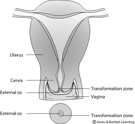
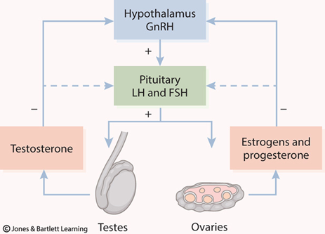
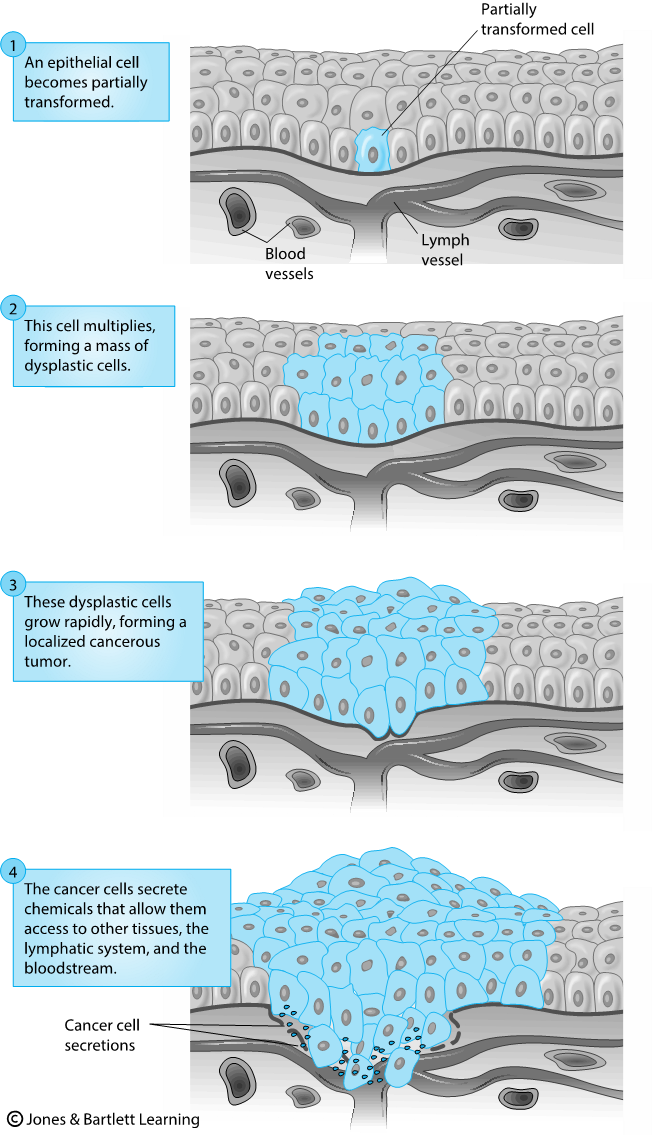
N: Not otherwise classified 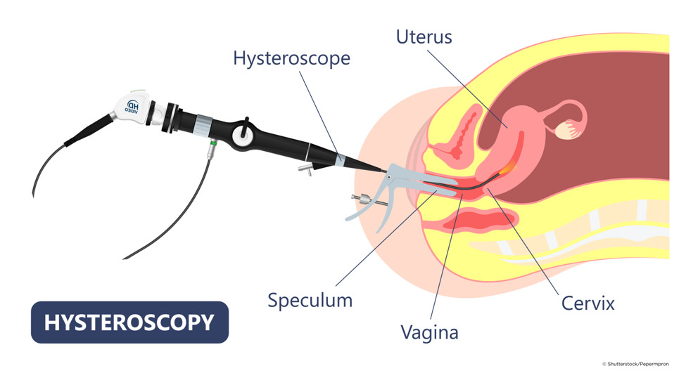
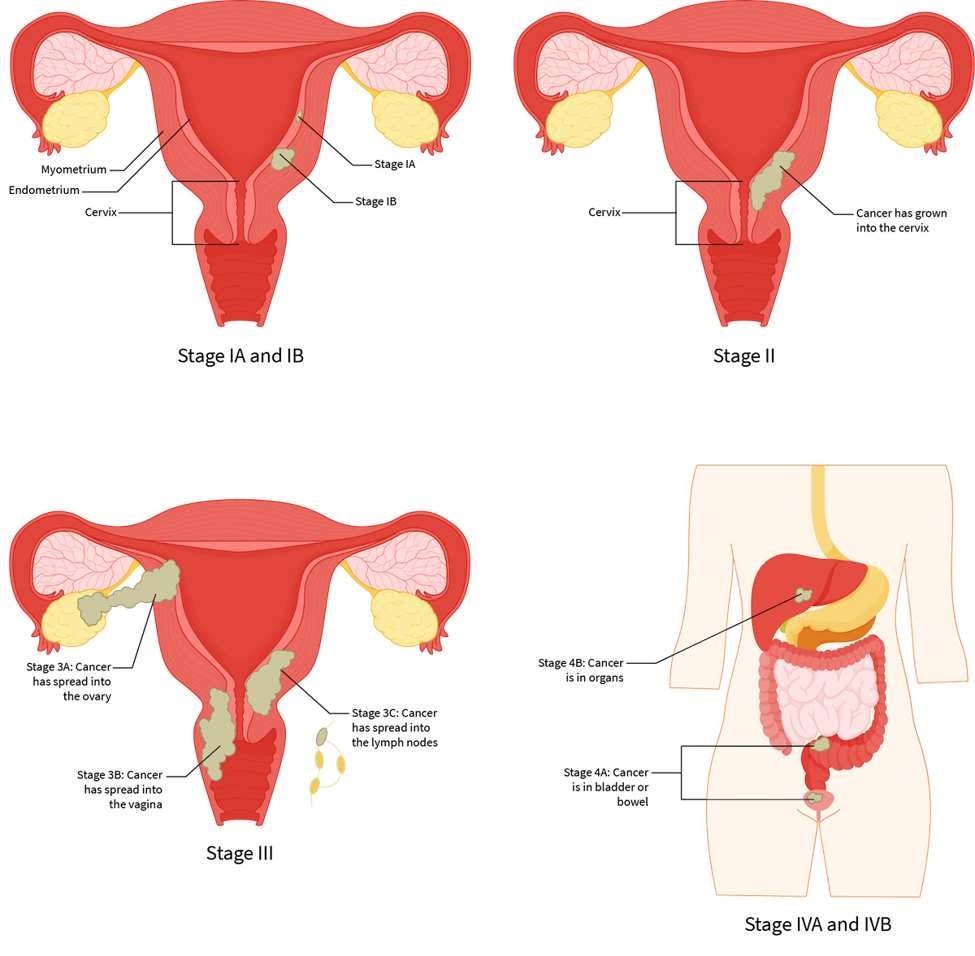
Single Course Cost: $19.00
Add to CartSave while Futhering your Education
Endometrial (Uterine) Cancer Nursing CE Course is offered in the packages below.- 69 ANCC Hours
- 24 Courses
- 554 ANCC Hours
- 196 Courses