About this course:
This module provides an overview of ovarian cancer, including its risk factors, signs and symptoms, and treatment options, to help diagnose females as early as possible and provide optimal care and support throughout the disease trajectory. Empowering nurses with the right tools and information can significantly impact the clinical outcomes of females affected by this disease.
Course preview
Ovarian Cancer
This module provides an overview of ovarian cancer, including its risk factors, signs and symptoms, and treatment options, to help diagnose females as early as possible and provide optimal care and support throughout the disease trajectory. Empowering nurses with the right tools and information can significantly impact the clinical outcomes of females affected by this disease.
By the completion of this module, the nurse should be able to:
- outline the epidemiology of ovarian cancer in the United States and common risk factors
- review the pathophysiology of ovarian cancer, identify the signs and symptoms of the disease, and discuss ovarian cancer subtypes
- review the diagnostic workup and staging criteria for ovarian cancer
- discuss treatment options, including surgery, chemotherapy, targeted therapy, immunotherapy, and hormonal therapy, and the most common side effects, monitoring, and components of patient education
Ovarian cancer is the second most common gynecologic malignancy in the United States, and it is the second most common cause of death from a female reproductive cancer in the United States, following uterine cancer. Ovarian cancer is a disease in which malignant (i.e., cancerous) cells originate within the ovaries, fallopian tubes, or peritoneum (primary peritoneal cancer [PPC]). These three cancers are grouped into one disease based on their anatomical proximity and similarities in risk factors and medical treatments. In this module, the term “ovarian cancer” is used to describe all three cancers unless otherwise specified. Nurses must be informed about the clinical features of ovarian cancer, as early diagnosis is essential for improving survival rates. Treatment options vary depending on the stage of the disease and the patients’ overall health. Nurses support patients and their families throughout the disease trajectory, providing education about the disease, treatment, and side effects and offering emotional support (Centers for Disease Control and Prevention [CDC], 2024; Chen & Berek, 2026a; National Comprehensive Cancer Network [NCCN], 2026b).
Epidemiology
According to the American Cancer Society (ACS, 2026a), there will be an estimated 21,010 new cases and 12,450 deaths from ovarian cancer this year, a decline from 2020 estimates of 21,750 new cases and 13,940 deaths. Although ovarian cancer is uncommon, accounting for only 1% of all new cancer diagnoses in the United States, it ranks fifth among cancer-related deaths in females and is the most lethal gynecological malignancy (Chen & Berek, 2026a; Liu et al., 2025b). Based on data from the National Cancer Institute’s (NCI, n.d.) in the Surveillance, Epidemiology, and End Results Program (SEER), the median age at diagnosis is 63; it is most frequently diagnosed in females aged 55 to 74 (23.9%). The average lifetime risk for developing ovarian cancer among the general US population is roughly 1 in 91, and the lifetime risk of dying is around 1 in 143 (ACS, 2026a). The median age at death is 71 years, with the highest percentage of deaths among females aged 65 to 74 (29.7%), followed by those aged 75 to 84 (26.7%). The 5-year overall survival rate for ovarian cancer is 52%, which drops to 31.5% for patients with distant metastases (i.e., cancer spread) at diagnosis (NCI, n.d.).
Incidence rates are highest for non-Hispanic American Indian/Alaska Native females (11.3 per 100,000), followed by non-Hispanic White and Hispanic (10.5 per 100,000), and non-Hispanic Asian/Pacific Islander (9.8 per 100,000). Based on the latest age-adjusted data from 2019 to 2023, Black females have the lowest incidence rate (9.4 per 100,000). Statistical analyses demonstrate declining new ovarian cancer diagnoses and deaths over the last decade. Between 2014 and 2023, the age-adjusted rates for new ovarian cancer diagnoses decreased on average by 1.4% each year, and death rates fell by 2.9% per year between 2015 and 2024. Non-Hispanic American Indian/Alaska Native females have the highest mortality (6.4 per 100,000), followed by non-Hispanic White females (6.0 per 100,000; NCI, n.d.).
Risk and Protective Factors
Although ovarian cancer is rare, all females are at risk, and the risk increases with age. Hormonal, environmental, and genetic risk factors all contribute to the development of the condition. Although most females are diagnosed without identifiable risk factors, several influences can increase or decrease a person’s risk of developing the disease. Table 1 describes the major risk and preventative factors (Chen & Berek, 2026a, 2026b).
Table 1
Ovarian Cancer Risk and Protective Factors
Risk Factors Anything that increases the chance of developing ovarian cancer | |
Factor | Description of Evidence |
Advancing age | Over 50% of all ovarian cancers are diagnosed in females aged 55 and older. |
Family history Refer to the next section on the inherited risk of ovarian cancer | ...purchase below to continue the course |
Pregnancy
| Nulliparity (i.e., those who have never carried a pregnancy to term) is one of the strongest risk factors for ovarian cancer since these females tend to have more ovulatory cycles. Females who had their first full-term pregnancy after age 35 are also at increased risk. |
Medical history | A medical history of endometriosis (i.e., a condition in which tissue from the lining of the uterus grows on other pelvic organs such as the ovaries or fallopian tubes) or breast cancer is associated with an increased risk of ovarian cancer. |
In vitro fertilization (IVF) | Although infertility treatments (particularly IVF) are linked to an increased risk for ovarian tumors known as “borderline” or “low malignant potential,” research has not demonstrated a causal relationship between IVF treatments and ovarian cancer. |
Protective Factors Anything that decreases the chance of developing ovarian cancer | |
Factor | Description of Evidence |
Pregnancy | Females who have their first full-term pregnancy before age 26 have a lower risk for ovarian cancer, which declines with each subsequent pregnancy. |
Breastfeeding | Breastfeeding is associated with a statistically significant reduced risk of invasive ovarian cancer across multiple studies (Chen & Berek, 2026a). Arcieri and colleagues (2026) found in a meta-analysis that breastfeeding was associated with a 40% overall reduced risk when combined with first birth. Breastfeeding’s protective effect was greatest after the first birth and the first six months after birth, with additional modest risk reductions for prolonged durations of breastfeeding. A systematic review of over six million females by Albezreh and colleagues (2026) validated that breastfeeding is protective against ovarian cancer, most significantly in premenopausal females. |
Oral contraceptive pills (OCPs) | Females who have taken OCPs have up to a 50% lower risk for ovarian cancer than females who never took OCPs, and the risk declines with increased duration of use. The protective effects of OCPs can persist for more than 15 years after discontinuation. In contrast, some studies suggest that females taking unopposed estrogen (without progesterone) for ten or more years may have an increased risk of ovarian cancer. The risk reduction for ovarian cancer with OCP use also applies to females with inherited variations in BRCA1 or BRCA2 genes. |
Prophylactic (preventative) surgery | A hysterectomy (i.e., uterus removal) reduces ovarian cancer risk by about one-third (33%). Risk-reducing salpingo-oophorectomy (RRSO) involves removing both ovaries and fallopian tubes. Research has demonstrated that RRSO in high-risk females (i.e., usually those with BRCA1 or BRCA2 gene variations) effectively lowers the risk of ovarian and fallopian tube cancers and death. RRSO can reduce the risk of BRCA-related ovarian cancer by 80% and all-cause mortality reduction of 68%. In premenopausal females with BRCA2, RRSO can reduce the risk of estrogen-driven breast cancers, but the same protective effect has not been demonstrated in BRCA1 carriers. RRSO does not reduce the risk of primary peritoneal cancer, although the risk is minimal. |
(ACS, 2026a, 2026b; Albezreh et al., 2026; Arshadi et al., 2024; CDC, 2025; Facing Our Risk of Cancer Empowered [FORCE], n.d.; Isaacs & Peshkin, 2026; Norquist, 2023b, 2026)
Inherited Risk
BRCA Variations
Approximately 15% to 20% of ovarian cancers are part of familial cancer syndromes resulting from inherited variations in specific genes. Approximately 7% of females with two or more affected first- or second-degree relatives with ovarian cancer will have a hereditary cancer syndrome (Isaacs & Peshkin, 2026; Peshkin & Isaacs, 2026a). Variations in the BRCA1 and BRCA2 (BRCA1/2) genes commonly lead to hereditary breast and ovarian cancer syndrome (HBOC), characterized by a heightened lifetime risk of developing breast and ovarian cancer. Under healthy conditions, the BRCA1/2 genes function as tumor suppressors, promoting the normal growth, development, and division of various cells in the body. When these genes are mutated, growth processes are unregulated, increasing the propensity toward cancer development. Variations in the BRCA1/2 genes are inherited in an autosomal dominant pattern; having one variant copy in each cell is sufficient to increase the risk of developing cancer. Although ovarian cancer is a sex-dependent disease, the altered gene can be inherited from either sex parent. Each child of a parent with a BRCA1 or BRCA2 variant has a 50% chance of inheriting the same variant (CDC, 2025, 2023c; Isaacs & Peshkin, 2026).
Approximately 1 in 400 females in the United States inherit a BRCA gene variant. Variations in BRCA1/2 are about 10 times more common in females of Ashkenazi Jewish descent than in the general US population. Roughly 15% of ovarian cancers are attributed to inherited variations in the BRCA1/2 genes. Patients with BRCA1/2 variations are more likely to be diagnosed with high-grade serous ovarian cancer than other histologic subtypes. Approximately 44% (BRCA1) and 17% (BRCA2) of females with an inherited BRCA1 or BRCA2 variation will be diagnosed with ovarian cancer by age 80. Females with BRCA1 variations tend to be diagnosed with ovarian cancer up to a decade younger than females with ovarian cancer not associated with a known genetic variation. By comparison, about 1.1% of females will be diagnosed with ovarian cancer sometime during their lifetime (CDC, 2024, 2025; Chen & Berek, 2026a; Isaacs & Peshkin, 2026; PDQ Screening and Prevention Editorial Board, 2025; NCI, n.d., 2025).
Hereditary Nonpolyposis Colorectal Cancer
Hereditary Nonpolyposis Colorectal Cancer (HNPCC), or Lynch syndrome, is known for its high-risk association with colorectal cancer; however, it also correlates with an increased risk of ovarian and endometrial (uterine) cancers. The lifetime risk of ovarian cancer in females with HNPCC is up to 13.4%, depending on genotype (e.g., MLH1, MSH2, MSH6, PMS2). The age at diagnosis is also less than in those with HNPCC, around 47 to 49 years, in contrast to 60 years in those without HNPCC. Although changes in the MLH1, MSH2, MSH6, or PMS2 genes are the most common culprits, many different genes can cause this syndrome. Under physiologic conditions, these genes repair potential errors during DNA replication (i.e., the process by which DNA is copied in preparation for cell division); collectively, they are known as the mismatch repair (MMR) genes. Since variations in any of these genes impede the cells’ ability to repair DNA replication errors, abnormal cells continue to divide. Over time, accumulated DNA replication errors lead to uncontrolled cell growth and the development of malignant tumors (Lu & Schmeler, 2025; US National Library of Medicine [NLM], 2021; Peshkin & Isaacs, 2026b). MutY DNA glycosylase (MUTYH)-associated polyposis (MAP) is a disorder caused by a variation in the MUTYH gene, which prevents cells from fixing DNA errors. As these errors accumulate, more polyps develop, increasing the likelihood of cancer. Pathogenic variations in the MUTYH gene are associated with a 6% to 14% lifetime risk for ovarian cancer with a median age at diagnosis of 51 years (Nielson et al., 2021). Additional genes linked to ovarian cancer with evolving clinical evidence include ATM, PTEN, STK11, PALB2, MSH2, MLH1, MSH6, PMS2, RAD51C, and RAD51D (Lu & Schmeler, 2025; Peshkin & Isaacs, 2026b).
Tumor Protein p53 (TP53)
Somatic (or acquired) variations occur during a person’s lifetime due to environmental exposures, such as sun ultraviolet (UV) radiation, free radicals, carcinogen exposure, ionizing background radiation, and chemical exposure. Somatic variations only occur in specific cells in the body. Unlike inherited variations, these changes do not appear in families and are not passed down from generation to generation. TP53 gene variations are common in ovarian cancer, occurring in at least half of diagnoses. Under healthy conditions, TP53 functions as a tumor suppressor gene, regulating cellular growth and division by preventing cells from growing and dividing too quickly or uncontrollably. TP53 prevents cells with mutated or damaged DNA from dividing, thereby preventing tumor formation. Variations in TP53 impair its ability to control cell proliferation, as it cannot trigger apoptosis (i.e., programmed cell death) in cells with mutated or damaged DNA. Consequently, DNA damage accumulates, cells divide uncontrollably, and tumors form (Liu et al., 2025b; NLM, 2020).
Pathophysiology
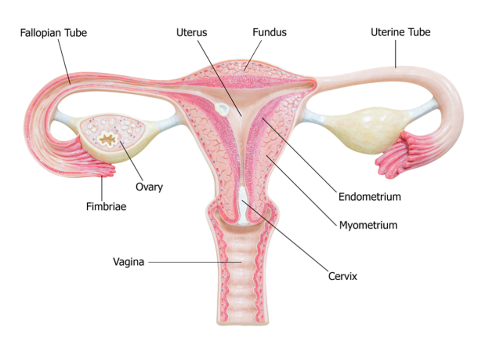
The ovaries and fallopian tubes are primary components of the female reproductive system. The ovaries, or female gonads, are a pair of organs located in the pelvis in a region called the ovarian fossa. The ovaries appear on either side of the uterus, which is the hollow, pear-shaped organ that carries a fetus (i.e., the womb). The ovaries are attached to the uterus by the ovarian ligament, which is embedded within the broad ligament. Figure 1 demonstrates the anatomical structures and positioning of the female reproductive system (Rogers & Brashers, 2023).
The ovaries serve two primary functions: the secretion of female sex hormones and the development and release of the female ova (i.e., eggs). Oocytes are immature egg cells that develop and mature within the ovarian follicles. The ovaries comprise three major cell types: epithelial, germ, and stromal. The tunica albuginea is a thin layer of dense connective tissue covering the ovary’s outermost aspect. The cortex appears directly beneath the tunica albuginea and consists of a framework of tissues called the ovarian stroma. The cortex forms the bulk of the ovary and nurtures the follicles at various stages of maturation. The medulla is the middle layer of the ovary; it comprises loose connective tissues, elastic fibers, and neurovascular structures, including the lymph and blood vessels. The hilum is the ovary’s innermost layer and provides the entry point for the blood vessels and nerves (Casanova et al., 2024; Rogers & Brashers, 2023).
Figure 1
Female Reproductive System
(iStock photo ID: 538949875)
The hypothalamic-pituitary-gonadal (HPG) axis is a tightly regulated feedback system that controls the production of female sex hormones and reproduction. The HPG forms a complex interplay between the organs of the female endocrine system, including the hypothalamus, pituitary gland, and ovaries. During puberty, the hypothalamus releases increasing levels of gonadotropin-releasing hormone (GnRH). In response, the anterior pituitary produces follicle-stimulating hormone (FSH) and luteinizing hormone (LH), prompting the ovaries to secrete increasing amounts of sex hormones, primarily estrogen and progesterone, along with a smaller quantity of testosterone (Casanova et al., 2024; Rogers & Brashers, 2023).
Ovarian follicles are tiny fluid-filled sacs composed of granulosa cells, theca cells, and oocytes, varying according to each oocyte’s maturation stage. Folliculogenesis (i.e., the maturation of ovarian follicles) involves several growth factors and receptors. This process primarily relies on the HPG axis, as coordinated interactions between granulosa and theca cells are necessary for estrogen synthesis. Granulosa cells form alongside the maturing follicle. FSH has a high affinity for granulosa cells, promoting follicle growth and maturation by stimulating the synthesis of androgens (e.g., testosterone) and estrogens. Theca cells are connective tissue cells and blood vessels surrounding the ovarian follicle. They produce estradiol from androgens in response to LH. Estradiol is an estrogen steroid hormone that binds to tissues in the body, serving as the body’s primary estrogen source during reproductive years. Estradiol influences secondary sex characteristics, including breast growth, body shape, muscle mass, fat deposition, body hair, and bone health. It also regulates the menstrual cycle and serves a vital role in pregnancy. When the maturation process is complete, the oocyte becomes an ovum. The pituitary gland releases a surge of LH, stimulating the follicle’s rupture and ova release (Casanova et al., 2024; Holesh et al., 2023).
The ovaries typically alternate, releasing a single mature egg each month; however, if an ovary is dysfunctional or absent, the other ovary will compensate. The ovaries change in size and appearance throughout a person’s lifetime. Their size and appearance depend on the individual’s menstrual status. Menopause denotes menstruation’s permanent cessation in females with an intact uterus, defined as 12 consecutive months without a menstrual cycle (Holesh et al., 2023). According to the Office on Women’s Health (OWH), the average age for menopause in the United States is 52 years. In premenopausal females (i.e., females of reproductive age who are menstruating), each ovary is comparable in size and shape to an almond, measuring approximately 0.8 inch (2 cm) wide and 1.4 inch (3.5 cm) long. Normal ovarian volume peaks at 0.3 oz (7.7 ml) around age 20 and then gradually decreases to approximately 0.1 oz (2.8 ml) in menopause. During menopause, reproductive hormone levels decline, egg production ceases, and the ovaries shrink as they undergo progressive involution (Gibson & Mahdy, 2023; Holesh et al., 2023; OWH, 2026).
The fallopian tubes are a pair of long, slim conduits on either side of the uterus that transport eggs from the ovaries to the uterus. The tubes attach to the uterus and curve up and over the ovaries, as shown in Figure 1. Each tube is 4.3 to 4.7 inch (11 to 12 cm) long, and the lumen is 0.04 inch (1 mm) in diameter; it flares into a bell shape at the ovarian end, called the infundibulum. The infundibulum contains fimbriae, which are swaying fringes that create a current to draw the ova into them. Once the ova enter the fallopian tube, cilia and peristalsis gradually propel them toward the uterus. The omentum is a layer of adipose (fatty) tissue that covers the intestines like an apron and is subdivided into the greater omentum (which drapes over the stomach) and the lesser omentum (which drapes over the liver). The peritoneum is a cellophane-like, moist sheet of tissue covering the surface of the organs in the abdomen and pelvis. It supports and protects the organs and consists of two layers: the visceral peritoneum and the parietal peritoneum. The visceral peritoneum is the inner layer of tissue lining the organs. The parietal peritoneum is the outer layer that adheres to the abdominal cavity wall. The space between the layers is called the peritoneal cavity, which contains a small amount of lubricating fluid (Han & Sadiq, 2023; Rogers & Brashers, 2023).
Ovarian Cancer
Many types of ovarian cancer initially develop in the distal portions of the fallopian tubes and spread to the ovary. Emerging research has found that the fallopian tubes may be the source of most hereditary ovarian cancers. PPC appears to start in the cells lining the fallopian tubes, spreading to the ovaries (ACS, 2025c). Ovarian cancer is an aggressive disease characterized by rapid growth, disseminated metastasis, and malignant ascites. Ascites is the abnormal buildup of fluid within the abdominal or pelvic cavity. Without an anatomic barrier surrounding the primary tumor, cancer cells easily infiltrate the peritoneal cavity, making the peritoneum and omentum the most common sites of initial metastases. Metastases often occur by direct extension as the cancer penetrates and ruptures the ovarian capsule, permitting invasion into surrounding structures. Ovarian cancer cells can survive after detaching from the primary tumor by forming multicellular spheroids. The spheroids can float in ascitic fluid, making the intraperitoneal cavity a predominant metastasis site. The continuous circulation of free peritoneal fluid facilitates the widespread dissemination of cancer cells, a process known as peritoneal seeding. As the disease advances, a higher volume of ascites accumulates, causing cancer to spread to distant sites in the abdomen (Boo et al., 2025; Casanova et al., 2024; Chen & Berek, 2026a). Ovarian cancer cells can also travel through the lymphatics, most commonly the pelvic or aortic lymph nodes. Some of the most common sites for distant metastases include the liver and pleura (i.e., the serous membrane lining the lungs; Chen & Berek, 2026a, 2026b; Ovarian Cancer Research Alliance [OCRA], 2021). Although PPC tends to spread along the surfaces of the pelvic and abdominal cavities, the precise origin is difficult to determine (ACS, 2025c; CDC, 2024).
Classification of Ovarian Cancer
Epithelial Ovarian Cancer
Ovarian cancer is classified as originating from epithelial, germ, or sex cord stromal cells, with 95% of cases being epithelial. Epithelial tumors are further divided into subtypes: high-grade serous, low-grade serous, endometrioid, mucinous, and clear cell (Table 2; Chen & Berek, 2025; National Ovarian Cancer Coalition [NOCC], 2023).
Table 2
Epithelial Ovarian Cancer Subtypes
Histologic Subtype | Clinical Features |
High-grade serous carcinoma (HGSC) |
|
Low-grade serous carcinoma (LGSC) |
|
Endometrioid carcinoma |
|
Clear cell carcinoma (CCC) |
|
Mucinous carcinoma |
|
(Chen & Berek, 2025; Köbel et al., 2023; Lewis et al., 2026; NOCC, 2023)
Less common types of ovarian cancer include germ cell tumors, sex-cord stromal cell tumors (SCSCTs), and small cell carcinoma of the ovary (Chen & Berek, 2025; Liu et al., 2025a)
Germ Cell Tumors
Germ cell tumors arise from cells that form ova, and most cases are benign. Mature teratomas are the most common type of benign germ cell tumors and are usually treated surgically. Malignant ovarian germ cell tumors only account for about 5% of ovarian cancers. Unlike epithelial ovarian cancers, these characteristically occur in adolescents and premenopausal females. They are often confined to an ovary and are usually curable. Immature teratomas are malignant tumors that primarily develop in females under 18. These can metastasize to other areas in the body. Immature teratomas are the most common type of malignant germ cell tumors. They usually affect females under 30, are cured with surgery and adjuvant chemotherapy, and have a long-term cure rate of more than 90% (ACS, 2025c; Chen & Berek, 2025; Gershenson, 2026a, 2026c; OCRA, n.d.).
Sex-Cord Stromal Cell Tumors
Ovarian SCSCTs are rare, accounting for under 8% of all malignant ovarian cancers. These tumors arise from stromal cells that support the ovary and produce female sex hormones. The most common subtype is the granulosa cell tumor, comprising 90% of SCSCTs. Patients with stromal tumors typically show signs of excess hormone production, like hirsutism (i.e., coarse hair growth on unexpected body areas such as the face or chest) and virilization. Virilization is a condition in which a female develops features of increased masculinization. Some of the most common symptoms include hair loss, acne, deepening of the voice, increased libido (i.e., sex drive), decreased size of the breasts, and menstrual irregularities, including cessation of menses (ACS, 2025c; Gershenson, 2026b; OCRA, n.d.).
Small Cell Carcinoma of the Ovary
Small cell carcinoma of the ovary (SCCO) is a rare, aggressive type of ovarian cancer, accounting for 0.1% of cases. It typically affects young females, with a median age of diagnosis of 23 years. The five-year survival rate is approximately 30% with a median survival time of 13 months (Liu et al., 2025a; OCRA, n.d.). Approximately 70% of patients have hypercalcemia and associated complications. Hypercalcemia of malignancy (HCM) is a complex metabolic disorder and oncologic emergency caused by bone destruction and increased renal calcium absorption. Three primary mechanisms cause HCM:
- excess secretion of parathyroid hormone-related protein
- bone metastases release osteoclast-activating factors, causing an imbalance between bone formation and bone resorption, leading to the excess release of calcium into the bloodstream
- overproduction of 1,25-dihydroxy vitamin D (Calcitriol; Horwitz, 2026)
HCM most commonly occurs in breast cancer, squamous cell lung cancer, and multiple myeloma. Symptoms can be vague, ranging from fatigue, anorexia, nausea, vomiting, constipation, polyuria, polydipsia, and lethargy to confusion, cognitive dysfunction, kidney failure, and cardiac arrhythmias. Treatment includes initial aggressive fluid administration, loop diuretics, and bisphosphonate therapy. Bisphosphonates are bone-modifying agents that lower the patient’s calcium level by inhibiting osteoclasts and stabilizing the bone matrix by binding to calcium phosphate. Following initial treatment, identifying and treating the underlying malignancy is critical for controlling the condition and preventing its recurrence (Dickens et al., 2022; Horwitz, 2026; Rothberg et al., 2022; Shane & Berenson, 2026).
Signs and Symptoms
Only approximately 20% of ovarian cancers are detected in the early stages. Due to the ovaries’ deep anatomical location within the pelvic cavity, diagnosing the condition at an early stage remains challenging. An ovarian tumor often grows undetected until it becomes large enough to cause symptoms or be detected on a pelvic exam. Most patients with early-stage ovarian cancer are asymptomatic. When symptoms do occur, they are often ambiguous and nonspecific, such as abdominal pain, bloating, pelvic pressure, low back discomfort, constipation, and fatigue. Symptoms are more common in advanced disease due to ascites and pressure on the bladder or rectum. These processes can lead to persistent abdominal pain and distention, heartburn, early satiety, nausea, weight loss, urinary frequency, menstrual cycle irregularities, and dyspareunia (i.e., pain with intercourse). In some females, a palpable mass may be present. The persistence of symptoms is crucial, and nurses should consider ovarian cancer in the differential diagnosis for any patient who reports the above symptoms for longer than 2 weeks (Boo et al., 2025; Chen & Berek, 2025; Goff, 2026; NOCC, n.d.).
Diagnostic Workup
Adnexa denote the region adjoining the uterus and include the ovary, fallopian tube, and associated structures (i.e., blood vessels, ligaments, and connective tissue). Masses in the adnexa may be symptomatic or discovered incidentally. Most are benign, some will regress spontaneously, and others require surgical removal. Some differential diagnoses for an adnexal mass include an ovarian cyst, ectopic pregnancy, abscess, uterine leiomyoma, or ovarian tumor (benign or malignant). Transvaginal ultrasound (TVUS) and serum cancer antigen-125 (CA-125) blood tests are often used to evaluate adnexal masses (Chen & Berek, 2025; Goff, 2026).
CA-125
CA-125 is a tumor marker for most ovarian cancers. Tumor markers are substances or proteins produced by cancer or by the body’s response to cancer’s presence, but they are also made in smaller quantities by healthy cells. Tumor markers can be detected in the blood or, less commonly, in the urine. Although they are considered nonspecific and not of significant value in isolation, they can be beneficial for establishing a baseline during the diagnostic workup, evaluating treatment response, and detecting cancer recurrence. Each tumor marker is specific to a distinct disease process (Chen & Berek, 2025, 2026b).
CA-125 is present on the surface of most ovarian cancer cells and can be easily measured in a blood test. However, not all females with ovarian cancer will have elevated CA-125 levels. Healthy tissues and some other cancer types produce small quantities of CA-125. Serum CA-125 is elevated in approximately 85% of females with epithelial ovarian cancer but in less than half of females with early disease. CA-125 may be an effective therapeutic tool in select patients. For example, if CA-125 levels are elevated at diagnosis and decline during treatment, the cancer is responding to the treatment. Following treatment, CA-125 levels may be monitored periodically to detect cancer recurrence (Chen & Berek, 2025; Li, 2026).
CA-125 levels may be elevated in benign conditions, such as endometriosis, uterine fibroids, pregnancy, infection, pelvic inflammatory disease, colitis, pancreatitis, cirrhosis, pericarditis, and systemic lupus erythematosus. CA-125 levels can also fluctuate with menstrual cycles (Carlson, 2026). According to the American Board of Internal Medicine (2026), the normal CA-125 level is below 35 U/mL. Although CA-125 is the most common and sensitive tumor marker for ovarian cancers, other tumor markers such as human chorionic gonadotropin (hCG), lactate dehydrogenase (LDH), or alpha-fetoprotein (AFP) may be useful for less common types of ovarian tumors (Chen & Berek, 2025).
Imaging
Ultrasound is a safe, noninvasive imaging modality that uses sound waves to generate images of internal body structures. Ultrasound does not use x-rays or ionizing radiation and is ordered for numerous diagnostic and therapeutic indications. Ultrasound images are obtained by placing a transducer (i.e., a small probe) on the skin and applying a layer of gel. The transducer produces sound waves at high frequencies, which exceed the threshold of human hearing. These high-frequency sound waves travel from the transducer through the gel into the body, generating images on a computer. Images are captured in real time, enabling evaluation of the structure and movement of the body’s internal organs. TVUS assesses the uterus, fallopian tubes, and ovaries by placing a transducer directly into the vaginal canal, thereby optimizing image quality due to its proximity to the ovaries. While TVUS can help identify a mass in or surrounding the ovary, it cannot indicate whether it is malignant or benign (National Institute of Biomedical Imaging and Bioengineering, 2023).
If the TVUS and the CA-125 demonstrate abnormalities, the NCCN (2026b) recommends a CT scan of the abdomen and pelvis—with contrast media unless contraindicated—to evaluate the extent of the disease and for signs of metastases. CT scans use a series of radiographs and computer technology to create cross-sections of the inside of the body, including bones, blood vessels, organs, and soft tissues. The x-ray beam circles the subject to generate various views of the identified structure, forming a complete picture in much greater detail than a conventional radiograph. CT scans use ionizing radiation to produce images, so patients should be counseled about the radiation exposure (International Atomic Energy Agency [IAEA], n.d.; Mafraji, 2025). For example, a CT scan of the abdomen and pelvis with IV contrast delivers a radiation dose of 15.4 millisieverts (mSv), which is approximately equivalent to 5.1 years of natural background radiation exposure (American College of Radiology, n.d.). CT scans are commonly used in the care of oncologic patients to evaluate treatment response and monitor for cancer progression or recurrence. Patients should be advised to lie flat and remain motionless on a table that slides into the scanning machine and resembles a large doughnut. The x-ray tube will rotate around the patient to capture all the necessary images. The test takes about 10 to 15 min to complete (IAEA, n.d.).
Paracentesis
Malignant ascites in the abdominal cavity is a classic feature of advanced ovarian cancer. Ascites can be detected on clinical exams and radiological imaging. It typically causes significant symptoms such as shortness of breath, abdominal distension, heartburn, bloating, nausea, anorexia, and early satiety. A paracentesis is an invasive procedure performed to provide short-term relief from symptoms. A needle is inserted into the abdominal cavity to drain the fluid into a collection bag. The fluid sample undergoes morphological assessment by a pathologist to evaluate for the presence of malignant cells and features of cancer. Sometimes, patients are diagnosed with ovarian cancer based on the cytology findings from a paracentesis (Boo et al., 2025).
The NCCN (2026b) ovarian cancer guidelines also support the following components of a diagnostic workup:
- MRI scan or positron emission tomography—CT (PET/CT) scan imaging may be indicated for indeterminate lesions if the results will alter the management
- chest CT or chest radiograph as clinically indicated
- CBC, chemistry profile, and liver function test (LFT)
- referral to a gynecologic oncologist for clinically suspicious lesions
- genetic risk evaluation, including germline testing, for all patients with ovarian cancer, fallopian tube cancer, or PPC
Ovarian Cancer Staging
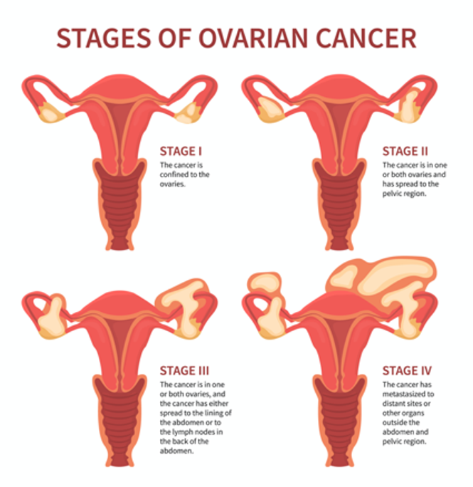
The cancer stage at diagnosis guides treatment options and strongly influences overall survival. There are detailed staging systems for ovarian cancer: the International Federation of Gynecology and Obstetrics (FIGO) system and the American Joint Committee on Cancer’s (AJCC) Tumor, Node, Metastasis (TNM) staging system, 8th edition. Both systems are essentially the same and describe specific characteristics to assign stages I through IV, as outlined in Table 3 and demonstrated in Figure 2. Since ovarian cancer is a surgically staged disease, the FIGO system has been adopted universally, and all patients with suspected ovarian malignancies should be promptly referred to an oncological gynecologist for surgical evaluation (AJCC, 2022; Berek et al., 2021; Bhatla & Denny, 2018; NCCN, 2026b; Olawaiye, n.d.; Renz et al., 2025).
Figure 2
Stages of Ovarian Cancer
(iStock illustration ID: 1249232483)
Cancer staging reflects the cell type, tumor grade, tumor location, and extent of malignancy. Within the TNM staging system, T denotes the size of the tumor and whether it has grown into nearby tissue, N refers to the presence of cancer in the lymph nodes, and M indicates if cancer has metastasized to other parts of the body beyond the origin site. Tumor grade measures how different the cancer cells look compared to healthy cells under the microscope. It is based on cell differentiation and varies from low grade (i.e., grade 1) to high grade (i.e., grade 3). Grade 1 is well differentiated and appears similar to healthy cells, whereas grade 3 is poorly differentiated (i.e., does not resemble healthy cells) and is most aggressive. The 5-year survival rate for FIGO stage 1 carcinoma of the ovary is 89%, whereas it decreases to only 20% when diagnosed in stage IV (AJCC, 2022; Bhatla & Denny, 2018; Goff, 2026; Olawaiye, n.d.; Sanchez et al., 2025).
Table 3
Cancer Staging
American Joint Committee on Cancer’s (AJCC) Tumor, Node, Metastasis (TNM) | International Federation of Gynecology and Obstetrics (FIGO) Stage | Description |
TX | — | primary tumor cannot be assessed |
T0 | — | no evidence of primary tumor |
T1 | I | tumor limited to one or both ovaries or fallopian tube(s) |
T1a | IA | tumor limited to one ovary (capsule intact) or fallopian tube; no tumor on ovarian or fallopian tube surface; no malignant cells in ascites or peritoneal washings |
T1b | IB | tumor limited to both ovaries (capsules intact) or fallopian tubes; no tumor on ovarian or fallopian tube surface; no malignant cells in ascites or peritoneal washings |
T1c
T1c1 T1c2 T1c3 | IC | tumor limited to one or both ovaries or fallopian tubes, with any of the following:
|
T2 | II | tumor limited to the pelvis; involves one or both ovaries or fallopian tubes with pelvic extension below the pelvic brim or primary peritoneal cancer (PPC) |
T2a | IIA | extension and/or implants on the uterus and/or fallopian tube(s) and/or ovaries |
T2b | IIB | extension to and/or implants on other pelvic tissues |
T3 | III | tumor involves one or both ovaries or fallopian tubes, or PPC, with microscopically confirmed peritoneal metastasis outside the pelvis and/or metastasis to the retroperitoneal (pelvic and/or para-aortic) lymph nodes |
T3a | IIIA2 | microscopic extra-pelvic (above the pelvic brim) peritoneal involvement with or without positive retroperitoneal lymph nodes |
T3b | IIIB | macroscopic peritoneal metastasis beyond the pelvis that is ≤2 cm in greatest dimension with or without metastasis to the retroperitoneal lymph nodes |
T3c | IIIC | macroscopic peritoneal metastasis beyond the pelvis that is >2 cm in the greatest dimension with or without metastasis to the retroperitoneal lymph nodes (includes an extension of the tumor to the capsule of the liver and spleen without parenchymal involvement of either organ) |
M1 | IV | distant metastasis, including pleural effusion with positive cytology; liver or splenic parenchymal metastasis; metastasis to extra-abdominal organs (including inguinal lymph nodes and lymph nodes outside the abdominal cavity); and transmural involvement of intestine |
| IVA | pleural effusion with positive cytology |
| IVB | liver or splenic parenchymal metastases; metastases to extra-abdominal organs (including the inguinal lymph nodes and lymph nodes outside the abdominal cavity); transmural involvement of the intestine |
(AJCC, 2022; Berek et al., 2021; Bhatla & Denny, 2018; Olawaiye, n.d.; Renz et al., 2025)
Cytoreduction
Research data demonstrate that primary assessment and cytoreduction (i.e., surgical debulking) by a gynecological oncologist leads to a survival advantage. Cytoreduction is a surgical procedure to remove, reduce, or eliminate cancerous tumors and should be performed by a trained gynecological oncologist. Some tumors may be removed entirely, whereas others may be reduced in size. Unlike other cancers, which are usually biopsied before surgery, ovarian cancers are typically staged during surgery. The surgical intervention provides tissue sampling for a definitive diagnosis and allows the surgeon to assess the extent of the disease during the cytoreductive procedure. Cytoreduction improves the effectiveness of subsequent chemotherapy and radiation; it is vital for females whose ovarian cancer has spread significantly throughout the abdomen at diagnosis. Optimal cytoreduction is defined as no residual cancer greater than 0.4 inch (1 cm) remaining after surgery. Studies have demonstrated survival is highest among females with no or only a small volume of residual disease, defined as less than 0.2 inch (0.5 cm; NCCN, 2026b; Salani & Cosgrove, 2026).
Treatment Modalities
Treatment for ovarian cancer is usually multifactorial, involving combined modalities, and primarily depends on the cancer stage. The NCCN (2026b) provides evidence-based treatment guidelines for ovarian cancer based on histopathological findings, genetics, staging, inheritance patterns, and other specific features, supported by rigorous clinical trial research, data compiled across institutions, and annual expert panel reviews. These guidelines are widely utilized in cancer care and guiding medical decision-making throughout the patient’s disease trajectory. Due to the aggressive biology of ovarian cancer, treatment is chronic, and most patients experience periods of remission and relapse. Many require maintenance therapy; all patients need close monitoring and surveillance after treatment completion to monitor for recurrence. This section will provide an overview of the most common treatment strategies (CDC, 2026; NCCN, 2026b).
Surgery
Surgery is the mainstay of treatment for most ovarian cancers. When ovarian cancer is diagnosed early, standard therapy includes surgical staging, cytoreduction, hysterectomy, and bilateral salpingo-oophorectomy (BSO). The procedure may or may not involve an omentectomy (i.e., removal of all or part of the omentum) and lymphadenectomy (i.e., removal of at least one lymph node to evaluate for cancer). Since most patients present with advanced disease at diagnosis, surgery is often followed by adjuvant (postsurgical) systemic therapy (Herzog & Armstrong, 2026; NCCN, 2026b).
Chemotherapy
Chemotherapy, cytotoxic or antineoplastic therapy, refers to high-risk, hazardous medications that destroy cancer cells throughout the body. Chemotherapy interferes with the normal cell cycle, impairing DNA synthesis and cell replication to prevent cancer cells from dividing and multiplying. Most chemotherapy agents attack broadly (i.e., they kill normal, healthy cells in the body together with the cancer cells). Chemotherapy is classified according to biochemical activity, mechanism of action, and phase of action during the cell cycle. Chemotherapy agents are divided into two categories: cell cycle-specific (CCS) and cell cycle-nonspecific (CCNS). CCS drugs exert cytotoxic effects on actively dividing cells at specific stages of the cell cycle. These drugs do not act against cancer cells during the resting phase (G0) and are schedule-dependent, so they are most effective when administered in divided doses or by continuous infusion. CCNS drugs have a broader impact on cancer cells, as they affect cells at any phase in the cell cycle, including G0. These agents are considered dose-dependent and are most effective when administered by bolus doses, as the number of cells affected is directly proportional to the amount of drug given (Konstantinopoulos & Bristow, 2026; Olsen et al., 2023; Priyadarshini, 2021; Song et al., 2024; Sun et al., 2021).
Chemotherapy is prominent in treating ovarian cancer and can be used at various time points during the disease trajectory. Neoadjuvant chemotherapy intends to reduce the tumor burden in preparation for surgery. Adjuvant chemotherapy (i.e., chemotherapy after surgery) aims to decrease cancer recurrence risk, reduce micro-metastases, and eradicate any remaining cancer cells. Palliative chemotherapy treats recurrent cancer, seeking to relieve or delay cancer symptoms, enhance comfort, reduce symptom burden, and improve quality of life. IV platinum-based chemotherapy agents (i.e., carboplatin [Paraplatin] or cisplatin [Platinol]) are highly effective against ovarian cancer, making them the first-line treatment unless contraindicated. They are typically combined with a mitotic inhibitor such as paclitaxel (Taxol) or docetaxel (Taxotere). Some of the most common chemotherapy agents for ovarian cancer include the following:
- Alkylating agents
- disrupt DNA, prevent DNA replication, and are CCNS
- carboplatin (Paraplatin)
- cisplatin (Platinol)
- ifosfamide (Ifex)
- cyclophosphamide (Cytoxan)
- Mitotic inhibitors
- inhibit cell division and keep enzymes from making the proteins necessary for the reproduction of cells, damaging all phases of cells
- paclitaxel (Taxol)
- docetaxel (Taxotere)
- Antitumor antibiotics
- bind to and unwind the DNA helix and are CCS (i.e., acting on the S-phase)
- doxorubicin (Adriamycin)
- liposomal doxorubicin (Doxil; ACS, 2025d; NCCN, 2026b; Konstantinopoulos & Bristow, 2026; Olsen et al., 2023; Sun et al., 2021)
Chemotherapy Side Effects
The side effects of chemotherapy vary depending on the drug type, dosage, treatment duration, and specific patient factors. Since cancer cells divide rapidly, chemotherapy impacts healthy and cancerous cells that divide quickly (e.g., in the GI tract, skin and hair cells, and bone marrow). As a group, the most common side effects include bone marrow suppression (e.g., anemia, thrombocytopenia, neutropenia), fatigue, nausea, anorexia, alopecia (i.e., hair loss), diarrhea, skin changes, and peripheral neuropathy (i.e., damage to the sensory nerves). Alopecia can cause significant emotional distress for females. Chemotherapy-induced hair loss starts about 2 to 3 weeks following the first chemotherapy dose. It results from damage to the dividing hair matrix cells, which causes the hair shaft to break at the follicular orifice or hair bulb. Although the degree of hair loss depends on the chemotherapy agent, dose, and administration schedule, paclitaxel (Taxol) and docetaxel (Taxotere) are well known for inducing alopecia. Nurses should reassure patients that their hair will begin to regrow within a few weeks after chemotherapy is discontinued, as permanent alopecia following chemotherapy is extremely rare (ACS, 2025a; Gale, 2026; Herzog & Armstrong, 2026; Olsen et al., 2023; Rugo & van den Hurk, 2026).
Chemotherapy-induced peripheral neuropathy (CIPN) is a common side effect of carboplatin (Paraplatin), cisplatin (Platinol), and paclitaxel (Taxol). It is often the dose-limiting toxicity (DLT) of these agents. DLTs are severe toxicities and side effects serious enough to warrant a reduction in the dose or discontinuation of the treatment. CIPN results from the demyelination of sensory and motor axons. Patients experience reduced nerve conduction velocity, leading to loss of deep tendon reflexes, paresthesia (numbness and tingling), weakness, and burning pain. Initially, CIPN often affects the most distal points of the extremities, such as the fingertips and toes, and progresses proximally toward the midline. In severe cases, patients may lose complete sensation in the fingers, hands, toes, and feet; this can cause significant disability, such as the inability to grasp or hold items, and gait disturbance, including imbalance and falls. CIPN is complex, as no single pathophysiologic process explains the neuropathies that occur following exposure to certain chemotherapies. CIPN is dose-dependent and progressive while a patient receives treatment, but can also have a cascading effect after treatment ends. During this cascading phenomenon, symptoms become more prominent after discontinuing the offending agent. Pain, sensory changes, and weakness that manifest during treatment generally lead to chemotherapy dose reductions, changes in treatment protocols, or termination of the therapeutic agent entirely. CIPN is associated with significant morbidity as it negatively impacts quality of life and impairs activities of daily living (Lee, 2026; Lustberg, 2026).
Currently, no medications or supplements are effective in preventing CIPN. Regular exercise, reducing alcohol intake, and treating preexisting medical conditions (e.g., diabetic neuropathy and vitamin B12 deficiency) may reduce the risk of CIPN. Management of CIPN is complex, and effective treatment options are limited. Pharmacologic treatment focuses on symptom relief, although many agents are not highly effective. Over-the-counter pain medications, menthol creams, capsaicin creams, or lidocaine patches may offer comfort. Some patients may be prescribed medications such as gabapentin (Neurontin), an anticonvulsant/anti-epileptic agent that also treats neuropathic pain. Some patients may find relief from selective serotonin-norepinephrine reuptake inhibitors (SNRIs) such as duloxetine (Cymbalta). Patients with CIPN must be counseled on ways to avoid injury (e.g., wearing supportive shoes and having regular foot examinations) and promoting home safety (e.g., using handrails on stairs and removing throw rugs). Patients must also be mindful of water temperatures, as decreased sensitivity to hot water increases their risk of burns when bathing or washing dishes. Improvement in function and symptom resolution often occurs gradually over time, but nerve damage may be permanent (Lee, 2026; Lustberg, 2026; Rogers & Brashers, 2023).
Hypersensitivity Reactions to Chemotherapy
A hypersensitivity reaction (HSR) occurs when the immune system becomes overstimulated by a foreign substance, leading to the production of antibodies that provoke an immune response. HSRs are commonly associated with several chemotherapy agents that are used widely in ovarian cancer treatment, such as paclitaxel (Taxol), docetaxel (Taxotere), and carboplatin (Paraplatin). HSR risk can be reduced by premedicating patients with corticosteroids, antihistamines, and/or acetaminophen (Tylenol). HSRs can occur during the initial chemotherapy infusion or after subsequent administrations of the same agent. Paclitaxel (Taxol) is well-known for its risk of a nearly immediate, acute HSR, whereas carboplatin (Paraplatin) more commonly induces an HSR after several cycles. Most HSRs occur during the first 15 min of the infusion. Initial signs and symptoms can include hives, urticaria, pruritus, swelling, back pain, facial flushing, rhinitis, abdominal cramping, chills, hypotension, and anxiety. Patients may require supplemental oxygen, fluid resuscitation, or other emergency medications as indicated. For life-threatening symptoms like bronchospasm, angioedema (e.g., swelling of the oral cavity, lips, or tongue), or anaphylaxis, epinephrine 0.5 mg (1:10,000 solution for adult patients) may be required (Campbell & Kelso, 2025; Castells et al., 2026; Olsen et al., 2023).
Intraperitoneal Chemotherapy
Chemotherapy for ovarian cancer may also be administered through the intraperitoneal (IP) route. Although IV chemotherapy is the standard of care for most patients with ovarian cancer, a select subset of patients may benefit from IP chemotherapy. IP chemotherapy is given through an implanted catheter and an access port inserted into the peritoneal space. The medication is instilled directly into the port to treat cancer within the peritoneal cavity. Several studies have demonstrated that the frequency and severity of toxic side effects are significantly higher in patients receiving IP chemotherapy. Common side effects include abdominal pressure, bloating, urinary frequency, nausea, vomiting, and anorexia. IP chemotherapy carries a risk of peritonitis (i.e., inflammation of the peritoneum), which can be life-threatening and lead to sepsis. IP chemotherapy is associated with prolonged bone marrow suppression and higher renal toxicity than IV chemotherapy. These factors limit widespread adoption even though IP chemotherapy has been shown to increase rates of survival in patients with ovarian cancer with an immune-hot phenotype (Markman & Olawaiye, 2025; Ogasawara et al., 2024). However, the NCCN (2026b) lists IP chemotherapy as an option for patients with optimally cytoreduced stage II to III disease. Based on expert consensus across various evidence-based guidelines, IP therapy is appropriate for patients with no or minimal residual disease following surgical cytoreduction (Ogasawara et al., 2024).
Targeted Therapy
Targeted therapies are novel treatment modalities that target specific parts of cancer cells to prevent tumor development or shrink existing tumors. Numerous proteins are located on the cellular membranes; these growth factor receptors connect the external and internal cellular environments and are essential for cell growth and development. Targeted agents can block or turn off chemical signals that tell cancer cells to grow and divide. They can also starve the tumor by cutting off its blood supply and preventing the formation of new blood vessels, or by delivering toxins or poisons directly to cancer cells, thereby killing them without harming healthy cells (Xu et al., 2023). Most ovarian cancer research over the last decade has focused on developing targeted drugs that block growth factor receptors and on identifying new targets. These drugs have revolutionized treatment options for patients with advanced and metastatic ovarian cancer by fighting against drug resistance and increasing survival. The next section provides an overview of the most commonly used targeted agents for ovarian cancer (NCCN, 2026b; NOCC, 2025; Xu et al., 2023).
Anti-Angiogenesis Agents
Vascular endothelial growth factor (VEGF) is a signaling protein that stimulates angiogenesis (i.e., new blood vessel formation) in healthy and cancerous cells. Blood vessels carry oxygen and nutrients to the tissue for growth and survival. Tumors need blood vessels to grow and spread. Anti-angiogenesis agents (i.e., VEGF inhibitors) cut off blood supply to cancer cells by interfering with the VEGF receptor; tumors stay small and eventually starve (Kuo, 2025; Olsen et al., 2023; Xu et al., 2023). Bevacizumab (Avastin) is a humanized monoclonal antibody that binds to and inhibits the activity of human VEGF to its receptors. It has tremendously impacted the management of ovarian cancer. Monk and colleagues (2025) purport that females with stage III suboptimal debulked disease and stage IV disease who received bevacizumab (Avastin) had significantly improved PFS and overall survival compared to those who received chemotherapy alone. The NCCN (2026b) added bevacizumab (Avastin) to its upfront chemotherapy guideline, alongside carboplatin (Paraplatin) and paclitaxel (Taxol), as a category 2B recommendation, reflecting consensus that the intervention is appropriate.
The side effects of bevacizumab (Avastin) include bleeding, headaches, hypertension, and proteinuria (i.e., protein excretion in the urine due to increased pressure within the kidneys). Many patients require concurrent antihypertensive treatment due to the medication’s elevation of blood pressure (Kuo, 2025; Olsen et al., 2023). VEGF inhibitors are contraindicated within 28 days of major surgery (preoperatively or postoperatively) due to the increased risk of major bleeding events, delayed wound healing, and fistula development (i.e., an abnormal connection between hollow spaces within the body). VEGF inhibitors have a black-box warning for bowel perforation (i.e., a hole in the intestines). Patients should be counseled to report the sudden onset of severe and diffuse abdominal pain, bloating, nausea, vomiting, or rectal bleeding (Kamath & Nair, 2026; Olsen et al., 2023).
Poly ADP-Ribose Polymerase Inhibitors
The poly ADP-ribose polymerase (PARP) enzyme plays a critical role in cell growth, cell regulation, and the repair of both healthy and cancer cells. It fixes DNA damage in cancer cells, helping them repair themselves and survive (Olsen et al., 2023). PARP inhibitors inhibit the PARP enzyme, preventing cancer cells with BRCA1/2 mutations from repairing DNA damage and inducing cell death. PARP inhibitors have transformed the treatment of BRCA–mutant cancers (Duranti et al., 2026). Olaparib (Lynparza), rucaparib (Rubraca), and niraparib (Zejula) were initially approved to treat BRCA1/2-mutant ovarian cancer. Due to emerging research demonstrating the effectiveness of oral PARP inhibitors in patients without a BRCA1/2 variation, the American Society of Clinical Oncology (ASCO) released a new recommendation: all females with epithelial ovarian cancer without a germline BRCA1/2 variation should undergo somatic tumor testing for BRCA1/2 (i.e., the tumor itself should be tested; Konstantinopoulos et al., 2020).
Olaparib (Lynparza) is administered at 300 mg PO twice daily, rucaparib (Rubraca) is administered at 600 mg PO twice daily, and niraparib (Zejula) is given at 200 to 300 mg PO once daily. The most common side effects of PARP inhibitors include anemia, neutropenia, fatigue, nausea, diarrhea or constipation, anorexia, and arthralgias. They are also associated with a low risk (under 7%) of myelodysplastic syndrome (MDS, a bone marrow failure disorder) or acute myeloid leukemia (AML, a type of blood cancer). They carry a slight risk (under 1%) of pneumonia and potential embryo-fetal toxicity. Niraparib (Zejula) has demonstrated more significant hematologic toxicities, including anemia and thrombocytopenia requiring transfusion support. PARP inhibitors have several considerable drug interactions, particularly with antifungal medications and certain antibiotics. Patients should alert their clinician before starting any new medications. Patients should avoid grapefruit and Seville oranges, as they can increase PARP inhibitors’ effects and toxicity (Kamath & Nair, 2026; NCCN, 2026b; Moore & Markham, 2026; Olsen et al., 2023; UpToDate Lexidrug, n.d.-b, n.d.-c, n.d.-e).
Immunotherapy
Immunotherapy stimulates the immune system to recognize and destroy cancer cells. Immunotherapy can produce antitumor effects by modifying the body’s natural host defense mechanisms, making them more sensitive to cancer cells. Immune-based treatments work differently from chemotherapy, with highly specialized activity. Immune checkpoint inhibitors block receptors that cancer cells use to inactivate immune cells (specifically T-cells). When this signal is blocked, T-cells can better differentiate between healthy and cancer cells, thereby augmenting the cancer cells’ immune response. Checkpoint inhibitors are divided into two categories: programmed cell death-1 (PD-1)/PD-ligand 1 (PD-L1) inhibitors and cytotoxic T-lymphocyte–associated antigen 4 (CTLA-4) inhibitors. PD-1 is a transmembrane checkpoint protein expressed on the surface of circulating immune cells. PD-1 usually acts as an “off switch” to keep the immune cells from attacking other cells in the body. When PD-1 binds to PD-L1, it signals the T-cell to leave the neighboring cells alone. Some cancer cells express high levels of PD-L1, which helps them evade immune attacks. PD-1 and PD-L1 inhibitors, monoclonal antibodies that target PD-1 or PD-L1, prevent this complex formation and boost the immune response against cancer cells. Pembrolizumab (Keytruda) is a humanized monoclonal antibody that binds to PD-1, preventing its interaction with PD-L1 and PD-L2 (Javed et al., 2024; Moore & Markham, 2026; Overman & Morse, 2026).
Immunotherapy’s role in treating ovarian cancers is less advanced than in other cancers, and clinical research is ongoing. Microsatellite instability (MSI) occurs in about 10% of endometrioid ovarian cancers. Therefore, microsatellite testing should be performed in all newly diagnosed endometrioid ovarian cancers (Evrard & Alexandre, 2021). ASCO recommends testing all females with clear cell, endometrioid, or mucinous ovarian cancer for MMR deficiency (dMMR). DMMR can be reported as microsatellite instability-high (MSI-H) or dMMR, but these terms have the same meaning (Konstantinopoulos et al., 2020). Initially, pembrolizumab (Keytruda) was the only agent used for metastatic ovarian tumors in this setting. It is approved by the US Food & Drug Administration (FDA) for MSI-H or dMMR ovarian tumors and unresectable or metastatic tumors with tumor variational burden-high (TMB-H) status, greater than or equal to 10 mutations/megabase (mut/Mb), who have progressed following prior treatment and who have no satisfactory alternative treatment options (Merck & Co., 2026). More recently, dostarlimab-gxly (Jemperli) was also approved for dMMR/MSI-H recurrent or advanced tumors and is similarly listed in the NCCN (2026b) guidelines alongside pembrolizumab (Keytruda) as an immunotherapy option for these patients (GlaxoSmithKline [GSK], 2025; Overman & Morse, 2026).
Pembrolizumab (Keytruda) and dostarlimab-gxly (Jemperli) have comparable side effect profiles. They are generally well-tolerated. Common side effects include fatigue, nausea, anorexia, coughing, diarrhea, skin rash, and itching. The most common adverse reactions (ARs) of pembrolizumab (Keytruda) include fatigue, musculoskeletal pain, rash, diarrhea, pyrexia, cough, decreased appetite, pruritus, dyspnea, constipation, pain, abdominal pain, nausea, and hypothyroidism (Merck & Co., 2026; UpToDate Lexidrug, n.d.-a, n.d.-d). The most common ARs with dostarlimab-gxly (Jemperli) include fatigue/asthenia, anemia, diarrhea, and nausea (GSK, 2025). However, both agents carry a risk for severe and possibly fatal autoimmune-related ARs. Although any organ system can be affected, the most commonly observed reactions include colitis, hepatitis, endocrinopathies (i.e., thyroid and adrenal dysfunction), pneumonitis, and skin rash progressing to Stevens-Johnson syndrome (Javed et al., 2024; UpToDate Lexidrug, n.d.-a, n.d.-d; Xu et al., 2023).
Antibody Drug Conjugates
Antibody drug conjugates (ADCs) are an emerging class of targeted agents that deliver chemotherapy directly to cancer cells via a linker attached to a monoclonal antibody (i.e., a protein). Once the linker binds to a specific target on the cancer cell, it releases a powerful cytotoxic drug into the cancer cell. ADCs aim to improve efficacy while minimizing systemic toxicity to normal tissue using a targeted delivery mechanism (Narayana & Gupta, 2025; Pettinato, 2021). Mirvetuximab soravtansine (Elahere) is an ADC targeting folate receptor alpha (FRa). FRa is a tumor-associated antigen overexpressed in over 80% of ovarian carcinomas and is associated with more poorly differentiated, aggressive tumors. FRa binds to folic acid, and its derivatives mediate cellular processes such as cell division, proliferation, and tumor growth. Since FRa is highly expressed in ovarian cancer and is readily measurable, it has become a novel therapeutic target for ovarian cancer patients (Ettorre et al., 2026; Mai et al., 2023). In the phase III MIRASOL trial, mirvetuximab soravtansine (Elahere) reduced the risk of disease progression or death by 35% compared with chemotherapy in patients with FRa-high platinum-resistant ovarian cancer (Moore et al., 2023). Mirvetuximab soravtansine (Elahere) was granted accelerated approval by the FDA in November 2022. It is the first biomarker-directed treatment to demonstrate an overall survival benefit in patients with platinum-resistant ovarian cancer (FDA, 2023).
Mirvetuximab soravtansine (Elahere) is administered as an IV infusion every 3 weeks. Before each dose, patients must be premedicated with the following medications to reduce the risk and severity of infusion reactions.
- corticosteroid (dexamethasone [Decadron])
- antihistamine (diphenhydramine [Benadryl])
- acetaminophen (Tylenol)
- antiemetic (to prevent nausea and vomiting)
The most common ARs include ocular toxicity (i.e., vision impairment, dry eye, keratopathy), fatigue, nausea, increased liver function enzymes, abdominal pain, peripheral neuropathy, diarrhea, constipation, hypomagnesemia, and bone marrow suppression (i.e., decreased leukocytes, neutrophils, and hemoglobin). Due to the risk of ocular toxicity, patients must undergo an ophthalmologic examination before starting treatment, before each subsequent dose, and promptly if eye problems arise. Treatment may need to be withheld or permanently discontinued based on the severity and potential improvement of the eye problems. Further, patients should administer eye drops containing corticosteroids into each eye six times daily, starting the day before each mirvetuximab soravtansine (Elahere) infusion, on the day of the infusion, and for 3 days after. On days 5 to 8, drops should be administered four times daily. Patients should also use preservative-free lubricant eye drops at least four times a day and avoid contact lens use during treatment with mirvetuximab soravtansine (Elahere). The lubricant eye drops should be administered at least 10 min after administering the corticosteroid eye drops. While mirvetuximab soravtansine (Elahere) has demonstrated therapeutic breakthroughs for patients with platinum-resistant ovarian cancer, they require substantial support, monitoring, and education to prevent, manage, and promptly treat ocular toxicities (AbbVie Inc., 2025; Moore et al., 2023; Narayana & Gupta, 2025).
Hormonal Therapy
Hormonal treatments block certain hormones from reaching cancer cells or prevent the body from producing these hormones (Gershenson & Birrer, 2026). Estrogen and estrogen receptors drive many types of ovarian cancers, making them amenable to hormone-blocking treatments to shrink or slow their growth. Hormonal therapy is also prescribed as maintenance therapy to reduce the risk of ovarian cancer recurrence. Currently, four types of hormonal therapy are used in ovarian cancer treatment: selective estrogen receptor modulators (SERMs), selective estrogen receptor downregulators (SERDs), luteinizing hormone-releasing hormone (LHRH) agonists, and aromatase inhibitors (AIs). These agents are outlined in Table 4 (Gershenson & Birrer, 2026; NCCN, 2026b).
Table 4
Hormonal Treatments for Ovarian Cancer
Drug and Dosing | Mechanism | Warnings and Precautions |
Selective estrogen receptor modulators (SERM) Tamoxifen (Soltamax) 20 mg PO daily |
|
|
Luteinizing hormone-releasing hormone (LHRH) Leuprolide acetate (Lupron) 7.5 mg IM injection once monthly |
|
|
Aromatase inhibitors (AIs) Anastrozole (Arimidex) 1 mg PO daily Letrozole (Femara) 2.5 mg PO daily Exemestane (Aromasin) 25 mg PO daily |
|
|
(ACS, 2025b; Gershenson & Birrer, 2026; Olsen et al., 2023)
The most common adverse effects of hormone treatment depend on the type of drug. They generally include hot flashes, night sweats, loss of libido, weight gain, joint aches or pains, mood changes, thinning or weakening of the bones (e.g., osteopenia or osteoporosis), and atrophic vaginitis (e.g., atrophy, loss of elasticity, dryness, and resulting irritation of the vaginal mucosa). Due to the impact of hormonal therapy on bone thinning, patients should be counseled on the importance of a calcium-rich diet, including supplements to achieve a total of 1,000 to 1,200 mg of calcium and 800 to 1,000 IU of vitamin D daily. Nurses should counsel patients on the importance of balance, flexibility, resistance, and strengthening exercises to preserve bone health. Exercise can also help reduce the severity of joint aches and pains associated with these medications (ACS, 2025b; Mineo et al., 2025; Olsen et al., 2023).
For a more detailed review of the principles of chemotherapy, immunotherapy, and specific oncology nursing implications, refer to the following NursingCE courses:
- Oncology Nursing CE Course Part 1
- Oncology Nursing CE Course Part 2
- Oncology Medication Administration Nursing CE Course for RNs and LPNs
- Oncology Medication Management Nursing CE Course for APRNs
Screening and Early Detection
Extensive research has focused on identifying screening tests for ovarian cancer without success. The need for a reliable method for the early detection of ovarian cancer among asymptomatic females remains a prominent issue and the subject of ongoing research. TVUS and CA-125 are not sensitive and specific enough to detect ovarian cancer at early, curable stages. Neither test has been shown to reduce morbidity or mortality associated with the disease. Ovarian cancer screening recommendations include females at average risk and those at high risk (Carlson, 2026).
Average-Risk Females
The US Preventive Services Task Force (USPSTF, 2018), ACS (2026b), and American College of Obstetricians and Gynecologists (ACOG, 2024) all recommend against screening for ovarian cancer in asymptomatic females without a known high-risk hereditary cancer syndrome. This recommendation is based on a higher risk-to-benefit ratio; harms associated with screening include false-positive results leading to unnecessary diagnostic testing, psychological damage, and surgical interventions in females who do not have cancer. The ACS (2026b) recommends that all females undergo routine health maintenance, including an annual physical exam and a pelvic examination with a bimanual rectovaginal exam. The USPSTF (2018) contrasts this recommendation with the following statement: “Current evidence is insufficient to assess the balance of benefits and harms of screening with a pelvic examination to detect a range of gynecologic conditions in asymptomatic, nonpregnant” patients (USPSTF, 2018, p. 590).
High-Risk Females
Ovarian cancer screening guidelines for high-risk females are more complex than those for average-risk females. The USPSTF (2018) classifies high-risk females as those with specific hereditary cancer syndromes or those with a strong family history of ovarian or breast cancer who may be at risk for a hereditary cancer syndrome. The USPSTF does not make specific recommendations regarding managing and screening females in this high-risk category. The ACS (2026b), USPSTF (2018), and ACOG (2017) collectively endorse referring all females with a family history suggestive of an inherited BRCA1/2 variant to a genetic counselor. Females with a family history of ovarian cancer who test negative for inherited gene variations may still be at increased risk relative to the general population (PDQ Screening and Prevention Editorial Board, 2025; Peshkin & Isaacs, 2026a).
Although it remains inconclusive whether screening leads to a decreased number of deaths in females with inherited risk, the NCCN (2026a) endorses the use of TVUS combined with serum CA-125 as screening modalities starting at 30 to 35 years in females who have not undergone risk-reducing salpingo-oophorectomy (RRSO). They do not provide further instructions regarding the frequency of these screening tests (NCCN, 2026a). Patients should be counseled about the limited effectiveness of these tests and the potential harms of screening (Peshkin & Isaacs, 2026a). The ACOG (2017) recommends that all females with BRCA1/2 variants or with another actionable deleterious variant predisposing them to ovarian cancer should be counseled on risk-reduction strategies, particularly RRSO (FORCE, n.d.; PDQ Screening and Prevention Editorial Board, 2025). The ACOG (2017) and NCCN (2026a) recommend RRSO between 35 and 40 years for BRCA1 carriers (or whenever childbearing is complete). Females with BRCA1 variations tend to be diagnosed with ovarian cancer approximately a decade younger than those without an identifiable genetic variation. Females with BRCA2 may consider delaying RRSO until 40 to 45 years due to the later onset of ovarian cancer. Salpingectomy alone remains under investigation as a risk-reduction method and is not the standard of care (NCCN, 2026a). The ACOG (2017) and NCCN (2026a) also recommend that females with BRCA1/2 variations or those who carry another actionable deleterious variation that predisposes to breast cancer should be offered risk-reducing bilateral mastectomy due to their heightened risk of breast cancer (Isaacs & Peshkin, 2026; PDQ Screening and Prevention Editorial Board, 2025).
References
AbbVie Inc. (2025). Highlights of prescribing information: Elahere (mirvetuximab soravtansine-gynx) injection, for intravenous use. https://www.elahere.com/pdf/prescribing-information.pdf
Albezreh, M. M., Alqahtani, F. M. D., & Al Masad, R. Y. A. (2026). Impact of breastfeeding duration on the risk of ovarian cancer: Systematic review. Saudi Journal of Medical and Pharmaceutical Sciences, 12(4), 218–227. https://doi.org/10.36348/sjmps.2026.v12i04.006
American Board of Internal Medicine. (2026). ABIM laboratory test reference ranges—January 2026. https://www.abim.org/media/e2wdwdqu/laboratory-reference-ranges.pdf
American Cancer Society. (2025a). Chemotherapy side effects. https://www.cancer.org/cancer/managing-cancer/treatment-types/chemotherapy/chemotherapy-side-effects.html
American Cancer Society. (2025b). Hormone therapy for ovarian cancer. https://www.cancer.org/cancer/types/ovarian-cancer/treating/hormone-therapy.html
American Cancer Society. (2025c). Ovarian cancer. https://www.cancer.org/cancer/ovarian-cancer.html
American Cancer Society. (2025d). Types of chemotherapy drugs. https://www.cancer.org/cancer/managing-cancer/treatment-types/chemotherapy/types-of-chemo-drugs.html
American Cancer Society. (2026a). Key statistics for ovarian cancer. https://www.cancer.org/cancer/ovarian-cancer/about/key-statistics.html
American Cancer Society. (2026b). Ovarian cancer early detection, diagnosis, and staging. https://www.cancer.org/cancer/types/ovarian-cancer/detection-diagnosis-staging.html
American College of Obstetricians and Gynecologists. (2017). Clinical management guidelines for obstetricians-gynecologists: Hereditary breast and ovarian cancer syndrome. Obstetrics & Gynecology, 130(3), e110–e126. https://www.sgo.org/wp-content/uploads/2012/09/PB-182.pdf
American College of Obstetricians and Gynecologists. (2024). The role of the obstetrician-gynecologist in the early detection of epithelial ovarian cancer in women at average risk (Committee Opinion Number 716). https://www.acog.org/clinical/clinical-guidance/committee-opinion/articles/2017/09/the-role-of-the-obstetriciangynecologist-in-the-early-detection-of-epithelial-ovarian-cancer-in-women-at-average-risk
American College of Radiology. (n.d.). Radiation dose to adults from common imaging examinations. Retrieved July 9, 2026, from https://edge.sitecorecloud.io/americancoldf5f-acrorgf92a-productioncb02-3650/media/ACR/Files/Clinical/Radiology-Safety/Dose-Reference-Card.pdf
American Joint Committee on Cancer. (2022). American Joint Committee on Cancer (AJCC): The cancer staging update process and new content format. Retrieved July 9, 2026, from https://www.facs.org/media/4vxbhojd/2022_ajcc-cancer-staging.pdf
Arcieri, M., Restaino, S., Crivellaro, N., Bogani, G., Pregnolato, S., Armenise, D., Paparcura, F., Bordin, F., Filippin, S., Del Pup, L., Driul, L., Peccatori, F. A., Ronsini, C., Cianci, S., Stabile, G., Perelli, F., Capozzi, V. A., Berretta, R., & Vizzielli, G. (2026). Modifiable lifestyle and environmental determinants of ovarian cancer risk: Implications for primary prevention. Healthcare, 14(9), 1215. https://doi.org/10.3390/healthcare14091215
Ashadi, M., Hesari, E., Ahmadinezhad, M., Yekta, E. M., Ebrahimi, F., Azizi, H., Esfarjani, S. V., Rostami, M., & Khodamoradi, F. (2024). The association between oral contraceptive pills and ovarian cancer risk: A systematic review and meta-analysis. Bulletin du Cancer, 111(10), 918–929. https://doi.org/10.1016/j.bulcan.2024.05.010
Berek, J. S., Renz, M., Kehoe, S., Kumar, L., & Friedlander, M. (2021). Cancer of the ovary, fallopian tube, and peritoneum: 2021 update. International Journal of Gynecology & Obstetrics, 155(S1), 61–68. https://doi.org/10.1002/ijgo.13878
Bhatla, N., & Denny, L. (2018). FIGO cancer report 2018. International Journal of Gynecology & Obstetrics, 143(S2), 2–3. https://doi.org/10.1002/Ijgo.12608
Boo, K. H., Lee, G., & Song, M. (2025). Malignant ascites in ovarian cancer: New advances and translational opportunities. Translational Oncology, 62, 102568. https://doi.org/10.1016/j.tranon.2025.102568
Campbell, R. L., & Kelso, J. M. (2025). Anaphylaxis: Emergency treatment. UpToDate. Retrieved July 13, 2026, from https://www.uptodate.com/contents/anaphylaxis-emergency-treatment
Carlson, K. J. (2026). Screening for ovarian cancer. UpToDate. Retrieved July 10, 2026, from https://www.uptodate.com/contents/screening-for-ovarian-cancer
Casanova, R., Connolly, A. M., Goepfert, A. R., Hueppchen, N. A., & Weiss, P. M. (2024). Beckman and Ling’s Obstetrics and Gynecology (9th ed.). Wolters Kluwer.
Castells, M., Matulonis, U. A., & Horton, T. M. (2026). Infusion reactions to systemic chemotherapy. UpToDate. Retrieved July 9, 2026, from https://www.uptodate.com/contents/infusion-reactions-to-systemic-chemotherapy
Centers for Disease Control and Prevention. (2024). Ovarian cancer basics. https://www.cdc.gov/ovarian-cancer/about/index.html
Centers for Disease Control and Prevention. (2025). Ovarian cancer risk factors. https://www.cdc.gov/ovarian-cancer/risk-factors/index.html
Centers for Disease Control and Prevention. (2026). Treatment of ovarian cancer. https://www.cdc.gov/ovarian-cancer/treatment/index.html
Chen, L.-M., & Berek, J. S. (2025). Epithelial carcinoma of the ovary, fallopian tube, and peritoneum: Clinical features and diagnosis. UpToDate. Retrieved July 9, 2026, from https://www.uptodate.com/contents/epithelial-carcinoma-of-the-ovary-fallopian-tube-and-peritoneum-clinical-features-and-diagnosis
Chen, L.-M., & Berek, J. S. (2026a). Epithelial carcinoma of the ovary, fallopian tube, and peritoneum: Incidence and risk factors. UpToDate. Retrieved July 9, 2026, from https://www.uptodate.com/contents/epithelial-carcinoma-of-the-ovary-fallopian-tube-and-peritoneum-incidence-and-risk-factors
Chen, L.-M., & Berek, J. S. (2026b). Overview of epithelial carcinoma of the ovary, fallopian tube, and peritoneum. UpToDate. Retrieved July 10, 2026, from https://www.uptodate.com/contents/overview-of-epithelial-carcinoma-of-the-ovary-fallopian-tube-and-peritoneum
Dickens, L. T., Derman, B., & Alexander, J. T. (2022). Endocrine Society hypercalcemia of malignancy guidelines. JAMA Oncology Clinical Guidelines Synopsis, 9(3), 430–431. https://doi.org/10.1001/jamaoncol.2022.7941
Duranti, S., Iacobelli, V., Trozzi, R., Camarda, F., Panfili, A., Fagotti, A., Fanfani, F., Marchetti, C., & Nero, C. (2026). Hereditary cancer syndromes with gynecological cancer risk: Focus on prevention strategies. Frontiers in Oncology, 16, 1811964. https://doi.org/10.3389/fonc.2026.1811964
Ettorre, V. M., Conca, B., Demirkiran, C., Bellone, S., Sethi, N., Hartwich, T. M. P., Niu, N., Buza, N., Angioli, R., Plotti, F., Palmieri, L., & Santin, A. D. (2026). Folate receptor alpha as a successful biomarker in the treatment of low-grade serous ovarian cancer patients using preclinical and clinical models. International Journal of Gynecological Cancer, 36(2), 102779. https://doi.org/10.1016/j.ijgc.2025.102779
Evrard, C., & Alexandre, J. (2021). Predictive and prognostic value of microsatellite instability in gynecologic cancer (endometrial and ovarian). Cancers, 13(10), 2434. https://doi.org/10.3390/cancers13102434
Facing Our Risk of Cancer Empowered. (n.d.). Hereditary ovarian cancer. Retrieved July 9, 2026, from https://www.facingourrisk.org/hereditary-fallopian-tube-ovarian-cancer-treatment-prevention
Gale, R. P. (2026). Management of adverse effects of cancer therapy. Merck Manual Professional Version. Retrieved July 13, 2026, from https://www.msdmanuals.com/professional/oncology/principles-of-cancer-therapy/management-of-adverse-effects-of-cancer-therapy
Gershenson, D. M. (2026a). Ovarian germ cell tumors: Pathology, epidemiology, clinical manifestations, and diagnosis. UpToDate. Retrieved July 10, 2026, from https://www.uptodate.com/contents/ovarian-germ-cell-tumors-pathology-epidemiology-clinical-manifestations-and-diagnosis
Gershenson, D. M. (2026b). Sex cord-stromal tumors of the ovary: Epidemiology, clinical features, and diagnosis in adults. UpToDate. Retrieved July 10, 2026, from https://www.uptodate.com/contents/sex-cord-stromal-tumors-of-the-ovary-epidemiology-clinical-features-and-diagnosis-in-adults
Gershenson, D. M. (2026c). Treatment of malignant germ cell tumors of the ovary. UpToDate. Retrieved July 13, 2026, from https://www.uptodate.com/contents/treatment-of-malignant-germ-cell-tumors-of-the-ovary
Gershenson, D. M., & Birrer, M. J. (2026). Management of low-grade, serous carcinomas of the ovary. UpToDate. Retrieved July 9, 2026, from https://www.uptodate.com/contents/management-of-low-grade-serous-carcinomas-of-the-ovary
Gibson, E., & Mahdy, H. (2023). Anatomy, abdomen and pelvis, ovary. In StatPearls [Internet]. StatPearls Publishing. Retrieved July 13, 2026, from https://www.ncbi.nlm.nih.gov/books/NBK545187/
GlaxoSmithKline. (2025). Highlights of prescribing information: Jemperli (dostarlimab-gxly) injection, for intravenous use. https://gskpro.com/content/dam/global/hcpportal/en_US/Prescribing_Information/Jemperli/pdf/JEMPERLI-PI-MG.PDF
Goff, B. (2026). Early detection of epithelial ovarian cancer: Role of symptom recognition. UpToDate. Retrieved July 13, 2026, from https://www.uptodate.com/contents/early-detection-of-epithelial-ovarian-cancer-role-of-symptom-recognition
Han, J., & Sadiq, N. M. (2023). Anatomy, abdomen and pelvis: Fallopian tube. In StatPearls [Internet]. StatPearls Publishing. Retrieved July 13, 2026, from https://www.ncbi.nlm.nih.gov/books/NBK547660/
Herzog, T. J., & Armstrong, D. K. (2026). First-line chemotherapy for advanced (stage II or IV) epithelial ovarian, fallopian tube, and peritoneal cancer. UpToDate. Retrieved July 9, 2026, from https://www.uptodate.com/contents/first-line-chemotherapy-for-advanced-stage-iii-or-iv-epithelial-ovarian-fallopian-tube-and-peritoneal-cancer
Holesh, J. E., Bass, A. N., & Lord, M. (2023). Physiology, ovulation. In StatPearls [Internet]. StatPearls Publishing. Retrieved July 9, 2026, from https://www.ncbi.nlm.nih.gov/books/NBK441996/
Horwitz, M. J. (2026). Hypercalcemia of malignancy: Mechanisms. UpToDate. Retrieved July 13, 2026, from https://www.uptodate.com/contents/hypercalcemia-of-malignancy-mechanisms
International Atomic Energy Agency. (n.d.). Computed tomography—What patients need to know. Retrieved July 9, 2026, from https://www.iaea.org/resources/rpop/patients-and-public/computed-tomography
Isaacs, C., & Peshkin, B. N. (2026). Cancer risks and management in BRCA 1/2 carriers. UpToDate. Retrieved July 13, 2026, from https://www.uptodate.com/contents/cancer-risks-and-management-in-brca1-2-carriers
Javed, S. A., Najmi, A., Ahsan, W., & Zoghebi, K. (2024). Targeting PD-1/PD-L-1 immune checkpoint inhibition for cancer immunotherapy: Success and challenges. Frontiers in Immunology, 15, 1383456. https://doi.org/10.3389/fimmu.2024.1383456
Kamath, S., & Nair, K. (2026). Clinical presentation and risk factors for chemotherapy-associated diarrhea, constipation, and intestinal perforation. UpToDate. Retrieved July 13, 2026, from https://www.uptodate.com/contents/clinical-presentation-and-risk-factors-for-chemotherapy-associated-diarrhea-constipation-and-intestinal-perforation
Köbel, M., Kang, E.-Y., Weir, A., Rambau, P. F., Lee, C.-H., Nelson, G. S., Ghatage, P., Meagher, N. S., Riggan, M. J., Alsop, J., Anglesio, M. S., Beckmann, M. W., Bisinotto, C., Boisen, M., Boros, J., Brand, A. H., Brooks-Wilson, A., Carney, M. E., Coulson, P., … Brenton, J. D. (2023). P53 and ovarian carcinoma survival: An ovarian tumor tissue analysis consortium study. Journal of Pathology Clinical Research, 9(3), 208–222. https://doi.org/10.1002/cjp2.311
Konstantinopoulos, P. A., Norquist, B., Lacchetti, C., Armstrong, D., Grisham, R. N., Goodfellow, P. J., Kohn, E. C., Levine, D. A., Liu, J. Y., Lu, K. H., Sparacio, D., & Annunziata, C. M. (2020). Germline and somatic tumor testing in epithelial ovarian cancer: ASCO guidelines. Journal of Clinical Oncology, 38(11), 1222–1245. https://doi.org/10.1200/JCO.19.02960
Konstantinopoulos, P. A., & Bristow, R. E. (2026). Patient selection and approach to neoadjuvant chemotherapy for newly diagnosed advanced ovarian cancer. UpToDate. Retrieved July 9, 2026, from https://www.uptodate.com/contents/patient-selection-and-approach-to-neoadjuvant-chemotherapy-for-newly-diagnosed-advanced-ovarian-cancer
Kuo, C. J. (2025). Overview of angiogenesis and angiogenesis inhibitors. UpToDate. Retrieved July 13, 2026, from https://www.uptodate.com/contents/overview-of-angiogenesis-and-angiogenesis-inhibitors
Lee, E. Q. (2026). Overview of neurologic complications of platinum-based chemotherapy. UpToDate. Retrieved July 9, 2026, from https://www.uptodate.com/contents/overview-of-neurologic-complications-of-platinum-based-chemotherapy
Lewis, M. E., Caricato, C., Roberts, H. L., Ganesan, S., Seksaf, N. A., Maniati, E., & Sideris, M. (2026). High-grade serous ovarian carcinoma In the genomics era: Current applications, challenges and future directions. International Journal of Molecular Sciences, 27(3), 1617. https://doi.org/10.3390/ijms27031617
Li, A. J. (2026). Adnexal mass: Role of serum biomarkers in diagnosing epithelial carcinoma of the ovary, fallopian tube, or peritoneum. UpToDate. Retrieved July 9, 2026, from https://www.uptodate.com/contents/adnexal-mass-role-of-serum-biomarkers-in-diagnosing-epithelial-carcinoma-of-the-ovary-fallopian-tube-or-peritoneum
Liu, H., Xiao, Z., Yang, Z., Zhou, H., Akerele, O. A., Okakuro, E., Ledet, E. M., Huang, M., & Shank, J. (2025a). Clinical characteristics, treatment, and survival outcomes in small cell ovarian cancer: A population-based analysis and review of the literature. Journal of Clinical Oncology, 43(S16), e17606. https://doi.org/10.1200/JCO.2025.43.16_suppl.e17606
Liu, Y., Zheng, Z., Ni, M., Mao, S., Xiao, Y., Zhao, Y., Tian, B., Wang, L., Xu, H., & Hua, Y. (2025b). Distinct functional heterogeneity of TP53 R175 mutations in platinum-resistant ovarian cancer: Unveiling molecular mechanisms and therapeutic targets. Cell Death & Disease, 16, 837. https://doi.org/10.1038/s41419-025-08172-0
Lu, K. H., & Schmeler, K. M. (2025). Lynch syndrome (hereditary nonpolyposis colorectal cancer): Screening and prevention of endometrial and ovarian cancer. UpToDate. Retrieved July 13, 2026, from https://www.uptodate.com/contents/lynch-syndrome-hereditary-nonpolyposis-colorectal-cancer-screening-and-prevention-of-endometrial-and-ovarian-cancer
Lustberg, M. B. (2026). Prevention and treatment of chemotherapy-induced peripheral neuropathy. UpToDate. Retrieved July 10, 2026, from https://www.uptodate.com/contents/prevention-and-treatment-of-chemotherapy-induced-peripheral-neuropathy
Mafraji, M. A. (2025). Computed tomography (CT). MSD Manual Professional Version. Retrieved July 13, 2026, from https://www.msdmanuals.com/professional/special-subjects/principles-of-radiologic-imaging/computed-tomography-ct
Mai, J., Wi, L., Yang, L., Sun, T., Liu, X., Yin, R., Jiang, Y., Li, J., & Li, Q. (2023). Therapeutic strategies targeting folate receptor a for ovarian cancer. Frontiers in Immunology, 14, 1254532. https://doi.org/10.3389/fimmu.2023.1254532
Markman, M., & Olawaiye, A. B. (2025). Intraperitoneal chemotherapy for treatment of ovarian cancer. UpToDate. Retrieved July 13, 2026, from https://www.uptodate.com/contents/intraperitoneal-chemotherapy-for-treatment-of-ovarian-cancer
Merck & Co. (2026). Highlights of prescribing information: Keytruda (pembrolizumab) injection, for intravenous use. https://www.merck.com/product/usa/pi_circulars/k/keytruda/keytruda_pi.pdf
Mineo, M. I., Arnaldi, G., & Guarnotta, V. (2025). Osteoporosis and fracture risk in ovarian cancer: Beyond the oncologic burden. Diagnostics, 15(23), 2966. https://doi.org/10.3390/diagnostics15232966
Monk, B. J., Lorusso, D., Fujiwara, K., & Sehouli, J. (2025). Optimal bevacizumab treatment strategy in advanced ovarian cancer: A review. Cancer Treatment Reviews, 137, 102945. https://doi.org/10.1016/j.ctrv.2025.102945
Moore, K. M., & Markham, M. J. (2026). Management of ovarian cancer associated with BRCA and other genetic mutations. UpToDate. Retrieved July 13, 2026, from https://www.uptodate.com/contents/management-of-ovarian-cancer-associated-with-brca-and-other-genetic-mutations
Moore, K. N., Angelergues, A., Konecny, G. E., Banerjee, S. N., Pignata, S., Colombo, N., Moroney, J. W., Cosgrove, C., Lee, J., Roszak, A., Breuer, S., Tromp, J. M., Bello-Roufai, D., Gilbert, L., Miller, R., Myers, T. K., Wang, Y., Berkenbilt, A., Lorusso, D., & Van Gorp, T. (2023). Phase III MIRASOL (GOG 3045/ENGOT-ov55) study: Initial report of mirvetuximab soravtansine vs. investigator’s choice of chemotherapy in platinum-resistant, advanced high-grade epithelial ovarian, primary peritoneal, or fallopian tube cancers with high folate receptor-alpha expression. Journal of Clinical Oncology, 41(17_Suppl), LBA5507. https://ascopubs.org/doi/pdf/10.1200/JCO.2023.41.17_suppl.LBA5507?role=tab
Narayana, R. V. L., & Gupta, R. (2025). Exploring the therapeutic use and outcome of antibody-drug conjugates in ovarian cancer treatment. Oncogene, 44, 2343–2356. https://doi.org/10.1038/s41388-025-03448-3
National Cancer Institute. (n.d.). SEER cancer stat facts: Ovarian cancer. https://seer.cancer.gov/statfacts/html/ovary.html
National Cancer Institute. (2025). Genetics of breast and gynecologic cancers (PDQ®)-Health professional version. https://www.cancer.gov/types/breast/hp/breast-ovarian-genetics-pdq#_88
National Comprehensive Cancer Network. (2026a). NCCN clinical practice guidelines in oncology (NCCN guidelines®) genetic/familial high-risk assessment: Breast, ovarian, and pancreatic: Version 3.2026. https://www.nccn.org/professionals/physician_gls/pdf/genetics_bopp.pdf
National Comprehensive Cancer Network. (2026b). NCCN clinical practice guidelines in oncology (NCCN guidelines®) ovarian cancer including fallopian tube cancer and primary peritoneal cancer: Version 4.2026, April 10, 2026. https://www.nccn.org/professionals/physician_gls/pdf/ovarian.pdf
National Institute of Biomedical Imaging and Bioengineering. (2023). Ultrasound. https://www.nibib.nih.gov/science-education/science-topics/ultrasound
National Ovarian Cancer Coalition. (n.d.). What are the signs & symptoms of ovarian cancer? Retrieved July 9, 2026, from https://ovarian.org/about-ovarian-cancer/signs-and-symptoms/
National Ovarian Cancer Coalition. (2023). Ovarian cancer types and stages. https://ovarian.org/about-ovarian-cancer/types-and-stages/
National Ovarian Cancer Coalition. (2025). Breakthroughs in ovarian cancer treatment & clinical trials. https://ovarian.org/teal-talk-breakthroughs-in-ovarian-cancer-treatment-clinical-trials/
Nielson, M., Infante, E., & Brand, R. (2021). MUTYH polyposis. In GeneReviews® [Internet]. University of Washington, Seattle. https://www.ncbi.nlm.nih.gov/books/NBK107219/#
Norquist, B. (2026). Risk-reducing salpingo-oophorectomy in patients at high risk of epithelial ovarian and fallopian tube cancer. UpToDate. Retrieved July 9, 2026, from https://www.uptodate.com/contents/risk-reducing-salpingo-oophorectomy-in-patients-at-high-risk-of-epithelial-ovarian-and-fallopian-tube-cancer
Office on Women’s Health. (2026). Menopause. https://www.womenshealth.gov/menopause
Ogasawara, A., Matsushita, H., Tan, T. Z., Shintani, D., Ye, J., Nagao, S., Demachi-Okamura, A., Muraoka, D., Kobayashi, Y., Kakimi, K., Yamaguchi, R., Matsuo, K., Yamamoto, K., Fujiwara, K., Huang, R. Y.-J., Tan, D. S. P., & Hasegawa, K. (2024). Immunological impact of intraperitoneal and intravenous chemotherapy in ovarian cancer, translational analyses of the Phase 3 iPocc trial. Gynecologic Oncology, 191, 124–131. https://doi.org/10.1016/j.ygyno.2024.09.023
Olawaiye, A. B. (n.d.). AJCC cancer staging 8th edition gynecologic cancers. Retrieved July 9, 2026, from https://www.facs.org/media/iomn5exr/gynecologic-cancers-8th-ed.pdf
Olsen, M., LeFebvre, K., Walker, S., & Dunphy, E. P. (2023). Chemotherapy and immunotherapy guidelines and recommendations for practice (2nd ed.). Oncology Nursing Society.
Ovarian Cancer Research Alliance. (n.d.). What is ovarian cancer? Retrieved July 9, 2026, from https://ocrahope.org/patients/about-ovarian-cancer/types-ovarian-cancer/#epithelial-ovarian-tumors-primary-peritoneal-tumors-fallopian-tube-tumors
Ovarian Cancer Research Alliance. (2021). Metastatic ovarian cancer. https://ocrahope.org/news/metastatic-ovarian-cancer/
Overman, M. J., & Morse, M. (2026). Overview of advanced unresectable and metastatic solid tumors with DNA mismatch repair deficiency or high tumor mutational burden. UpToDate. Retrieved July 13, 2026, from https://www.uptodate.com/contents/overview-of-advanced-unresectable-and-metastatic-solid-tumors-with-dna-mismatch-repair-deficiency-or-high-tumor-mutational-burden
PDQ Screening and Prevention Editorial Board. (2025). Ovarian, fallopian tube, and primary peritoneal cancer prevention (PDQ®) health professional version. National Cancer Institute. https://www.ncbi.nlm.nih.gov/books/NBK65921/
Peshkin, B. N., & Isaacs, C. (2026a). Genetic testing for hereditary breast and ovarian cancer syndromes. UpToDate. Retrieved July 9, 2026, from https://www.uptodate.com/contents/genetic-testing-for-hereditary-breast-and-ovarian-cancer-syndromes
Peshkin, B. N., & Isaacs, C. (2026b). Overview of hereditary breast, ovarian, pancreatic and prostate cancer syndromes. UpToDate. Retrieved July 9, 2026, from https://www.uptodate.com/contents/overview-of-hereditary-breast-ovarian-pancreatic-and-prostatic-cancer-syndromes
Pettinato, M. C. (2021). Introduction to antibody-drug conjugates. Antibodies (Basel), 10(4), 42. https://doi.org/10.3390/antib10040042
Priyadarshini, R. (2021). General principles of cancer chemotherapy. In A. Paul, N. Anandabaskar, J. Mathaiyan, & G. M. Raj (Eds.), Introduction to basics of pharmacology and toxicology. Springer. https://doi.org/10.1007/978-981-33-6009-9_62
Renz, M., Friedlander, M., & Berek, J. (2025). Cancer of the ovary, fallopian tube, and peritoneum: 2025 update. International Journal of Obstetrics & Gynecology, 171(Suppl. 1), 6–35. https://doi.org/10.1002/Ijgo.70282
Rogers, J. L., & Brashers, V. L. (Eds.). (2023). McCance and Huether’s pathophysiology: The biologic basis for disease in adults and children (9th ed.). Elsevier.
Rothberg, B. E. G., Quest, T. E., Yeung, S.-C. J., Pelosof, L. C., Gerber, D. E., Seltzer, J. A., Bischof, J. J., Thomas, C. R., Jr., Akhter, N., Mamtani, M., Stutman, R. E., Baugh, C. W., Anatharaman, V., Pettit, N. R., Klotz, A. D., Gibbs, M. A., & Kyriacou, D. N. (2022). Oncologic emergencies and urgencies: A comprehensive review. CA: A Cancer Journal for Clinicians, 72(6), 570–593. https://doi.org/10.3322/caac.21727
Rugo, H. S., & van den Hurk, C. (2026). Alopecia related to systemic cancer therapy. UpToDate. Retrieved July 13, 2026, from https://www.uptodate.com/contents/alopecia-related-to-systemic-cancer-therapy
Salani, R., & Cosgrove, C. M. (2026). Cancer of the ovary, fallopian tube, and peritoneum: Surgical cytoreduction. UpToDate. Retrieved July 9, 2026, from https://www.uptodate.com/contents/cancer-of-the-ovary-fallopian-tube-and-peritoneum-surgical-cytoreduction
Sanchez, P., Groves, C., & Adamo, M. (2025). SEER program coding and staging manual 2026. National Cancer Institute, U.S. Department of Health and Human Services. https://seer.cancer.gov/manuals/2026/SPCSM_2026_MainDoc.pdf
Shane, E., & Berenson, J. R. (2026). Treatment of hypercalcemia. UpToDate. Retrieved July 13, 2026, from https://www.uptodate.com/contents/treatment-of-hypercalcemia
Song, G., Liu, J., Tang, X., Zhong, J., Zheng, Y., Zhang, X., Zhou, J., Zhou, J., Cao, L., Zhang, Q., & Li, Y. (2024). Cell cycle checkpoint revolution: Targeted therapies in the fight against malignant tumors. Frontiers in Pharmacology, 15, 1459057. https://doi.org/10.3389/fphar.2024.1459057
Sun, Y., Liu, Y., Ma, X., & Hu, H. (2021). The influence of cell cycle regulation on chemotherapy. International Journal of Molecular Sciences, 22(13), 6923. https://doi.org/10.3390/Ijms22136923
UpToDate Lexidrug. (n.d.-a). Dostarlimab: Drug information. UpToDate. Retrieved July 13, 2026, from https://www.uptodate.com/contents/dostarlimab-drug-information
UpToDate Lexidrug. (n.d.-b). Niraparib: Drug information. UpToDate. Retrieved July 13, 2026, from https://www.uptodate.com/contents/niraparib-drug-information
UpToDate Lexidrug. (n.d.-c). Olaparib: Drug information. UpToDate. Retrieved July 13, 2026, from https://www.uptodate.com/contents/olaparib-drug-information
UpToDate Lexidrug. (n.d.-d). Pembrolizumab: Drug information. UpToDate. Retrieved July 13, 2026, from https://www.uptodate.com/contents/pembrolizumab-drug-information
UpToDate Lexidrug. (n.d.-e). Rucaparib: Drug information. UpToDate. Retrieved July 13, 2026, from https://www.uptodate.com/contents/rucaparib-drug-information
US Food & Drug Administration. (2023). FDA D.I.S.C.O burst edition: FDA approval of elahere (mirvetuximab soravtansine-gynx) for FRα positive, platinum-resistant epithelial ovarian, fallopian tube, or peritoneal cancer. https://www.fda.gov/drugs/resources-information-approved-drugs/fda-disco-burst-edition-fda-approval-elahere-mirvetuximab-soravtansine-gynx-fra-positive-platinum
US National Library of Medicine. (2020). TP53 gene: Tumor protein p53. Retrieved July 9, 2026, from https://medlineplus.gov/genetics/gene/tp53/#conditions
US National Library of Medicine. (2021). Lynch syndrome. https://ghr.nlm.nih.gov/condition/lynch-syndrome
US Preventive Services Task Force. (2018). Screening for ovarian cancer: US Preventive Task Force recommendation statement. JAMA, 319(6), 558–594. https://doi.org/10.1001/jama.2017.21926
Xu, M., Han, X., Xiong, H., Gao, Y., Xu, B., Zhu, G., & Li, J. (2023). Cancer nanomedicine: Emerging strategies and therapeutic potentials. Molecules, 28(13), 5145. https://doi.org/10.3390/molecules28135145
Powered by Froala Editor